. 2009 Nov;16(11):1128-33.
doi: 10.1038/nsmb.1666. Epub 2009 Oct 11.

Rates of in situ transcription and splicing in large human genes

Affiliations

Rates of in situ transcription and splicing in large human genes

Jarnail Singh et al. Nat Struct Mol Biol. 2009 Nov.

Abstract

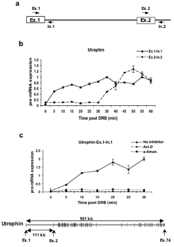
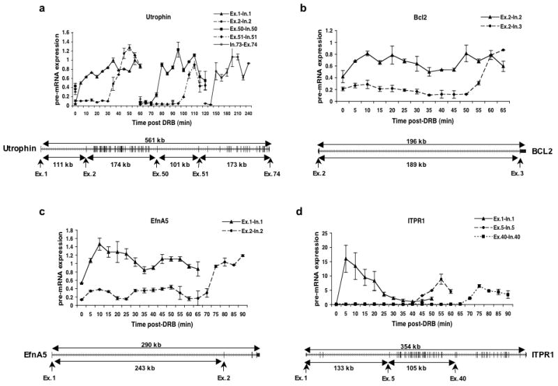
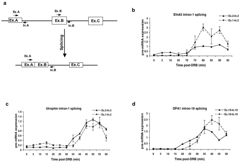
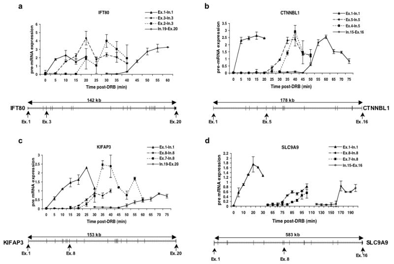
Transcription and splicing must proceed over genomic distances of hundreds of kilobases in many human genes. However, the rates and mechanisms of these processes are poorly understood. We have used the compound 5,6-dichlorobenzimidazole 1-beta-D-ribofuranoside (DRB), which reversibly blocks gene transcription in vivo, combined with quantitative RT-PCR to analyze the transcription and RNA processing of several long human genes. We found that the rate of RNA polymerase II transcription over long genomic distances is about 3.8 kb min(-1) and is similar whether transcribing long introns or exon-rich regions. We also determined that co-transcriptional pre-mRNA splicing of U2-dependent introns occurs within 5-10 min of synthesis, irrespective of intron length between 1 kb and 240 kb. Similarly, U12-dependent introns were co-transcriptionally spliced within 10 min of synthesis, confirming that these introns are spliced within the nuclear compartment. These results show that the expression of large genes is unexpectedly rapid and efficient.


Figures

Figure 1
Figure 1. DRB reversibly inhibits new transcription by RNA polymerase II
a) Schematic diagram showing the primer sets used to amplify the exon-intron junctions of exons 1 and 2 of the Utrophin gene. b) Quantitative RT-PCR was used to measure the expression levels of Utrophin pre-mRNA in the exon 1 and exon 2 regions of the gene. Cells were first treated with 100μM of DRB for 3hrs and then fresh medium was added after DRB removal. The cells were harvested at 5 minute intervals for RNA isolation and qRT-PCR. The expression values are plotted relative to the expression level of the no treatment control which is set to 1 in all experiments. c) Effect of actinomycin D (Act. D) and α-amanitin (α-Aman.) on transcription of the first exon-intron junction of the Utrophin gene after DRB treatment. Cells were treated with DRB for 3hrs and either actinomycin D or α-amanitin was added 30 minutes prior to the removal of DRB. Cells were released from DRB but maintained in the other drugs. Cells were harvested at 5 minute intervals and quantitative RT-PCR was performed as above to measure the levels of expression of the exon1-intron 1 region of the Utrophin gene. Shown at the bottom is the exon/intron structure of the Utrophin gene. Transcription is from left to right and the exons are represented as larger vertical bars.
Figure 2
Figure 2. Kinetics of RNAPII dependent transcription elongation
Cells were treated with DRB and transcription was analyzed as in Figure 1b. (a-d) Quantitative real time RT-PCR was performed using primer sets specific for different parts of the indicated genes to measure the levels of pre-mRNA expression. The gene structure is shown below each graph with arrows indicating the exon-intron junctions that were analyzed.
Figure 3
Figure 3. Kinetics of splicing of U2-dependent introns
a) Schematic diagram showing the locations of primers used to measure the levels of partially spliced mRNA. Primer sets A and B detect the unspliced pre-mRNA containing the upstream and downstream exons flanking the intron respectively. The Ex-A and In-B primer pair detects partially spliced RNA in which intron A has been spliced out but intron B remains unspliced. RNA samples from DRB treated cells were analyzed by qRT-PCR using primer pairs that detect the transcription of the exon downstream of the indicated intron or the ligated exon product of splicing. b) Transcription of exon 2 (Ex.2-In.2) and splicing of intron 1 (Ex.1-In.2) of the Ephrin A5 gene. c) Transcription of exon 2 (Ex.2-In.2) and splicing of intron 1 (Ex.1-In.2) of the Utrophin gene. d) Transcription of exon 19 (Ex.19-In.19) and splicing of intron 18 (Ex.18-In.19) of the OPA1 gene.
Figure 4
Figure 4. Kinetics of splicing of U12-dependent introns
RNA samples from DRB treated cells were analyzed by qRT-PCR using primer pairs that detect the transcription of the exon downstream of the indicated intron or the ligated exon product of splicing. a) Transcription of exons 1 (Ex.1-In.1), 3 (Ex.3-In.3) and 20 (In.19-Ex.20) as well as splicing of intron 2 (Ex.2-In.3) of the IFT80 gene. b) Transcription of exons 1 (Ex.1-In.1), 5 (Ex.5-In.5) and 16 (In.15-Ex.16) as well as splicing of intron 4 (Ex.4-In.5) of the CTNNBL1 gene. c) Transcription of exons 1 (Ex.1-In.1), 8 (Ex.8-In.8) and 20 (In.19-Ex.20) as well as splicing of intron 7 (Ex.7-In.8) of the KIFAP3 gene. d) Transcription of exons 1 (Ex.1-In.1), 8 (Ex.8-In.8) and 16 (In.15-Ex.16) as well as splicing of intron 7 (Ex.7-In.8) of the SLC9A9 gene.

Comment in

References

    1. Rasmussen EB, Lis JT. In vivo transcriptional pausing and cap formation on three Drosophila heat shock genes. Proc Natl Acad Sci U S A. 1993;90:7923–7927. - PMC - PubMed
    1. Proudfoot NJ, Furger A, Dye MJ. Integrating mRNA processing with transcription. Cell. 2002;108:501–512. - PubMed
    1. Neugebauer KM. On the importance of being co-transcriptional. J Cell Sci. 2002;115:3865–3871. - PubMed
    1. Bentley DL. Rules of engagement: co-transcriptional recruitment of pre-mRNA processing factors. Curr Opin Cell Biol. 2005;17:251–256. - PubMed
    1. Hirose Y, Manley JL. RNA polymerase II is an essential mRNA polyadenylation factor. Nature. 1998;395:93–96. - PubMed

Publication types

MeSH terms