Review
. 2009 Nov;7(11):813-21.
doi: 10.1038/nrmicro2219. Epub 2009 Oct 12.

Humans and evolutionary and ecological forces shaped the phylogeography of recently emerged diseases

Affiliations
Review

Humans and evolutionary and ecological forces shaped the phylogeography of recently emerged diseases

Paul S Keim et al. Nat Rev Microbiol. 2009 Nov.

Abstract

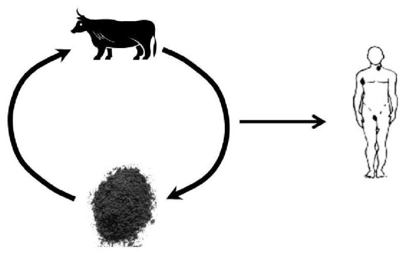
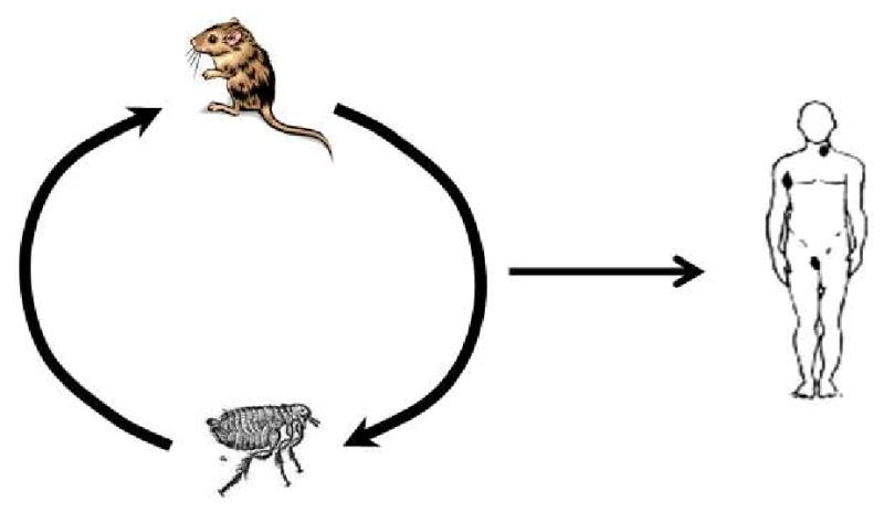
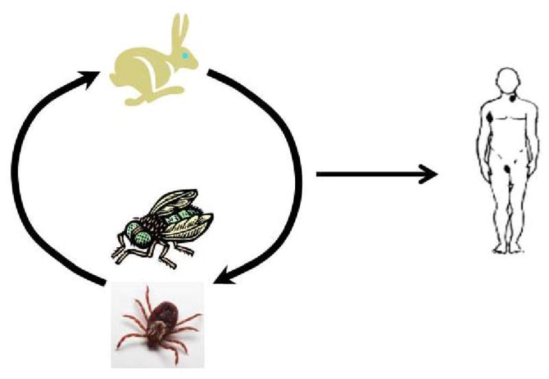
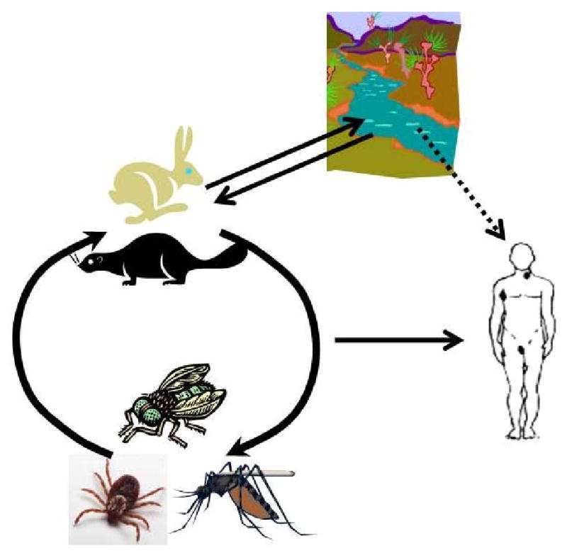
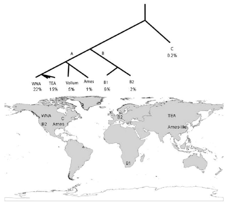
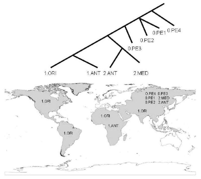
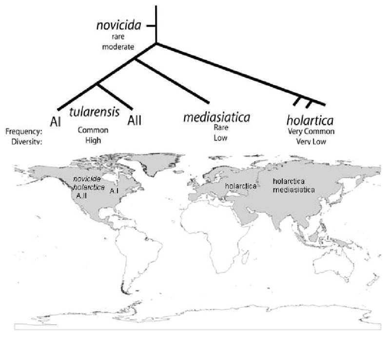
The development of human civilizations and global commerce has led to the emergence and worldwide circulation of many infectious diseases. Anthrax, plague and tularaemia are three zoonotic diseases that have been intensely studied through genome characterization of the causative species and phylogeographical analyses. A few highly fit genotypes in each species represent the causative agents for most of the observed disease cases. Together, mutational and selective forces create highly adapted pathogens, but this must be coupled with ecological opportunities for global expansion. This Review describes the distributions of the bacteria that cause anthrax, plague and tularaemia and investigates the forces that created clonal structures in these species.


Figures

Figure 1
Figure 1
Figure 1a. Infection cycle of Bacillus anthracis. Cattle and other ungulates serve as hosts for Bacillus anthracis, which causes anthrax. They inhale spores from the soil while grazing and, once ingested, these spores germinate. The bacteria then ultimately kill the host. Humans can be exposed to spores from the environment or contaminated animal products, such as meat or skin. Figure 1b. Infection cycle of Yersinia pestis. Rodents are the hosts of Yersinia pestis, the causative agent of plague, and fleas are the vectors that spread the organism between hosts. Y. pestis continuously cycles between hosts and vectors without ever persisting in the environment. Humans are most commonly infected from the bite of an infected flea but can also become infected through contact with an infected rodent or other host. Figure 1c. Infection cycle of Francisella tularensis subsp. tularensis. Type A tularaemia, caused by Francisella tularensis subsp. tularensis, is thought to be primarily a disease of lagomorphs (rabbits and hares) and arthropods. Lagomorphs are the hosts for the bacterium, and ticks and flies serve as vectors that transmit the disease between infected and naive hosts and may also serve as long-term reservoirs for the bacteria. Humans are typically infected by the bite of an infected tick or fly or by handling a diseased animal. Figure 1d. Infection cycle of Francisella tularensis subsp. holarctica. Francisella tularensis subsp. holarctica has a more complex life cycle. A variety of mammals are thought to serve as hosts, including hares, rabbits and beavers. Vectors include blood-feeding mosquitoes, tabanid flies and ticks, and ticks are also possibly long-term reservoirs for the pathogen. Water probably serves as one route of infection for mammals, which may seed aquatic areas. Humans become infected primarily by contact with infected hosts or vectors, but water may also be a source of infection.
Figure 1
Figure 1
Figure 1a. Infection cycle of Bacillus anthracis. Cattle and other ungulates serve as hosts for Bacillus anthracis, which causes anthrax. They inhale spores from the soil while grazing and, once ingested, these spores germinate. The bacteria then ultimately kill the host. Humans can be exposed to spores from the environment or contaminated animal products, such as meat or skin. Figure 1b. Infection cycle of Yersinia pestis. Rodents are the hosts of Yersinia pestis, the causative agent of plague, and fleas are the vectors that spread the organism between hosts. Y. pestis continuously cycles between hosts and vectors without ever persisting in the environment. Humans are most commonly infected from the bite of an infected flea but can also become infected through contact with an infected rodent or other host. Figure 1c. Infection cycle of Francisella tularensis subsp. tularensis. Type A tularaemia, caused by Francisella tularensis subsp. tularensis, is thought to be primarily a disease of lagomorphs (rabbits and hares) and arthropods. Lagomorphs are the hosts for the bacterium, and ticks and flies serve as vectors that transmit the disease between infected and naive hosts and may also serve as long-term reservoirs for the bacteria. Humans are typically infected by the bite of an infected tick or fly or by handling a diseased animal. Figure 1d. Infection cycle of Francisella tularensis subsp. holarctica. Francisella tularensis subsp. holarctica has a more complex life cycle. A variety of mammals are thought to serve as hosts, including hares, rabbits and beavers. Vectors include blood-feeding mosquitoes, tabanid flies and ticks, and ticks are also possibly long-term reservoirs for the pathogen. Water probably serves as one route of infection for mammals, which may seed aquatic areas. Humans become infected primarily by contact with infected hosts or vectors, but water may also be a source of infection.
Figure 2
Figure 2. Phylogeography of B. anthracis
The population structure of B. anthracis revolves around three major groups (A, B, and C). The A group is found in all parts of the world and is very common, whereas the B1, B2, and C groups are relative rare and mostly restricted to subcontinental locations. Highly successful clonal lineage exist even within the A-radiation.
Figure 3
Figure 3. Phylogeography of Y. pestis
There are currently three major branches within the Y. pestis phylogeny (0, 1, and 2), with eight molecular groups. Six of these groups are only found in Central Asia, where Y. pestis likely evolved from its Y. pseudotuberculosis ancestor. The 1.ANT group is found in just a small region of Africa. The 1.ORI group is widely distributed and currently occurs on all continents except Australia and Antarctic. This group was spread around the world within the last 150 years by humans during the third plague pandemic.
Figure 4
Figure 4. Phylogeography of F. tularensis
The subspecies of F. tularensis are clonal, with differential success on a global scale. F. t. subsp. tularensis is differentiated into two subpopulations locally restricted to parts of North America. The subspecies novicida and mediasiatica are the rarest and restricted mostly to North America and Central Asia, respectively. F.t. subsp. holarctica appears to be highly fit and highly mobile with a circumpolar distribution in the northern hemisphere.

Similar articles

Cited by

References

    1. Pearson T, et al. Phylogenetic discovery bias in Bacillus anthracis using single-nucleotide polymorphisms from whole-genome sequencing. PNAS. 2004;101:13536–13541. - PMC - PubMed
    1. Girard JM, et al. Differential plague-transmission dynamics determine Yersinia pestis population genetic structure on local, regional, and global scales. Proc Natl Acad Sci U S A. 2004;101:8408–13. - PMC - PubMed
    1. Vogler AJ, et al. Effect of repeat copy number on variable-number tandem repeat mutations in Escherichia coli O157:H7. J Bacteriol. 2006;188:4253–63. - PMC - PubMed
    1. Price LB, et al. In vitro selection and characterization of Bacillus anthracis mutants with high-level resistance to ciprofloxacin. Antimicrob Agents Chemother. 2003;47:2362–5. - PMC - PubMed
    1. Vogler AJ, et al. Molecular analysis of rifampin resistance in Bacillus anthracis and Bacillus cereus. Antimicrob Agents Chemother. 2002;46:511–3. - PMC - PubMed

Publication types

MeSH terms