. 2009 Oct;47(10):1103-9.
doi: 10.1007/s11517-009-0537-x.

Time domain algorithm for accelerated determination of the first order moment of photo current fluctuations in high speed laser Doppler perfusion imaging

Affiliations

Time domain algorithm for accelerated determination of the first order moment of photo current fluctuations in high speed laser Doppler perfusion imaging

Matthijs Draijer et al. Med Biol Eng Comput. 2009 Oct.

Abstract

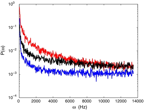
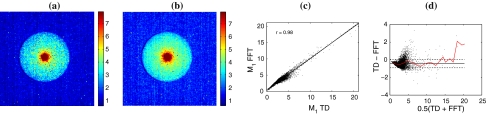
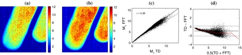
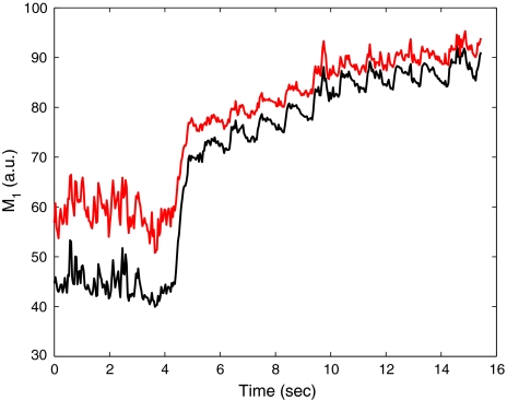
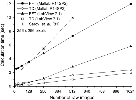
Advances in optical array sensor technology allow for the real time acquisition of dynamic laser speckle patterns generated by tissue perfusion, which, in principle,allows for real time laser Doppler perfusion imaging(LDPI). Exploitation of these developments is enhanced with the introduction of faster algorithms to transform photo currents into perfusion estimates using the first moment of the power spectrum. A time domain (TD)algorithm is presented for determining the first-order spectral moment. Experiments are performed to compare this algorithm with the widely used Fast Fourier Transform(FFT). This study shows that the TD-algorithm is twice as fast as the FFT-algorithm without loss of accuracy.Compared to FFT, the TD-algorithm is efficient in terms of processor time, memory usage and data transport.


Figures

Fig. 1
Fig. 1
The power spectra of Delrin (blue line), Intralipid (red line) and human tissue (black line) averaged over an area of 5 × 5 pixels
Fig. 2
Fig. 2
128 × 128 pixel images of the perfusion obtained from a cylindrical piece of Delrin of 40 mm in diameter with a hole of 4 mm in diameter, and placed on green surgery paper; the hole was filled with IntraLipid 20% (imaged area approx. 7 × 7 cm). Perfusion plots with a the TD-algorithm, b the FFT-algorithm and c scatter plot of FFT-results versus TD-results (solid line: linear fit; dashed line: TD=FFT) and d Bland–Altman plot of both perfusion plots, the red line indicates the average value of TD-FFT over horizontal intervals of 1,000
Fig. 3
Fig. 3
128 × 128 pixel images of the perfusion of the finger tips of the hand of a volunteer (imaged area approx. 4 × 4 cm). a Perfusion plot with the TD-algorithm, b perfusion plot with the FFT-algorithm, c the scatter plot of FFT-results versus TD-results (solid line: linear fit; dashed line: TD=FFT) and d Bland–Altman plot of both perfusion plots; the red line indicates the average value of TD-FFT over horizontal intervals of 1,000
Fig. 4
Fig. 4
Perfusion as a function of time during occlusion and reperfusion calculated with the FFT-algorithm (black line), and the TD-algorithm (red line)
Fig. 5
Fig. 5
Calculation time for the TD-algorithm and the FFT-algorithm for an image of 256 × 256 pixels for two software packages, as a function of the number of raw images

References

    1. Aizu Y, Asakura T. Bio-speckle phenomena and their application to the evaluation of blood flow. Opt Laser Technol. 1991;23(4):205–219. doi: 10.1016/0030-3992(91)90085-3. - DOI
    1. Bland JM, Altman DG. Statistical methods for assessing agreement between two methods of clinical measurement. Lancet. 1986;1:307–310. - PubMed
    1. Bland JM, Altman DG. Comparing methods of measurement: why plotting difference against standard method is misleading. Lancet. 1995;346:1085–1087. doi: 10.1016/S0140-6736(95)91748-9. - DOI - PubMed
    1. Bonner R, Nossal R. Model for laser Doppler measurements of blood flow in tissue. Appl Opt. 1981;20(12):2097–2107. doi: 10.1364/AO.20.002097. - DOI - PubMed
    1. Bray R, Forrester K, Leonard C, McArthur R, Tulip J, Lindsay R. Laser Doppler imaging of burn scars: a comparison of wavelength and scanning methods. Burns. 2003;29(3):199–206. doi: 10.1016/S0305-4179(02)00307-8. - DOI - PubMed

Publication types

LinkOut - more resources