. 2009 Dec;50(6):1861-70.
doi: 10.1002/hep.23214.

Sorafenib inhibits signal transducer and activator of transcription-3 signaling in cholangiocarcinoma cells by activating the phosphatase shatterproof 2

Affiliations

Sorafenib inhibits signal transducer and activator of transcription-3 signaling in cholangiocarcinoma cells by activating the phosphatase shatterproof 2

Boris R A Blechacz et al. Hepatology. 2009 Dec.

Abstract

The Janus kinase/signal transducer and activator of transcription (JAK/STAT) pathway is one of the key signaling cascades in cholangiocarcinoma (CCA) cells, mediating their resistance to apoptosis. Our aim was to ascertain if sorafenib, a multikinase inhibitor, may also inhibit JAK/STAT signaling and, therefore, be efficacious for CCA. Sorafenib treatment of three human CCA cell lines resulted in Tyr(705) phospho-STAT3 dephosphorylation. Similar results were obtained with the Raf-kinase inhibitor ZM336372, suggesting sorafenib promotes Tyr(705) phospho-STAT3 dephosphorylation by inhibiting Raf-kinase activity. Sorafenib treatment enhanced an activating phosphorylation of the phosphatase SHP2. Consistent with this observation, small interfering RNA-mediated knockdown of phosphatase shatterproof 2 (SHP2) inhibited sorafenib-induced Tyr(705) phospho-STAT3 dephosphorylation. Sorafenib treatment also decreased the expression of Mcl-1 messenger RNA and protein, a STAT3 transcriptional target, as well as sensitizing CCA cells to tumor necrosis factor-related apoptosis-inducing ligand (TRAIL)-mediated apoptosis. In an orthotopic, syngeneic CCA model in rats, sorafenib displayed significant tumor suppression resulting in a survival benefit for treated animals. In this in vivo model, sorafenib also decreased tumor Tyr(705) STAT3 phosphorylation and increased tumor cell apoptosis.

Conclusion: Sorafenib accelerates STAT3 dephosphorylation by stimulating phosphatase SHP2 activity, sensitizes CCA cells to TRAIL-mediated apoptosis, and is therapeutic in a syngeneic rat, orthotopic CCA model that mimics human disease.


Conflict of interest statement

Potential conflict of interest: Nothing to report.

Figures

Fig. 1
Fig. 1
Sorafenib interferes with IL-6 signaling by loss of Tyr705 phospho-STAT3. HuCCT-1 cells were treated in the presence or absence of sorafenib (10 µM) for 12 hours. (A) Inhibition of known STAT3 activating signaling pathways. HuCCT-1 cells were treated with JAK-1/2, ERK-1/2, Src and EGFR inhibitors at the indicated doses for 4 hours followed by immunoblot analysis of whole-cell lysates for Tyr705 phospho-STAT3. (B) Effect of sorafenib on IL-6 secretion (left panel) and on the IL-6 receptor complex (right panel). IL-6 secretion by HuCCT-1 cells into the supernatant was quantified by enzyme-linked immunosorbent assay. The IL-6 receptor complex was evaluated by SDS-PAGE and immunoblot analysis of protein (50 µg) obtained from whole-cell lysates. Bands were cut and combined (separated by dotted line) from the same radiograph. (C) Immunoblot analysis of sorafenib on total and Tyr705 phospho-STAT3 in HuCCT-1 cells. (D) Immunoblot analysis of sorafenib on total and Tyr705 phospho-STAT3 in KMCH-1 and Mz-Cha-1 cells. (E) Immunoblot analysis of the Raf-inhibitor ZM336372 on total and Tyr705 phospho-STAT3 in HuCCT-1 cells.
Fig. 2
Fig. 2
Sorafenib inhibits transcriptional activity of STAT3 and results in loss of cellular Mcl-1. HuCCT-1 cells were treated with sorafenib (10 µM) followed by analysis of STAT3 cellular localization and Mcl-1 expression. (A) Sorafenib-treated and untreated cells were subjected to immunocytochemistry for anti-STAT3 antibody and analyzed by confocal microscopy. DAPI staining and overlay demonstrate nuclear accumulation of STAT3 in untreated, but not in sorafenib-treated cells. (B) Percentages of cells with nuclear STAT3 fluorescence were quantitated by imaging total nuclei labeled with DAPI (10 µg/mL). (C) Immunoblot analysis for Mcl-1 protein expression. As assessed by immunoblot analysis, Mcl-1 protein levels decreased as early as 2 hours following sorafenib treatment. (D) Quantitative real-time polymerase chain reaction for Mcl-1 mRNA. Cellular RNA was isolated from sorafenib-treated and untreated cells 2 hours after treatment, and analyzed for Mcl-1 mRNA as described in Materials and Methods section. Note the significant reduction (P < 0.05) in Mcl-1 mRNA levels.
Fig. 3
Fig. 3
Sorafenib-induced Tyr705 STAT3 dephosphorylation and Mcl-1 reduction is mediated by Raf-kinase inhibition and depends on phosphatase activity. (A) HuCCT-1 cells were treated with sorafenib (10 µM) in the absence or presence of the phosphatase inhibitor pervanadate at the indicated concentrations. Four hours after treatment, whole-cell lysates were obtained and 50 µg of proteins analyzed by immunoblot analysis for Tyr705 phospho-STAT3 and total STAT3. (B) HuCCT-1 cells were cotreated with the b-Raf and c-Raf inhibitor ZM336372 (0.1 µM) and the phosphatase inhibitor pervanadate at the indicated concentrations. Four hours after treatment, whole-cell lysates were obtained and 50 µg of proteins analyzed by immunoblot analysis for Tyr705 phosphoSTAT3 and total STAT3. (C) HuCCT-1 cells were cotreated with sorafenib (10 µM) and the phosphatase inhibitor pervanadate (100 µM). At 12 hours after treatment, whole-cell lysates were obtained and 50 µg protein was analyzed by immunoblot analysis for Tyr705 phospho-STAT3, total STAT3, Mcl-1, and actin.
Fig. 4
Fig. 4
Sorafenib-induced Tyr705 STAT3 dephosphorylation is mediated by phosphatase SHP2. (A) HuCCT-1 cells were treated for 2 hours with sorafenib at the indicated concentrations followed by immunoblot analysis for Tyr580 phospho-SHP2, total STAT3, and actin. The relative increase in Tyr580 phospho-SHP2 was quantified by densitometric analysis. (B) HuCCT-1 cells were transfected with scrambled or SHP2-specific siRNA followed by treatment with sorafenib (10 µM) for 4 hours. Subsequently, whole-cell lysates were obtained and 50 µg of proteins analyzed by immunoblot analysis for Tyr705 phospho-STAT3, total STAT3 and SHP2. Bands were cut and combined (separated by dotted line) from the same radiograph.
Fig. 5
Fig. 5
Sorafenib sensitizes CCA cells to TRAIL-induced apoptosis. Effects of sorafenib on KMCH-1 (left) and HuCCT-1 cells (right). Shown are average values with standard deviations (n = 3). (A) Cells were treated with sorafenib (10 µM) and/or TRAIL (2.5 ng/mL) followed DAPI-staining and fluorescence microscopic analysis. (B) Cells were treated for 2 hours with sorafenib (10 µM), followed by addition of TRAIL (2.5 ng/mL). Note the significant (P < 0.05) increase in caspase 3/7 activity in cells treated with combination treatment. (C) Dose-dependency of sorafenib mediated sensitization to TRAIL-induced apoptosis. HuCCT-1 and KMCH-1 cells were treated with 2.5, 5, and 10 µM of sorafenib in the presence of 2.5 ng/mL TRAIL followed by quantification of apoptotic nuclei using DAPI-staining and fluorescence microscopic analysis. (D) Untreated, scramble siRNA and SHP2 siRNA transfected HuCCT-1 cells were treated with sorafenib (10 µM) and TRAIL (2.5 ng/mL) followed DAPI-staining and fluorescence microscopic analysis.
Fig. 6
Fig. 6
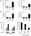
Sorafenib monotherapy induces apoptosis in TFK-1 cells. TFK-1 cells were treated for 6 hours in the presence or absence of Sorafenib (10 µM), followed by analysis for apoptosis induction and Tyr705 p-STAT3 dephosphorylation. (A) Caspase-3/7 activity (left) and quantification of apoptotic nuclei using DAPI-staining and fluorescence microscopic analysis (right) following sorafenib treatment. (B) Immunoblot analysis for Tyr705 phospho-STAT3 and total STAT3 in whole-cell lysates obtained after 6 hours incubation in the presence or absence of sorafenib. The relative decrease in Tyr705 p-STAT3 was quantified by densitometry of three separate experiments.
Fig. 7
Fig. 7
Sorafenib is tumor suppressive in a rat, syngeneic, orthotopic rodent model of CCA. BDEneu cells were implanted in left hepatic duct-ligated, male Fischer rats. One week after surgery, animals were treated daily with sorafenib (10 mg/kg body weight) or vehicle by intraperitoneal injection for 7 days (n = 9 animals/treatment group). One week after completion of treatment, all vehicle-treated animals had died, and remaining animals were sacrificed and analyzed. (A) Macroscopic analysis of hepatic tumors. Depicted are photographs of livers of normal, and sorafenib-treated or vehicle-treated tumor-bearing rats. There was almost complete replacement of the liver by tumor tissue in vehicle-treated versus sorafenib-treated animals resulting in significant increase of liver weight in tumor-bearing rats versus normal rats. (B) Comparison of survival at day 21 after tumor implantation. None of the vehicle-treated animals had survived at day 21 versus 60% animal survival in the sorafenib treatment group. (C) Immunohistochemical analysis for Tyr705 phospho-STAT3 in BDEneu tumor-tissue of sorafenib-treated and vehicle-treated rats. (D) TUNEL assay for evaluation and quantification of apoptosis in BDEneu tumor sections of sorafenib-treated and vehicle-treated animals.

Similar articles

Cited by

References

    1. Blechacz B, Gores GJ. Cholangiocarcinoma: advances in pathogenesis, diagnosis, and treatment. Hepatology. 2008;48:308–321. - PMC - PubMed
    1. Bollrath J, Phesse TJ, von Burstin VA, Putoczki T, Bennecke M, Bateman T, et al. gp130-mediated Stat3 activation in enterocytes regulates cell survival and cell-cycle progression during colitis-associated tumorigenesis. Cancer Cell. 2009;15:91–102. - PubMed
    1. Grivennikov S, Karin E, Terzic J, Mucida D, Yu GY, Vallabhapurapu S, et al. IL-6 and Stat3 are required for survival of intestinal epithelial cells and development of colitis-associated cancer. Cancer Cell. 2009;15:103–113. - PMC - PubMed
    1. Yu H, Jove R. The STATs of cancer–new molecular targets come of age. Nat Rev Cancer. 2004;4:97–105. - PubMed
    1. Aggarwal BB, Sethi G, Ahn KS, Sandur SK, Pandey MK, Kunnumakkara AB, et al. Targeting signal-transducer-and-activator-of-transcription-3 for prevention and therapy of cancer: modern target but ancient solution. Ann N Y Acad Sci. 2006;1091:151–169. - PubMed

Publication types

MeSH terms