. 2009 Nov 4;131(43):15729-38.
doi: 10.1021/ja903879w.

Structural examination of the transient 3-aminotyrosyl radical on the PCET pathway of E. coli ribonucleotide reductase by multifrequency EPR spectroscopy

Affiliations

Structural examination of the transient 3-aminotyrosyl radical on the PCET pathway of E. coli ribonucleotide reductase by multifrequency EPR spectroscopy

Mohammad R Seyedsayamdost et al. J Am Chem Soc. .

Abstract

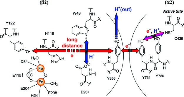
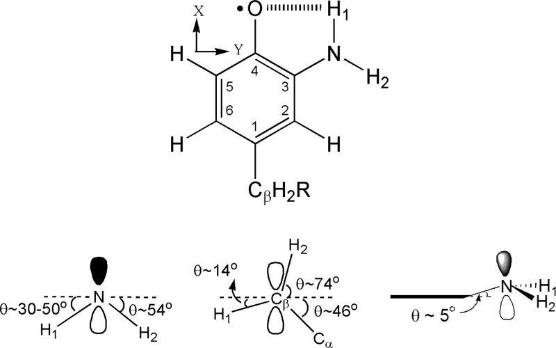
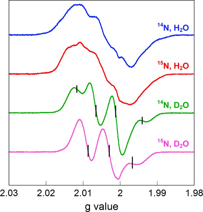
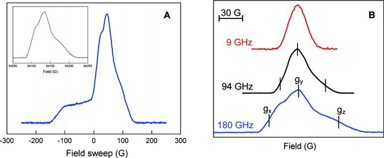
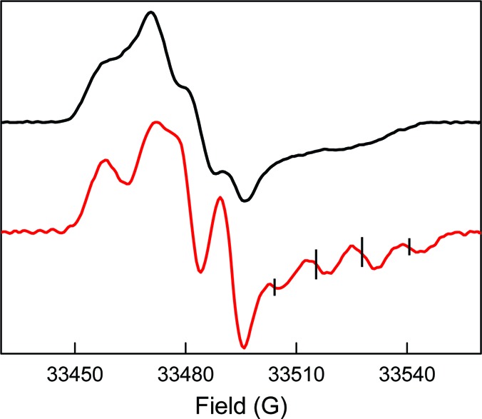
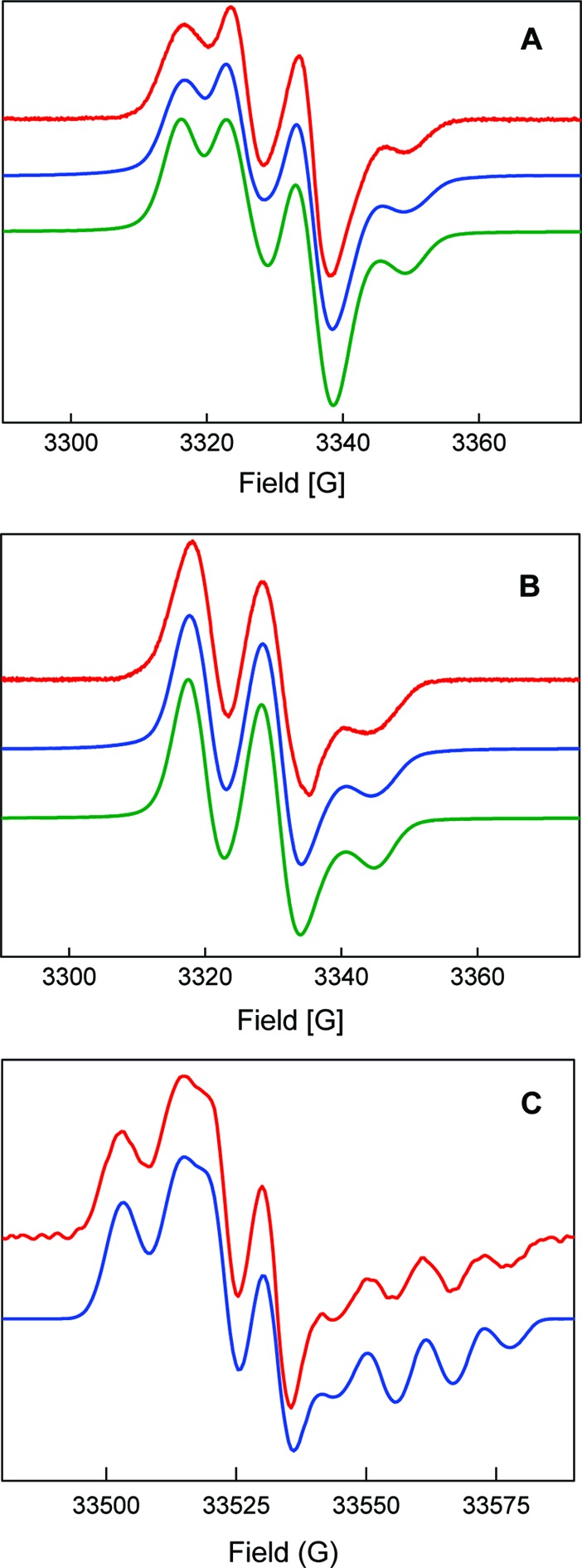
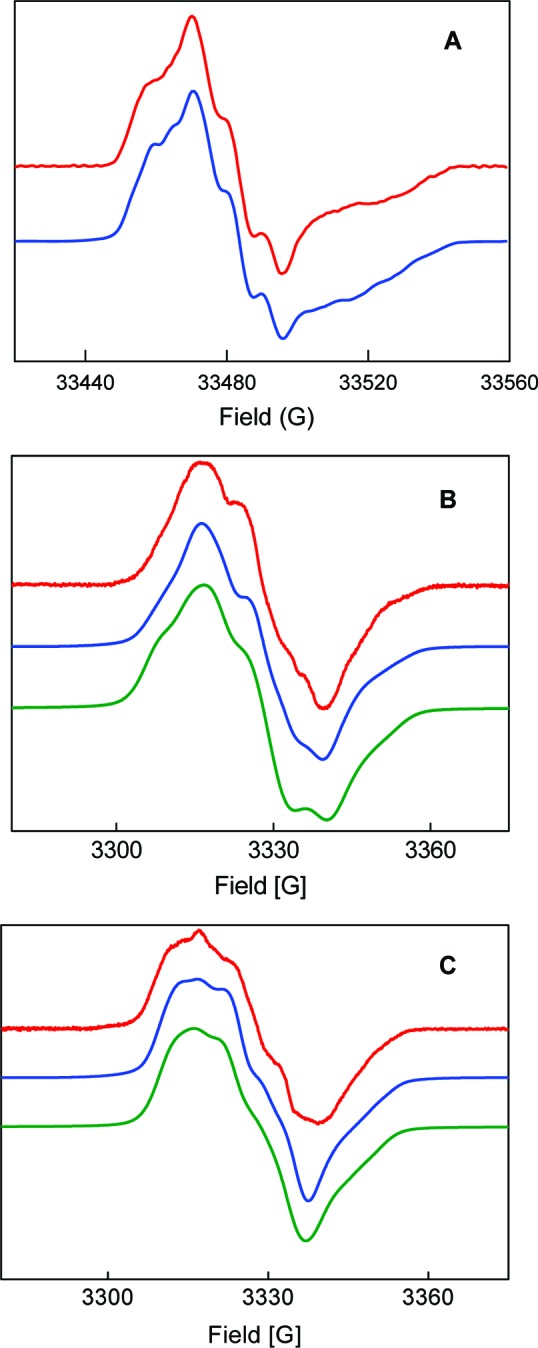
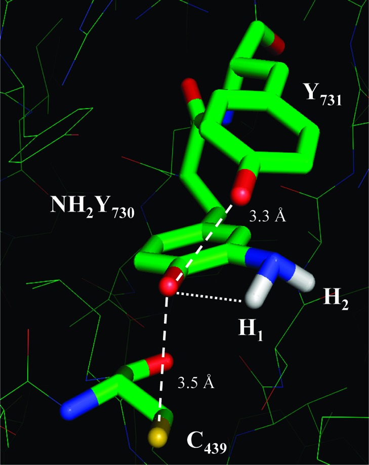
E. coli ribonucleotide reductase (RNR) catalyzes the conversion of nucleotides to deoxynucleotides, a process that requires long-range radical transfer over 35 A from a tyrosyl radical (Y(122)*) within the beta2 subunit to a cysteine residue (C(439)) within the alpha2 subunit. The radical transfer step is proposed to occur by proton-coupled electron transfer via a specific pathway consisting of Y(122) --> W(48) --> Y(356) in beta2, across the subunit interface to Y(731) --> Y(730) --> C(439) in alpha2. Using the suppressor tRNA/aminoacyl-tRNA synthetase (RS) methodology, 3-aminotyrosine has been incorporated into position 730 in alpha2. Incubation of this mutant with beta2, substrate, and allosteric effector resulted in loss of the Y(122)* and formation of a new radical, previously proposed to be a 3-aminotyrosyl radical (NH(2)Y*). In the current study [(15)N]- and [(14)N]-NH(2)Y(730)* have been generated in H(2)O and D(2)O and characterized by continuous wave 9 GHz EPR and pulsed EPR spectroscopies at 9, 94, and 180 GHz. The data give insight into the electronic and molecular structure of NH(2)Y(730)*. The g tensor (g(x) = 2.0052, g(y) = 2.0042, g(z) = 2.0022), the orientation of the beta-protons, the hybridization of the amine nitrogen, and the orientation of the amino protons relative to the plane of the aromatic ring were determined. The hyperfine coupling constants and geometry of the NH(2) moiety are consistent with an intramolecular hydrogen bond within NH(2)Y(730)*. This analysis is an essential first step in using the detailed structure of NH(2)Y(730)* to formulate a model for a PCET mechanism within alpha2 and for use of NH(2)Y in other systems where transient Y*s participate in catalysis.


Figures

Figure 1
Figure 1
Proposed radical initiation pathway in E. coli class Ia RNR. Radical transfer occurs by orthogonal PCET within β2, where long-distance ET within the pathway is coupled to short-distance off-pathway PT, and by collinear PCET (or hydrogen atom transfer) within α2. Note that the position of Y356 is structurally unknown.
Figure 2
Figure 2
Structure of NH2Y730•. (Top) Numbering scheme (inside numbers), axis system, and the intramolecular H-bond represented by dashed lines. (Bottom) NH2Y730• viewed along the C3-N and the phenol C2−C3 bond within the aromatic plane. Dihedral angles for the amino protons (left) and the Cβ-protons (middle) of NH2Y730• with respect to the phenol plane, which is indicated by dashed lines. (Right) Tetrahedral nature of the NH2 moiety and the 5° tilt of the C3−N bond away from the aromatic plane, which is indicated by a bold line.
Figure 3
Figure 3
Normalized EPR spectra of isotopically substituted NH2Y730•. Spectra were acquired with Y730NH2Y-α2/β2 containing [14N]-NH2Y or [15N]-NH2Y in H2O or D2O assay buffer in the presence of CDP/ATP. The ticks on the deuterated traces indicate the positions of the quartet and triplet peaks with [14N]- and [15N]-NH2Y730•, respectively. See text for details.
Figure 4
Figure 4
Multifrequency EPR spectra of NH2Y730• in the presence of CDP/ATP in H2O assay buffer. (A) 180 GHz spectrum at 6 K consisting of Y122• and NH2Y730• signals. Inset: 180 GHz spectrum at 70 K containing only the NH2Y730• signal. (B) Spectrum of NH2Y730• at 9, 94, and 180 GHz, demonstrating the higher g resolution with increasing frequency. The spectrum at 180 GHz yields g values of gx = 2.00520, gy = 2.00420, and gz = 2.00220. The error in these measurements was ±0.0001.
Figure 5
Figure 5
94 GHz pulsed derivative EPR spectra of NH2Y730• in H2O (black trace) and D2O (red trace). Experimental parameters: echo sequence: π/2 = 32 ns, τ = 248 ns (in H2O), τ = 260 ns (in D2O), π = 64 ns; trep = 5 ms; shots/point = 50; scans = 700; T = 70 K. The ticks on the deuterated trace indicate the positions of the quartet. See text for details.
Figure 6
Figure 6
Simulations (blue = SimFonia; green = EasySpin) of the first derivative EPR spectra (red) of [14N]-NH2Y730• and [15N]-NH2Y730• in D2O assay buffer. (A) [14N]-NH2Y730• at 9 GHz. (B) [15N]-NH2Y730• at 9 GHz. (C) [14N]-NH2Y730• at 94 GHz. See Table 1 for parameters. The simulations obtained with EasySpin and the parameters of Table 1 are in agreement with those from SimFonia.
Figure 7
Figure 7
Simulations (blue = SimFonia; green = EasySpin) of the first derivative EPR spectra (red) of [14N]-NH2Y730• and [15N]-NH2Y730• in H2O assay buffer. (A) [14N]-NH2Y730• at 94 GHz. (B) [14N]-NH2Y730• at 9 GHz. (C) [15N]-NH2Y730• at 9 GHz. See Table 1 for parameters. The simulations with EasySpin appear slightly broader due to the effect of forbidden transitions.
Figure 8
Figure 8
Model of the Y731−NH2Y730−C439 pathway in NH2Y730-α2. NH2Y has been modeled into residue 730 of α2 using the coordinates from ref (14). The pz orientation and Cβ- and amino proton dihedral angles are based on results herein. The distance between the NH1 and the phenol O is indicated. The distances between the essential residues around NH2Y730 are illustrated as discussed in the text.

Similar articles

Cited by

References

    1. Stubbe J.; van der Donk W. A. Chem. Rev. 1998, 98, 705. - PubMed
    1. Jordan A.; Reichard P. Annu. Rev. Biochem. 1998, 67, 71. - PubMed
    1. Brown N. C.; Canellakis Z. N.; Lundin B.; Reichard P.; Thelander L. Eur. J. Biochem. 1969, 9, 561. - PubMed
    1. Thelander L. J. Biol. Chem. 1973, 248, 4591. - PubMed
    1. Stubbe J. J. Biol. Chem. 1990, 265, 5329. - PubMed

Publication types

MeSH terms

LinkOut - more resources