. 2010 Apr;25(4):769-81.
doi: 10.1359/jbmr.091017.

Estrogens attenuate oxidative stress and the differentiation and apoptosis of osteoblasts by DNA-binding-independent actions of the ERalpha

Affiliations

Estrogens attenuate oxidative stress and the differentiation and apoptosis of osteoblasts by DNA-binding-independent actions of the ERalpha

Maria Almeida et al. J Bone Miner Res. 2010 Apr.

Abstract

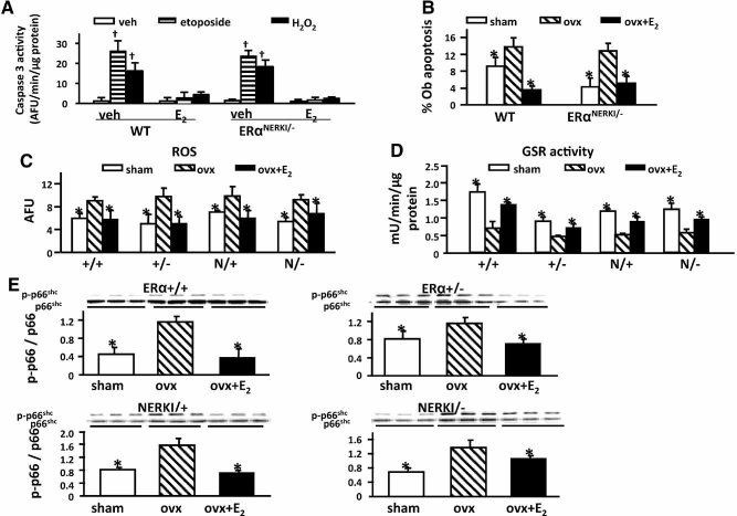
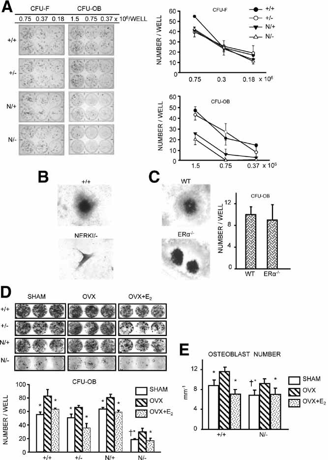
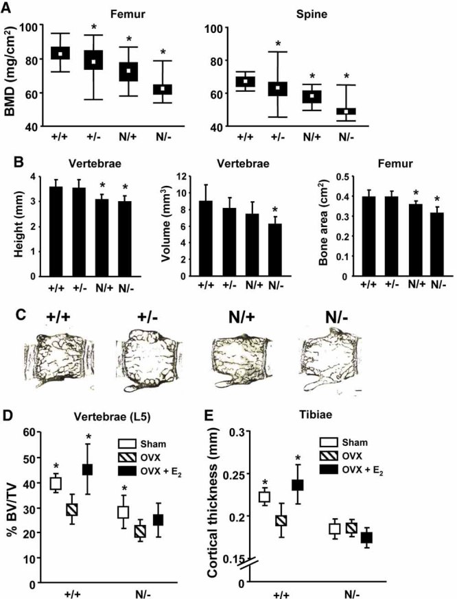
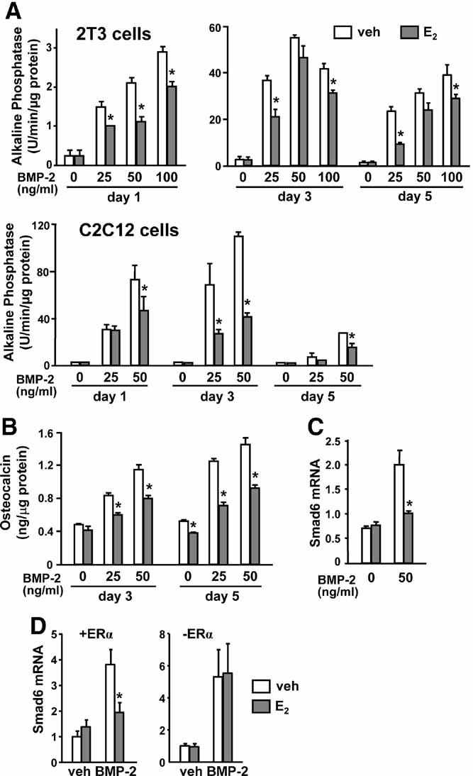
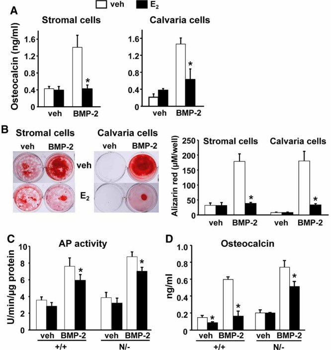
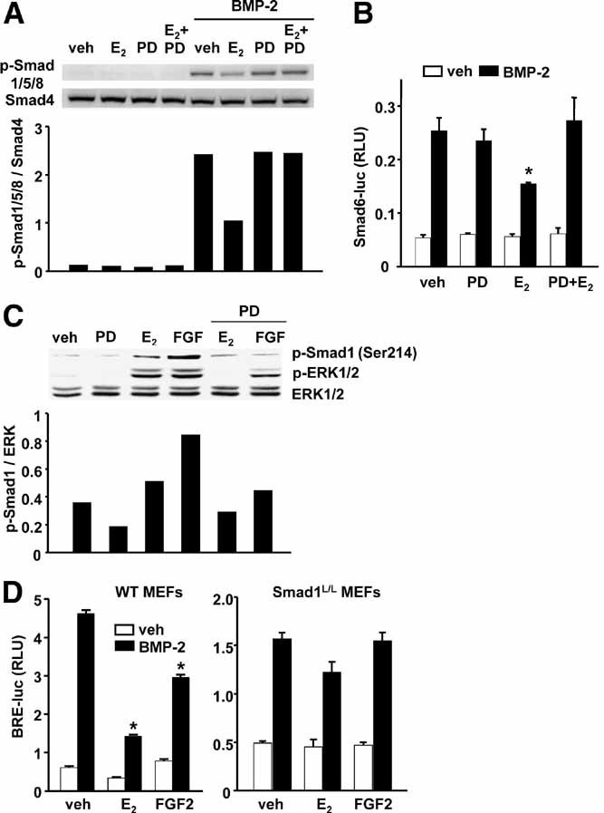
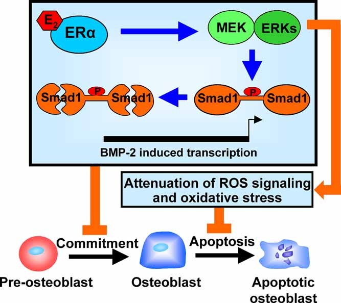
Estrogens diminish oxidative stress in bone and bone marrow, attenuate the generation of osteoblasts, and decrease the prevalence of mature osteoblast apoptosis. We have searched for the molecular mechanism of these effects using as tools a mouse model bearing an estrogen receptor alpha (ERalpha) knock-in mutation that prevents binding to DNA (ERalpha(NERKI/-)) and several osteoblast progenitor cell models expressing the wild-type ERalpha or the ERalpha(NERKI/-). We report that the ability of estrogens to diminish the generation of reactive oxygen species, stimulate the activity of glutathione reductase, and decrease the phosphorylation of p66(shc), as well as osteoblastogenesis and osteoblast number and apoptosis, were fully preserved in ERalpha(NERKI/-) mice, indicating that the DNA-binding function of the ERalpha is dispensable for all these effects. Consistent with the attenuation of osteoblastogenesis in this animal model, 17beta-estradiol attenuated bone morphogenetic protein 2 (BMP-2)-induced gene transcription and osteoblast commitment and differentiation in murine and human osteoblastic cell lines. Moreover, 17beta-estradiol attenuated BMP-2-induced differentiation of primary cultures of calvaria- or bone marrow-derived osteoblastic cells from ERalpha(NERKI/-) mice as effectively as in cells from wild-type littermates. The inhibitory effect of the hormone on BMP-2 signaling resulted from an ERalpha-mediated activation of ERKs and the phosphorylation of Smad1 at the linker region of the protein, which leads to proteasomal degradation. These results illustrate that the effects of estrogens on oxidative stress and the birth and death of osteoblasts do not require the binding of ERalpha to DNA response elements, but instead they result from the activation of cytoplasmic kinases.


Figures

Fig. 1
Fig. 1
E2 effects on osteoblast apoptosis, ROS levels, GSR activity, and p66shc phosphorylation are preserved in ERαNERKI/− mice. (A) Caspase-3 activity in calvaria cells isolated from wild-type (WT) control or ERαNERKI/− mice pretreated with vehicle (veh) or E2 (10−8 M) for 1 hour. Cells then were treated with veh, etoposide (5 × 10⁻⁵ M), or H2O2 (5 × 10⁻⁵ M) for 6 hours. (B) Osteoblast apoptosis determined by in situ end labeling in sections of undecalcified vertebral bone (L1–5) of 5-month-old WT or ERαNERKI/− mice sham operated or OVX. OVX animals received veh or E2 replacement for 6 weeks (n = 4 to 6 animal/group). (C) ROS levels and (D) GSR activity in bone marrow cells from 5-month-old WT, ERα+/−, ERαNERKI/+, and ERαNERKI/− mice sham operated or OVX and treated as described in B (n = 4 animals/group). (E) Phosphorylation of p66shc determined by Western blot analyses in vertebral lysates from the same mice as in C; each lane represents one animal. p < .05 versus respective vehicle; *p < .05 versus OVX.
Fig. 2
Fig. 2
Osteoblastogenesis is decreased in ERαNERKI/− mice. (A) CFU-Fs or CFU-OBs in the bone marrow from femora of intact mice of the indicated genotypes. Cells from three mice were pooled and plated in duplicate at three different densities for each genotype. CFU-Fs were stained for alkaline phosphatase after 10 days, and CFU-OBs were stained with von Kossa to detect mineral after 25 days (left panel). The graphs on the right represent the quantification of CFUs depicted on the left. (B) Photomicrographs show representative CFU-OB colonies (50×) obtained from WT (ERα+/+) or ERαNERKI/− mice or (C) from WT or ERα−/− mice; +/+ and WT refer to the respective littermate controls, as detailed in “Materials and Methods.” The graph on the right represents the quantification of CFU-OBs depicted on the right. (D) CFU-OBs obtained from femora of mice used in the experiment described in Fig. 1C. Cells from three mice were pooled and plated in triplicate at 106 cells per well. The graph on the bottom represents the quantification of CFU-OBs depicted on the top. (E) Osteoblast numbers on longitudinal undecalcified sections of L1–4 vertebrae from mice used in the experiment described in Fig. 1C (n = 6 animals per group). *p < .05 versus OVX; p < .05 versus +/+ sham.
Fig. 3
Fig. 3
ERαNERKI/− mice have low bone mass and lose cancellous bone following OVX. (A) Femoral and spinal BMD determined in 5-month-old mice of the indicated genotypes using Piximus (n = 26 to 31 per group). The box indicates the interquartile range around the median (white square inside the box), and the vertical lines represent values plus or minus 1.5 times the interquartile range. (B) Vertebral dimensions and femoral bone area were determined in mice described in A using a micrometer and Piximus, respectively (n = 7 to 11 per group). (C) Representative photomicrographs of lumbar vertebrae from three mice per genotype, unstained and viewed at 25× without coverslips. (D) Vertebral cancellous bone volume (BV/TV) and (E) cortical thickness of tibiae as determined by µCT in ERα+/+ or ERαNERKI/− mice from the experiment described in 1C (n = 7 to 11 per group). *p < .05 versus +/+; p < .05 versus OVX.
Fig. 4
Fig. 4
E2 attenuates BMP-2-induced osteoblast differentiation and target gene expression. (A) Alkaline phosphatase activity in 2T3 and C2C12 cells incubated with vehicle or the indicated doses of BMP-2 in the presence of E2 (10−8 M) for 1, 3, and 5 days. (B) Osteocalcin levels in the culture medium of C2C12 cells treated as described earlier for 3 and 5 days. (C) Smad6 mRNA levels determined by quantitative RT-PCR in C2C12 cells treated as described earlier for 3 days or (D) in U2OS cells pretreated with vehicle or E2 (10−7 M) for 1 hour and treated with or without BMP-2 (100 ng/mL) for 2 hours in the presence (left panel) or absence (right panel) of doxycycline. *p < .05 versus BMP-2.
Fig. 5
Fig. 5
BMP-2-induced osteoblast differentiation and mineralization is decreased by E2 in primary osteoblastic cell cultures. (A) Osteocalcin levels in the medium and (B) mineralized matrix visualized following alizarin red staining (left panel) and quantified after extraction (right panel) of bone marrow– or calvaria-derived cells treated with BMP-2 (25 ng/mL), E2 (10−8 M), or E2 and BMP-2 in the presence of β-glycerophosphate for 18 days. (C) Alkaline phosphatase activity in parallel cultures of calvaria-derived osteoblasts from WT controls (ERα+/+) and ERαNERKI/− mice incubated with BMP-2 (25 ng/mL), E2 (10−8 M), or E2 and BMP-2 for 3 days. (D) Osteocalcin levels in the culture medium of same cells as in C treated as described for 10 days. *p < .05 versus BMP-2 alone. The same results were reproduced in a second experiment.
Fig. 6
Fig. 6
ERKs are required for the inhibitory actions of E2 on BMP-2-induced transcription. (A) Smad1/5/8 phosphorylation by Western blot analysis in C2C12 cells pretreated with vehicle, PD98049 (25 µM), or E2 (10−8 M) for 1 hour followed by vehicle or BMP-2 (50 ng/mL) for 1 hour. Bar graph represents the quantification of the intensity of the bands with an imaging system. (B) Luciferase activity in C2C12 cells transfected with a Smad6-Luc reporter construct and pretreated as in A, followed by treatment with vehicle or BMP-2 for 24 hours. (C) ERK1/2 and Smad1 (Ser214) phosphorylation by Western blot analysis in C2C12 cells pretreated with vehicle or PD98049 for 1 hour followed by E2 or FGF2 (5 ng/mL) for 15 minutes in serum-free medium. (D) Luciferase activity in MEFs transfected with a BMP response element reporter construct and pretreated for 1 hour with E2 or FGF2, followed by treatment with vehicle or BMP-2 (25 ng/mL) for 24 hours. *p < .05 versus BMP-2 alone.
Fig. 7
Fig. 7
DNA-binding-independent actions of ERα on osteoblasts. Estrogens attenuate BMP-2-induced transcription by promoting the phosphorylation of Smad1 at its linker region, which, in turn, increases the proteasomal degradation of Smad1. This latter effect of the ERα results from the activation of ERKs. This mechanism contributes to the suppressive effect of estrogens on osteoblastogenesis and thereby osteoblast number and bone formation. A similar ERK-dependent decrease of ROS by estrogens is responsible for the antiapoptotic effect of these sex steroids on osteoblasts. The unleashing of this inhibitory effect, on loss of estrogens (e.g., menopause), is responsible for the increased osteoblast number and bone formation as well as the increase in osteoblast/osteocyte apoptosis that ensues following acute estrogen deficiency.

Similar articles

Cited by

References

    1. Manolagas SC. Birth and death of bone cells: basic regulatory mechanisms and implications for the pathogenesis and treatment of osteoporosis. Endocr Rev. 2000;21:115–137. - PubMed
    1. Manolagas SC, Kousteni S, Jilka RL. Sex steroids and bone. Recent Prog Horm Res. 2002;57:385–409. - PubMed
    1. Manolagas SC, Kousteni S, Chen JR, Schuller M, Plotkin L, Bellido T. Kinase-mediated transcription, activators of nongenotropic estrogen-like signaling (ANGELS), and osteoporosis: a different perspective on the HRT dilemma. Kidney Int Suppl. 2004;91:S41–49. - PubMed
    1. Jilka RL, Hangoc G, Girasole G, et al. Increased osteoclast development after estrogen loss: mediation by interleukin-6. Science. 1992;257:88–91. - PubMed
    1. DiGregorio G, Yamamoto M, Ali A, et al. Attenuation of the self-renewal of transit amplifying osteoblast progenitors in the murine bone marrow by 17β-estradiol. J Clin Invest. 2001;107:803–812. - PMC - PubMed

Publication types

MeSH terms

Substances