. 2010 Mar;25(3):627-39.
doi: 10.1359/jbmr.091008.

Estrogen augments shear stress-induced signaling and gene expression in osteoblast-like cells via estrogen receptor-mediated expression of beta1-integrin

Affiliations

Estrogen augments shear stress-induced signaling and gene expression in osteoblast-like cells via estrogen receptor-mediated expression of beta1-integrin

Chiuan-Ren Yeh et al. J Bone Miner Res. 2010 Mar.

Abstract

Estrogen and mechanical forces are positive regulators for osteoblast proliferation and bone formation. We investigated the synergistic effect of estrogen and flow-induced shear stress on signal transduction and gene expression in human osetoblast-like MG63 cells and primary osteoblasts (HOBs) using activations of extracellular signal-regulated kinase (ERK) and p38 mitogen-activated protein kinase (MAPK) and expressions of c-fos and cyclooxygenase-2 (I) as readouts. Estrogen (17beta-estradiol, 10 nM) and shear stress (12 dyn/cm(2)) alone induced transient phosphorylations of ERK and p38 MAPK in MG63 cells. Pretreating MG63 cells with 17beta-estradiol for 6 hours before shearing augmented these shear-induced MAPK phosphorylations. Western blot and flow cytometric analyses showed that treating MG63 cells with 17beta-estradiol for 6 hrs induced their beta(1)-integrin expression. This estrogen-induction of beta(1)-integrin was inhibited by pretreating the cells with a specific antagonist of estrogen receptor ICI 182,780. Both 17beta-estradiol and shear stress alone induced c-fos and Cox-2 gene expressions in MG63 cells. Pretreating MG63 cells with 17beta-estradiol for 6 hrs augmented the shear-induced c-fos and Cox-2 expressions. The augmented effects of 17beta-estradiol on shear-induced MAPK phosphorylations and c-fos and Cox-2 expressions were inhibited by pretreating the cells with ICI 182,780 or transfecting the cells with beta(1)-specific small interfering RNA. Similar results on the augmented effect of estrogen on shear-induced signaling and gene expression were obtained with HOBs. Our findings provide insights into the mechanism by which estrogen augments shear stress responsiveness of signal transduction and gene expression in bone cells via estrogen receptor-mediated increases in beta(1)-integrin expression.


Figures

Fig. 1
Fig. 1
Estrogen augments shear stress–induced ERK and p38 MAPK phosphorylations in MG63 cells. MG63 cells were kept as controls or stimulated with 17β-estradiol (E2; 10 nM) (A) or shear stress (12 dyn/cm2) (B) for the times indicated, and their ERK and p38 MAPK phosphorylations were determined by using Western blot analysis. (C) Cells were treated with 17β-estradiol for 6 hours and then kept in static condition or exposed to shear stress for 10 minutes. (D) Cells were subjected to simultaneous stimuli with shear stress and 17β-estradiol for 10 minutes in the absence of 17β-estradiol pretreatment. The amounts of phosphorylated ERK and p38 MAPK in stimulated cells are presented as band densities (normalized to the total protein levels) relative to those in unstimulated control cells. The results are mean ± SEM from three independent experiments. *p < .05 versus unstimulated control cells. #p < .05 versus sheared cells without 17β-estradiol pretreatment (C).
Fig. 2
Fig. 2
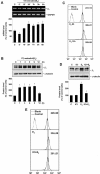
Estrogen induces β1-integrin expression in MG63 cells through ERs. MG63 cells were kept as controls or stimulated with 17β-estradiol (E2; 10 nM) for the times indicated (AC). In some experiments, MG63 cells were kept as controls (C) or pretreated with ICI 182,780 (ICI; 1 µM) for 1 hr and then stimulated with 17β-estradiol (E2; 10 nM) for 6 hrs (ICI + E2) (D, E). The β1 mRNA and protein expressions were determined by using RT-PCR (A), Western blot (B, D), and flow cytometric (C, E) analyses, respectively. The results are shown as mean ± SEM from three separate experiments. Data in panels A, B, and D are presented as percentage changes in band density from unstimulated control cells and normalized to GAPDH (A) or α-tubulin protein level (B, D). In panels C and E, MG63 cells incubated with FITC-conjugated antibody alone were used as blanks. Numbers are mean ± SEM of mean fluorescent intensity for all experiments determined by comparison with corresponding blanks. *p < .05 versus unstimulated control cells. #p < .05 versus E2-treated cells (D).
Fig. 3
Fig. 3
Estrogen-mediated augmentation of shear stress–induced ERK and p38 MAPK activations in MG63 cells is regulated by ERs and β1-integrin. MG63 cells were kept as controls or pretreated with ICI 182,780 (ICI; 1 µM) for 1 hr and then stimulated with 17β-estradiol (E2; 10 nM) for 6 hrs, followed by exposure to shear stress (S; 12 dyn/cm2) for 10 minutes (E2 + S) (A). In parallel experiments, MG63 cells were transfected with control (siCL) or β1-integrin-specific siRNA (siβ1) for 48 hours prior to 17β-estradiol and shear stress stimuli (B). (C) Cells were transfected with control or β1-integrin-specific siRNA for 48 hrs and then kept as controls or treated with 17β-estradiol for 15 minutes (for ERK) or 1 hr (for p38 MAPK). The phosphorylations of ERK and p38 MAPK in these cells were determined by Western blot analysis. Data are presented as the amounts (band densities normalized to the total protein levels) of phosphorylated ERK and p38 MAPK proteins relative to those in static control cells pretreated with vehicle control (A) or transfected with control siRNA (B, C) and are shown as mean ± SEM from three independent experiments. *p < .05 versus unstimulated control cells. #p < .05 versus sheared cells without 17β-estradiol pretreatment. §p < .05 versus 17β-estradiol/shear stress–stimulated cells pretreated with vehicle control (A) or transfected with control siRNA (B, C).
Fig. 4
Fig. 4
Estrogen augments shear stress–induced c-fos and Cox-2 expressions in MG63 cells. MG63 cells were kept as controls (C), exposed to shear stress at 12 dyn/cm2 (S) (A), or stimulated with 17β-estradiol (E2; 10 nM) (C) for the times indicated, and their c-fos and Cox-2 mRNA expressions were determined by quantitative real-time PCR. In some experiments, MG63 cells were pretreated with vehicle control DMSO or specific inhibitor of ERK (PD98058; 30 µM) or p38 MAPK (SB203580; 10 µM) or a combination of these inhibitors for 1 hr before exposure to shear stress for 15 minutes (for c-fos) or 1 hour (for Cox-2) (B). In parallel experiments, the cells were stimulated with 17β-estradiol for 6 hrs and then exposed to shear stress for 15 minutes (for c-fos) or 1 hour (for Cox-2) (E2 + S) (D). Prior to 17β-estradiol and shear stress treatments, the cells were preincubated with PD98058 or SB203580 or a combination of these inhibitors for 1 hour (F). (E) MG63 cells were subjected to simultaneous stimuli with shear stress and 17β-estradiol for 15 and 30 minutes without 17β-estradiol pretreatment. Data are presented as percentage changes relative to the unstimulated control cells (normalized to β-actin gene expression) and shown as mean + SEM from three or four separate experiments. *p < .05 versus unstimulated control cells. #p < .05 versus sheared cells without inhibitor pretreatment (B). §p < .05 versus sheared cells without 17β-estradiol pretreatment (D, F). δp < .05 versus sheared/E2-treated cells without inhibitor pretreatment (F).
Fig. 5
Fig. 5
Estrogen-mediated augmentation of shear stress–induced c-fos and Cox-2 expressions is mediated by both ERs and β1-integrin. MG63 cells were kept as controls or pretreated with ICI 182,780 (ICI; 1 µM) for 1 hr (A) and then stimulated with 17β-estradiol (E2; 10 nM) for 6 hours, followed by exposure to shear stress (S; 12 dyn/cm2) for 15 minutes (for c-fos) or 1 hour (for Cox-2) (A). In parallel experiments, MG63 cells were transfected with control (siCL) or specific siRNA of β1-integrin (siβ1) for 48 hours prior to 17β-estradiol and shear stress stimuli (B). The expressions of c-fos and Cox-2 mRNA were determined by quantitative real-time PCR. Data are presented as the expression (normalized to the β-actin gene expression) of c-fos or Cox-2 relative to those in static control cells pretreated with vehicle control (A) or transfected with control siRNA (B) and are shown as mean ± SEM from three independent experiments. *p < .05 versus unstimulated control cells. #p < .05 versus sheared cells without 17β-estradiol pretreatment. §p < .05 versus 17β-estradiol/shear stress–stimulated cells pretreated with vehicle control (A) or transfected with control siRNA (B).
Fig. 6
Fig. 6
Confirmation on the augmented effect of estrogen on shear stress–induced signaling and gene expression in primary HOBs. Primary HOBs were kept as controls or stimulated with 17β-estradiol (E2; 10 nM) for 6 hrs and then exposed to shear stress at 12 dyn/cm2 for 10 minutes (for MAPK) (A), 15 minutes (for c-fos), or 1 hr (for Cox-2) (B). The phosphorylations of ERK and p38 MAPK (A) and expressions of c-fos and Cox-2 (B) in these cells were determined by using Western blot and quantitative real-time PCR, respectively. In parallel experiments, HOBs were transfected with control (siCL) or specific siRNA (40 nM) of β1-integrin (siβ1) for 48 hrs prior to 17β-estradiol and/or shear stress stimuli (D, E). (C) Flow cytometric analysis of β1-integrin surface expression on HOBs treated with 17β-estradiol for 6 hrs. In some experiments, HOBs were pretreated with ICI 182,780 (ICI; 1 µM) for 1 hr prior to 17β-estradiol stimulation. HOBs incubated with FITC-conjugated antibody alone were used as blanks. Data in panels A, B, D, and E are presented as percentage changes relative to the unstimulated control cells [normalized to the MAPK total protein levels (A, D) or β-actin gene expression (B, E)] and shown as mean ± SEM from three separate experiments. Numbers in panel C are mean ± SEM of mean fluorescent intensity for three experiments determined by comparison with corresponding blanks. *p < .05 versus unstimulated control cells. #p < .05 versus sheared cells without 17β-estradiol pretreatment. §p < .05 versus 17β-estradiol/shear stress–stimulated cells transfected with control siRNA (D, E).

Similar articles

Cited by

References

    1. Hillam RA, Skerry TM. Inhibition of bone resorption and stimulation of formulation by mechanical loading of the modeling rat ulna in vivo. J Bone Miner Res. 1995;10:683–689. - PubMed
    1. Hughes-Fulford M. Signal transduction and mechanical stress. Sci STKE. 2004;249:RE12. - PubMed
    1. Sikavitsas VI, Temenoff JS, Mikos AG. Biomaterials and bone mechanotransduction. Biomaterials. 2001;22:2581–2593. - PubMed
    1. Kapur S, Baylink DJ, Lau KH. Fluid flow shear stress stimulates human osteoblast proliferation and differentiation through multiple interacting and competing signal transduction pathways. Bone. 2003;32:241–251. - PubMed
    1. Pavalko FM, Chen NX, Turner CH, et al. Fluid shear-induced mechanical signaling in MC3T3-E1 osteoblasts requires cytoskeleton-integrin interactions. Am J Physiol. 1998;275:C1591–1601. - PubMed

Publication types

MeSH terms