. 2009;11(5):R74.
doi: 10.1186/bcr2411.

BU-32: a novel proteasome inhibitor for breast cancer

Affiliations

BU-32: a novel proteasome inhibitor for breast cancer

Joseph K Agyin et al. Breast Cancer Res. 2009.

Abstract

Introduction: Proteasome inhibition provides an attractive approach to cancer therapy and may have application in the treatment of breast cancer. However, results of recent clinical trials to evaluate the effect of the proteasome inhibitor Bortezomib (Velcade, also called PS-341) in metastatic breast cancer patients have shown limited activity when used as a single agent. This underscores the need to find new and more efficacious proteasome inhibitors. In this study, we evaluate the efficacy of the novel proteasome inhibitor BU-32 (NSC D750499-S) using in vitro and in vivo breast cancer models.

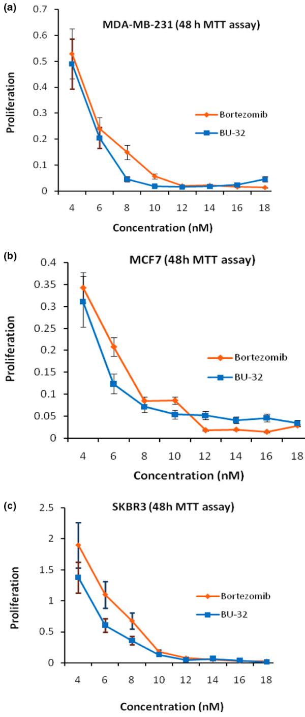
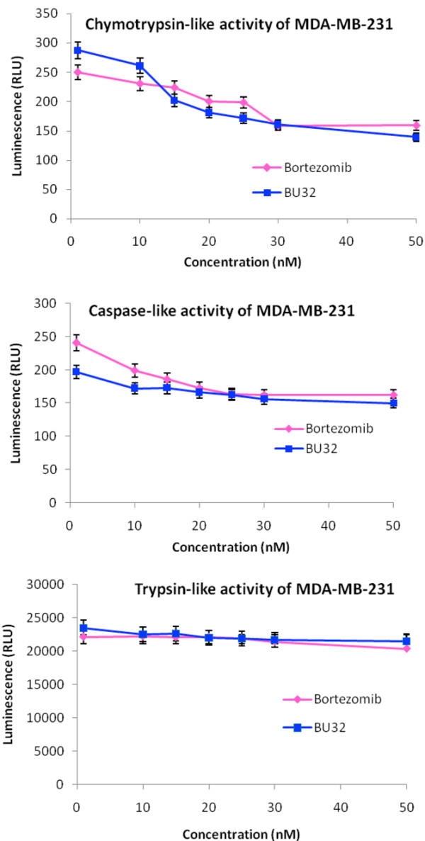
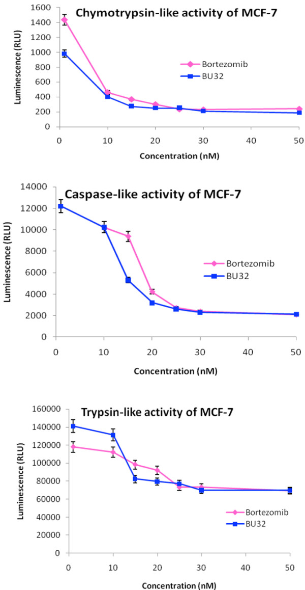
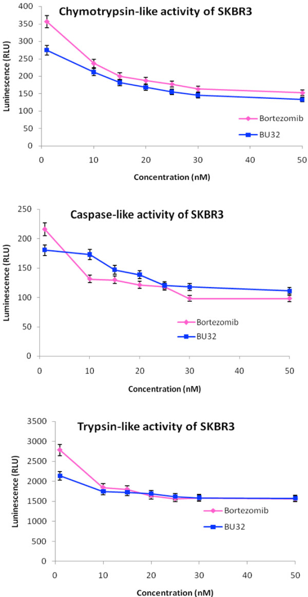
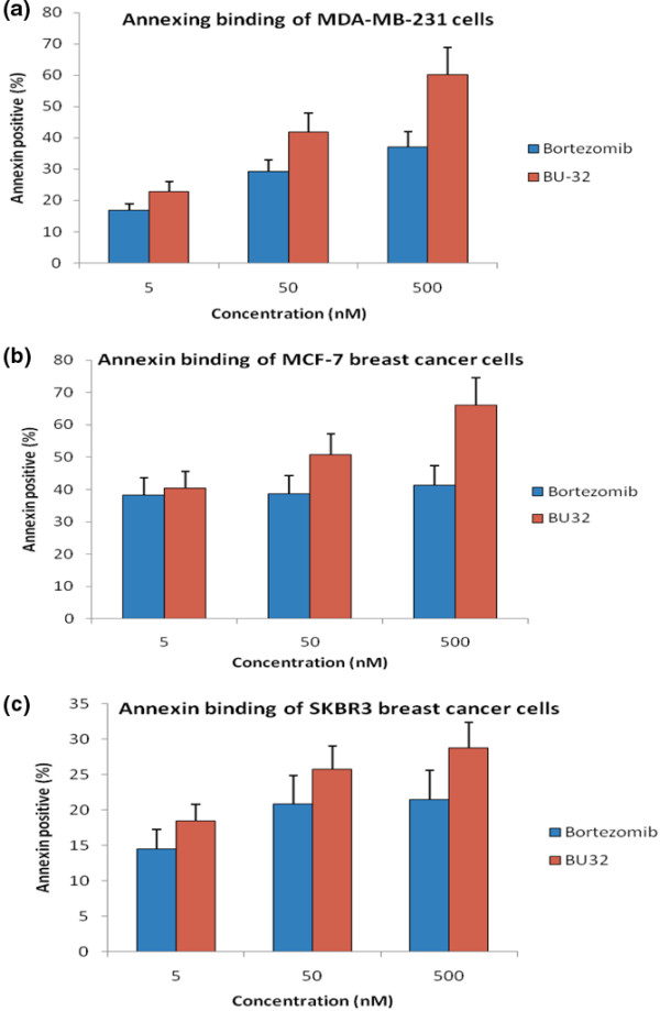
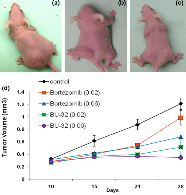
Methods: We have recently synthesized a novel proteasome inhibitor (BU-32) and tested its growth inhibitory effects in different breast cancer cells including MCF-7, MDA-MB-231, and SKBR3 by in vitro cytotoxicity and proteasomal inhibition assays. The apoptotic potential of BU32 was tested using flow cytometry and analyzing cell cycle regulatory proteins. In vivo tumor xenograft studies for solid tumor as well as tumor metastasis were conducted using MDA-MB-231-GFP cells.

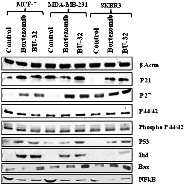
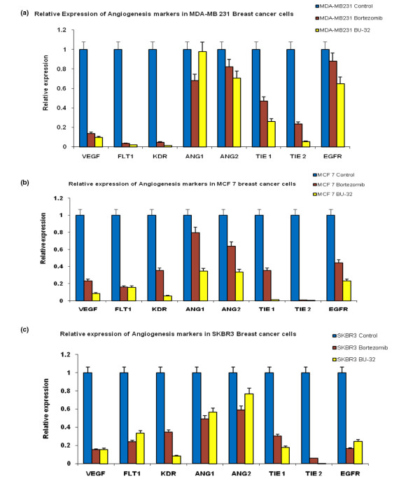
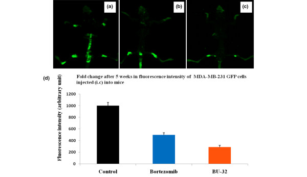
Results: We report for the first time that BU-32 exhibits strong cytotoxicity in a panel of cell lines: MDA-MB-231 (IC50 = 5.8 nM), SKBR3 (IC50 = 5.7 nM) and MCF-7 cells (IC50 = 5.8 nM). It downregulates a wide array of angiogenic marker genes and upregulates apoptotic markers, including Bid and Bax. Incubation of MDA-MB-231 cells with BU-32 results in the accumulation of cell cycle inhibitor proteins p21 and p27 and stabilization of the tumor suppressor protein p53. Studies in in vivo solid tumor and metastasis models show significant effect with a 0.06 mg/kg dose of BU-32 and marked reduction in tumor burden in the skeleton.

Conclusions: We have shown that BU-32 is effective in cultured breast cancer cells and in breast cancer xenografts. The results suggest its potential benefit in breast cancer treatment.


Figures

Figure 1
Figure 1
Effect of proteasome inhibitor on proliferation. 3-(4,5-Dimethylthiazol-2-yl)-2,5-diphenyltetrazolium bromide (MTT) assay of (a) MDA-MB231, (b) MCF7 and (c) SKBR3 breast cancer cells treated with different concentrations (4 to 18 nM) of Bortezomib and BU-32 for 48 hours. Results are mean of three independent experiments.
Figure 2
Figure 2
Effect of BU-32 on proteasomal catalytic activity of MDA-MB-231 cells. Proteasome inhibition by Bortezomib and BU-32 in the MDA-MB-231 breast cancer cell line was measured. The cells were treated with different concentrations of proteasome inhibitor from 1 to 50 nM for 24 hours, and were then analyzed for the inhibition of chymotryptic-like, caspase-like, and tryptic-like intracellular proteasome activities. Results are mean of three individual experiments. RLU: Relative Luminescence Units.
Figure 3
Figure 3
Effect of BU-32 on proteasomal catalytic activity of MCF-7 cells. Proteasome inhibition by Bortezomib and BU-32 in the MCF-7 breast cancer cell line was measured. The cells were treated with different concentrations of proteasome inhibitor from 1 to 50 nM for 24 hours, and were then analyzed for the inhibition of chymotryptic-like, caspase-like, and tryptic-like intracellular proteasome activities. Results are mean of three individual experiments. RLU: Relative Luminescence Units.
Figure 4
Figure 4
Effect of BU-32 on proteasomal catalytic activity of SKBR3 cells. Proteasome inhibition by Bortezomib and BU-32 in the SKBR3 breast cancer cell line was measured. The cells were treated with different concentrations of proteasome inhibitor from 1 to 50 nM for 24 hours, and were then analyzed for the inhibition of chymotryptic-like, caspase-like, and tryptic-like intracellular proteasome activities. Results are mean of three individual experiments. RLU: Relative Luminescence Units.
Figure 5
Figure 5
BU-32 upregulates apoptosis and downregulates NF-κB expression. BU-32 exposure induces accumulation of proapoptotic markers, cell-cycle-dependent kinase inhibitors and the p53 tumor suppressor gene, and downregulates NF-κB expression in breast cancer cells. Western blot analysis of total cell extracts of all three breast cancer cell lines shows the effect of treatment with Bortezomib and BU-32 on the expression level of the cyclin-dependent kinase inhibitor proteins p21 and p27, tumor suppressor p53, proapoptotic genes Bid and Bax, anti-apoptotic NF-κB, and cell cycle regulatory protein p44/42, phospho p44/42. Breast cancer cell lines were examined after 24 hours of exposure to the proteasome inhibitors (5 nM) and a series of western blots using specific antibodies was performed to detect relative levels of proteins. Results are the average of three independent measurements.
Figure 6
Figure 6
BU-32 downregulates the expression of genes involved in angiogenesis. Expression levels of vascular endothelial growth factor (VEGF), VEGF-receptor 1 (FLT1), kinase-insert domain-containing receptor (KDR), Ang1, Ang2, Tie1, Tie2, and epidermal growth factor receptor (EGFR) genes were measured by RT-PCR in (a) MDA-MB-231, (b) MCF7 and (c) SKBR3 breast cancer cell lines exposed to proteasome inhibitors BU-32 and Bortezomib, as evaluated by real-time RT-PCR. The RT-PCR was performed on 200 ng total RNA extracted with Trizol reagent from breast cancer cell lines and was cultured for 24 hours with 5 nM concentrations of Bortezomib and BU-32. Results are mean ± standard deviation of three independent determinations.
Figure 7
Figure 7
Exposure to BU-32 induces apoptosis in tumor cell lines. Annexin staining was conducted with the use of a kit (Annexin V--FITC Apoptosis detection kit; BD Pharmingen (San Jose, CA, USA)). The breast cancer cells were treated with 5 nM, 50 nM, and 500 nM Bortezomib and BU-32 for 24 hours. Cells were stained with annexin V--FITC and propydium iodide according to the manufacturer's protocol before analysis by flow cytometry. Results are mean ± standard deviation of three independent experiments for (a) MDA-MB-231, (b) MCF7 and (c) SKBR3 cell lines.
Figure 8
Figure 8
Effect of proteasome inhibitor on established human breast cancer cell (MDA-MB-231-GFP) tumor xenograft. Subcutaneous tumors were generated by injecting 1 × 10⁷ MDA-MB-231-GFP cells into the flanks of nude mice. Tumors were established for 10 days before the initiation of therapy. Representative images were taken under microscope at the end of week 3 of therapy and show results for (a) vehicle-treated controls, (b) Bortezomib-treated mice and (c) BU-32-treated mice. Doses of 0.02 and 0.06 mg/kg body weight were used for Bortezomib and BU-32. (d) Tumor volumes were measured 1, 2, and 3 weeks after initiation of therapy. Results are mean ± standard deviation (n = 10).
Figure 9
Figure 9
Inhibition of breast cancer metastasis in bone by proteasome inhibitor. Mice were injected intracardially with 1 × 10⁵ MDA-MB-231-GFP cells. The mice were then treated with Bortezomib and BU-32 using a dose of 0.06 mg/kg body weight or with vehicle PBS as control. Whole-body high-resolution fluorescence images of MDA-MB231-GFP-expressing breast cancer metastatic cells in the skeleton of live intact animals were measured after 5 weeks. Representative fluorescence images are shown for (a) the control group, (b) Bortezomib-treated mice and (c) BU-32-treated mice. (d) The fluorescence intensities were quantified. GFP, green fluorescent protein; i.c., intracisternal.

Similar articles

Cited by

References

    1. Ciechanover A. Proteolysis: from the lysosome to ubiquitin and the proteasome. Nat Rev Mol Cell Biol. 2005;6:79–87. doi: 10.1038/nrm1552. - DOI - PubMed
    1. Dalton WS. The proteasome. Semin Oncol. 2004;31:3–9. doi: 10.1053/j.seminoncol.2004.10.012. discussion 33. - DOI - PubMed
    1. Kisselev AF, Goldberg AL. Proteasome inhibitors: from research tools to drug candidates. Chem Biol. 2001;8:739–758. doi: 10.1016/S1074-5521(01)00056-4. - DOI - PubMed
    1. Groll M, Berkers CR, Ploegh HL, Ovaa H. Crystal structure of the boronic acid-based proteasome inhibitor bortezomib in complex with the yeast 20S proteasome. Structure. 2006;14:451–456. doi: 10.1016/j.str.2005.11.019. - DOI - PubMed
    1. Orlowski M, Cardozo C, Michaud C. Evidence for the presence of five distinct proteolytic components in the pituitary multicatalytic proteinase complex. Properties of two components cleaving bonds on the carboxyl side of branched chain and small neutral amino acids. Biochemistry. 1993;32:1563–1572. doi: 10.1021/bi00057a022. - DOI - PubMed

Publication types

MeSH terms