. 2009 Oct 13:5:60.
doi: 10.1186/1744-8069-5-60.

Morphine modulation of pain processing in medial and lateral pain pathways

Affiliations

Morphine modulation of pain processing in medial and lateral pain pathways

Jin-Yan Wang et al. Mol Pain. .

Abstract

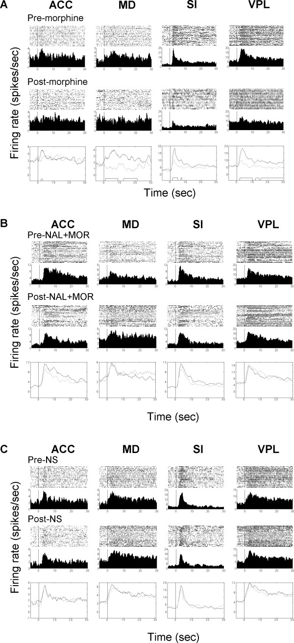
Background: Despite the wide-spread use of morphine and related opioid agonists in clinic and their powerful analgesic effects, our understanding of the neural mechanisms underlying opioid analgesia at supraspinal levels is quite limited. The present study was designed to investigate the modulative effect of morphine on nociceptive processing in the medial and lateral pain pathways using a multiple single-unit recording technique. Pain evoked neuronal activities were simultaneously recorded from the primary somatosensory cortex (SI), ventral posterolateral thalamus (VPL), anterior cingulate cortex (ACC), and medial dorsal thalamus (MD) with eight-wire microelectrode arrays in awake rats.

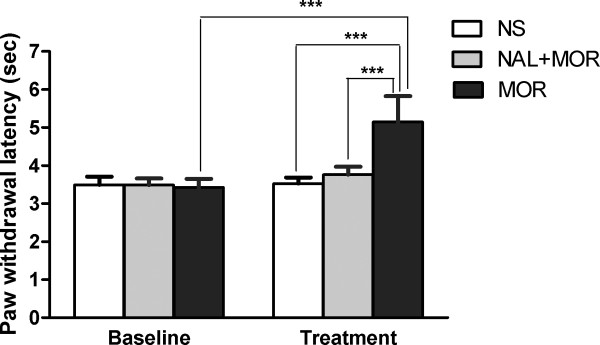
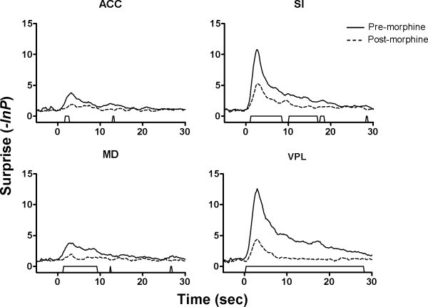
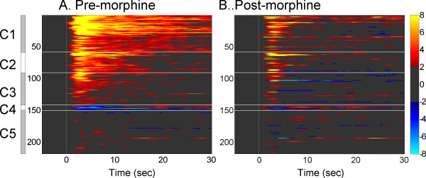
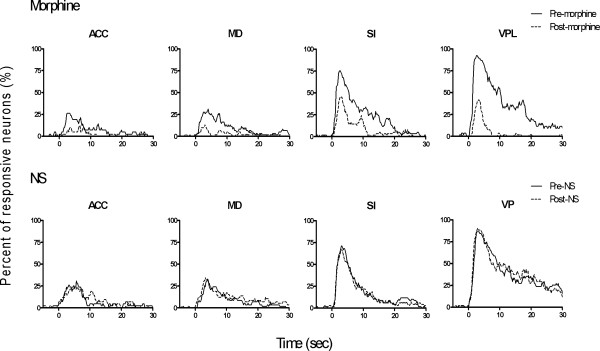
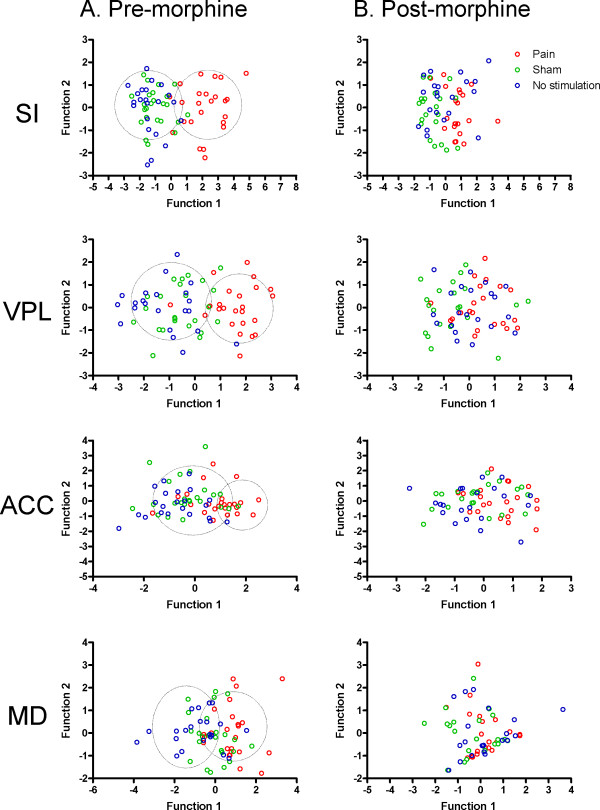
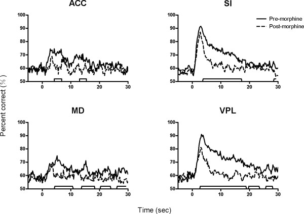
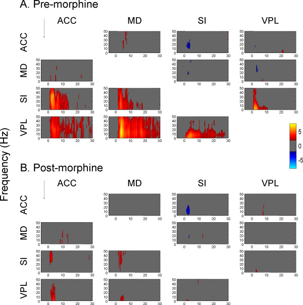
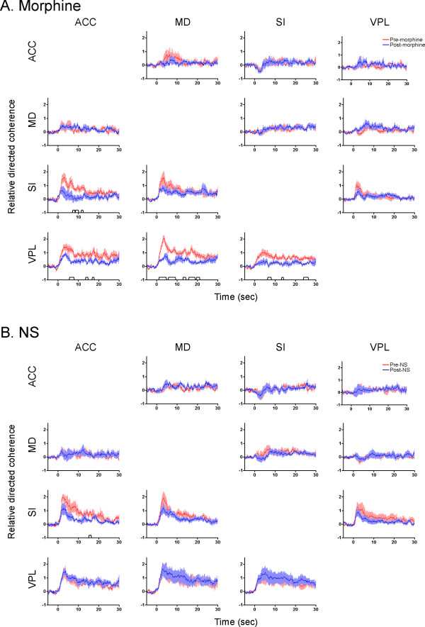
Results: The results showed that the noxious heat evoked responses of single neurons in all of the four areas were depressed after systemic injection of 5 mg/kg morphine. The depressive effects of morphine included (i) decreasing the neuronal response magnitude; (ii) reducing the fraction of responding neurons, and (iii) shortening the response duration. In addition, the capability of cortical and thalamic neural ensembles to discriminate noxious from innocuous stimuli was decreased by morphine within both pain pathways. Meanwhile, morphine suppressed the pain-evoked changes in the information flow from medial to lateral pathway and from cortex to thalamus. These effects were completely blocked by pre-treatment with the opiate receptor antagonist naloxone.

Conclusion: These results suggest that morphine exerts analgesic effects through suppressing both sensory and affective dimensions of pain.


Figures

Figure 1
Figure 1
Effect of morphine pretreatment on the thermal pain thresholds in rats. Noxious radiant heat was used as painful stimulation, which was randomly applied to the plantar surface of the rats' hindpaws. Baseline paw withdrawal latency was measured. Morphine (5 mg/kg) or equivalent volume of NS (n = 8) was injected intraperitoneally and pain-related activities were recorded 10 min later. For NAL + MOR treatment, rats were pre-treated with 4 mg/kg naloxone 10 min prior to administration of morphine (n = 4). The ANOVA revealed that pre-treatment with morphine produced significantly longer withdrawal latency than that of any other session (P < 0.001). The effect of morphine was antagonized by naloxone pretreatment prior to morphine administration. NAL: naloxone; MOR: morphine; NS: normal saline.
Figure 2
Figure 2
Changes of neuronal response magnitude following morphine (A), NAL + MOR (B) or NS (C) treatment. Raster and perievent histograms show the noxious induced responses in the four recorded areas. Figures in the top two rows in each panel illustrate typical excitatory responses before and after drug administration in each brain regions. Figures in the bottom row show the results of comparison of the pain-evoked neural activities before (black lines) and after (gray lines) morphine, NAL + MOR or NS administration. The trapezoid markers along the x-axis indicate the statistically significant difference between two sessions (Student's t-test, P < 0.01). Time = 0 on the axis corresponds to the time of noxious stimulation start. Note that a single dose of 5 mg/kg morphine significantly reduced the noxious heat induced activation in all the recorded areas (A), which can be reversed by naloxone pretreatment (B). No change was observed after NS injection (C). NAL: naloxone; MOR: morphine; NS: normal saline.
Figure 3
Figure 3
Comparison of neuronal response magnitude before and after the administration of morphine. Neuronal responses to noxious stimulation were evaluated using a sliding window technique, in which a 1-s time window was slid through the entire period of a trial at 1-bin step. The bin counts of each window were compared with those of a baseline window by Student's t-test. Only a change of firing rates from the baseline exceeds the limit of P < 0.01 for at least three consecutive windows were taken as a response. The 'P values' were then converted into the information theory concept surprise by performing logarithmic transformation, i.e., -ln P. The surprise values were also compared using the sliding window method (1-s time window, 0.1-s step, P < 0.01 for three consecutive windows). Significant differences (trapezoid markers along the x-axis) between the response magnitude before (solid lines) and after (dashed lines) morphine administration were observed in all of the recorded areas.
Figure 4
Figure 4
Cluster plot depicted changes of the temporal distribution patterns of neural activity before (A) and after morphine administration (B). A clustering analysis was performed to classify neuronal responses depending on the similarities in patterns of excitation or inhibition around stimulation events. The firing rates were transferred into z-scores and neurons with z-scores > 2 were accepted as significantly excited and < -2 as significantly inhibited. Colorbar indicates z-scores (light yellow for highest and light blue for lowest). Each line of the image represents normalized activity of one neuron. C1-C5 represent different categories according to the response patten. The cluster analysis revealed that the cortical and thalamic areas contain units with five clusters of coding patterns in response to the noxious radiant heat (A). All of the excitatory responses were weakened by systemic injection of morphine (B).
Figure 5
Figure 5
Comparison of percentage of neurons responding to noxious stimulation before (solid lines) and after (dashed lines) morphine or NS delivery. Chi-square tests were used to detect the percentage differences between different sessions over time. Consistent with the result of neuronal response magnitude, the number of responding neurons in all recorded areas was also decreased after injection of morphine. By contrast, NS treatment did not affect the nociceptive related responses.
Figure 6
Figure 6
Scatter plots depicted the capability of neural ensembles in each brain area to discriminate noxious, sham and no stimulation before (A) and after morphine administration (B). The linear discriminant analysis (LDA) was used to investigate whether morphine administration affected the capability of neural ensembles to discriminate different types of sensory stimulation. The noxious thermal evoked firing rates were chosen for discriminant analysis. The firing rates of multiple principle components around noxious, sham, or no-stimulation (randomly selected points where no events occurred within 30 seconds around) events were calculated and the discriminant function coefficients were estimated. As can be seen, the three categories can be well separated before the delivery of morphine (A), as indicated by the dashed line circles. In contrast, the three categories are mixed up after morphine injection (B).
Figure 7
Figure 7
Effects of morphine on the temporal distribution of discriminant capability of neural ensembles in ACC, MD, SI and VPL. The correct percentage of neural ensembles to differentiate three types of sensory inputs was compared before and after morphine injection. As can be seen, the ability of neural ensembles to discriminate pain from non-pain stimuli significantly decreased after morphine administration. The markers along the x-axis indicate the statistically significant difference in disciminant performance before (solid lines) and after (dashed lines) injection of morphine.
Figure 8
Figure 8
Partial directed coherence (PDC) among different brain regions induced by noxious stimuli before (A) and after (B) injection of morphine. The PDC values were normalized to z-scores relative to the mean and variance of baseline PDC (i.e., before noxious stimulation). The normalized PDCs exceeding 95% confident interval of the baseline were displayed in pseudo color (yellow and red for the increase and cyan and blue for the decrease). Arrows indicate the direction of information flow. (A) Before morphine injection, the amount of directed coherence from medial to lateral pathway significantly increased with a latency of 0.5 sec. (B) No significant change was observed in the PDC following morphine administration
Figure 9
Figure 9
Comparison of averaged PDC across all frequencies between pre- (red lines) and post-treatment (blue lines) of morphine (A) or NS (B). For each frequency band, the PDC values were normalized to z-scores relative to its mean and variance of baseline PDC. Then the PDC values of all 50 frequency bands were averaged over time. As can be seen in this figure, following morphine injection, the amount of directed coherence was significantly decreased in each direction in comparison to the pre-morphine condition. Data are presented as mean ± S.E.

Similar articles

Cited by

References

    1. Lorenz J, Beck H, Bromm B. Cognitive performance, mood and experimental pain before and during morphine-induced analgesia in patients with chronic non-malignant pain. Pain. 1997;73:369–375. doi: 10.1016/S0304-3959(97)00123-1. - DOI - PubMed
    1. Kalliomäki J, Luo XL, Yu YB, Schouenborg J. Intrathecally applied morphine inhibits nociceptive C fiber input to the primary somatosensory cortex (SI) of the rat. Pain. 1998;77:323–329. doi: 10.1016/S0304-3959(98)00115-8. - DOI - PubMed
    1. da Fonseca Pacheco D, Klein A, de Castro Perez A, da Fonseca Pacheco CM, de Francischi JN, Duarte ID. The mu-opioid receptor agonist morphine, but not agonists at delta- or kappa-opioid receptors, induces peripheral antinociception mediated by cannabinoid receptors. Br J Pharmacol. 2008;154:1143–1149. doi: 10.1038/bjp.2008.175. - DOI - PMC - PubMed
    1. Reichert JA, Daughters RS, Rivard R, Simone DA. Peripheral and preemptive opioid antinociception in a mouse visceral pain model. Pain. 2001;89:221–227. doi: 10.1016/S0304-3959(00)00365-1. - DOI - PubMed
    1. Jones AK, Friston KJ, Qi LY, Harris M, Cunningham VJ, Jones T, Feinman C, Frackowiak RS. Sites of action of morphine in the brain. Lancet. 1991;338:825. doi: 10.1016/0140-6736(91)90717-4. - DOI - PubMed

Publication types