. 2009 Dec 11;284(50):34490-505.
doi: 10.1074/jbc.M109.057315. Epub 2009 Oct 12.

ATP released by electrical stimuli elicits calcium transients and gene expression in skeletal muscle

Affiliations

ATP released by electrical stimuli elicits calcium transients and gene expression in skeletal muscle

Sonja Buvinic et al. J Biol Chem. .

Abstract

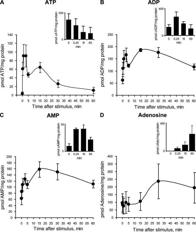
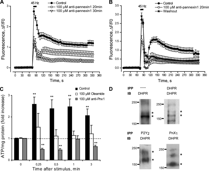
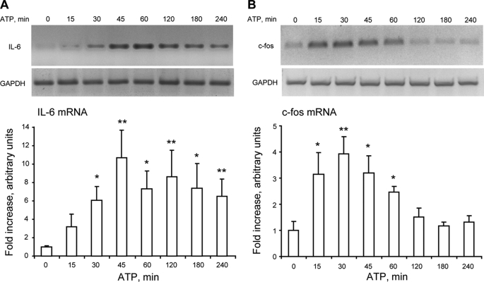
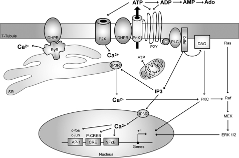
ATP released from cells is known to activate plasma membrane P2X (ionotropic) or P2Y (metabotropic) receptors. In skeletal muscle cells, depolarizing stimuli induce both a fast calcium signal associated with contraction and a slow signal that regulates gene expression. Here we show that nucleotides released to the extracellular medium by electrical stimulation are partly involved in the fast component and are largely responsible for the slow signals. In rat skeletal myotubes, a tetanic stimulus (45 Hz, 400 1-ms pulses) rapidly increased extracellular levels of ATP, ADP, and AMP after 15 s to 3 min. Exogenous ATP induced an increase in intracellular free Ca(2+) concentration, with an EC(50) value of 7.8 +/- 3.1 microm. Exogenous ADP, UTP, and UDP also promoted calcium transients. Both fast and slow calcium signals evoked by tetanic stimulation were inhibited by either 100 mum suramin or 2 units/ml apyrase. Apyrase also reduced fast and slow calcium signals evoked by tetanus (45 Hz, 400 0.3-ms pulses) in isolated mouse adult skeletal fibers. A likely candidate for the ATP release pathway is the pannexin-1 hemichannel; its blockers inhibited both calcium transients and ATP release. The dihydropyridine receptor co-precipitated with both the P2Y(2) receptor and pannexin-1. As reported previously for electrical stimulation, 500 mum ATP significantly increased mRNA expression for both c-fos and interleukin 6. Our results suggest that nucleotides released during skeletal muscle activity through pannexin-1 hemichannels act through P2X and P2Y receptors to modulate both Ca(2+) homeostasis and muscle physiology.


Figures

FIGURE 1.
FIGURE 1.
Molecular and pharmacological determination of P2Y/P2X receptors in rat skeletal myotubes. A, mRNA for several P2Y and P2X receptor subtypes is expressed in skeletal myotubes derived from newborn rat primary cultures. Total RNA was extracted from differentiated skeletal myotubes, and the mRNA for all the P2X/P2Y receptor subtypes was assessed by RT-PCR and detected in 1.5% agarose gels on the basis of their estimated molecular weight. A representative gel of three different RNA extractions is presented. B–E, natural agonists for P2Y/P2X receptors evoke calcium transients in skeletal myotubes. The effect of ATP, ADP, UTP and UDP (500 μm) over intracellular calcium changes was assessed in skeletal myotubes. A sequence of images was taken with the charge-coupled device camera attached to the epifluorescence microscope side port, which was equipped with the correct filters to capture fluo3-AM fluorescence to monitor the intracellular Ca+2 level. The analyzed regions of interest were from whole myotubes, considering both cytosolic and nuclear components. Stimuli were applied where indicated by an arrow and maintained throughout the recording period. Traces correspond to mean ± S.E. For each condition, 20–50 cells coming from 4–10 independent coverslips were quantified. In B, ATP was assessed either in regular Krebs buffer (solid circles, 1 mm Ca+2) or in calcium-free Krebs buffer (open circles, 0 Ca+2 plus 2 mm EGTA). All other nucleotides were assessed in regular Krebs buffer.
FIGURE 2.
FIGURE 2.
Quantitation and kinetics of calcium transients evoked by ATP in skeletal myotubes. ATP increases intracellular Ca2+ in a concentration-dependent manner in skeletal myotubes. Cells were incubated with 0.001 μm-1 mm ATP, and calcium transients were measured as described in the legend for Fig. 2. From the calcium signals we analyzed the maximal fluorescence reached (A), the percentage of activated cells (B), and the time to peak (C). Values were expressed as mean ± S.E. Each coverslip was used to assay only one ATP concentration. Each bar represents values obtained in 12–25 cells from three independent coverslips.
FIGURE 3.
FIGURE 3.
Blockade of the P2Y/P2X receptor signaling reduces calcium transients evoked by tetanic electrical stimulation. A, fast and slow calcium transients evoked by tetanus (45 Hz, 400 1-ms pulses) are strongly reduced after general P2Y/P2X blockade using 100 μm suramin for 20 min prior to and during the protocol. A sequence of images shows fluo3 fluorescence to monitor the intracellular Ca+2 level (representative of n = 33–43 cells, five coverslips, three different cultures). B, a small reduction in calcium transients evoked by tetanus is observed when a specific P2Y1 receptor antagonist (MRS2179) was added for 20 min prior to and during the protocol (n = 107–149 cells, six coverslips, three different cultures). C, apyrase reduced in a time-dependent manner the calcium transients evoked by tetanus (n = 13–47 cells, four coverslips, three different cultures). A–C, experiments were performed using the epifluorescence microscope. For each coverslip, calcium transients evoked by tetanus before and after the inhibitor were tested. Values are expressed as mean ± S.E. D, to accurately detect changes in the fast calcium signal, we used a single pulse protocol (0.33 Hz, 1-ms pulses) and acquired fluorescence data every 1.9 ms by line scan confocal microscopy. As shown in the representative tracing, there was an evident decrease in the fast signal amplitude after 20 min of incubation with apyrase. E, isolated fibers from mouse FDB muscle loaded with fluo3-AM were stimulated with a couple of platinum electrodes with a tetanic protocol (45 Hz, 400 0.3-ms pulses). Fluorescence images were acquired in a confocal microscope every 1.8 s and analyzed frame by frame. Calcium transients evoked by tetanus were assessed before and after incubation with 2 units/ml apyrase for 20 min. Apyrase treatment evoked a significant reduction in both fast and slow calcium transients promoted by tetanus (n = 3 fibers, two different cultures). Values are expressed as mean ± S.E.
FIGURE 4.
FIGURE 4.
ATP release from skeletal myotubes after titanic electrical stimulation or K+-evoked depolarization. A, skeletal myotubes were depolarized by electrical stimulation (45 Hz, 400 1-ms pulses). Aliquots of the extracellular medium were removed at the indicated times after the stimulus. ATP at the samples was measured by luciferin/luciferase assay and quantitated using a calibration standard curve. B, skeletal myotubes were depolarized by 70 mm KCl, and extracellular ATP changes were evaluated as in A. Values are expressed as mean ± S.E. In A and B, enlargements of the x axis during the first 5 min are shown in the insets. C, conditioned saline from electrically stimulated myotubes evokes calcium transients in fluo3-loaded myotubes. Extracellular medium derived from nonstimulated myotubes (control) or from myotubes stimulated electrically (45 Hz, 400 1-ms pulses) in the presence and absence of apyrase (2 units/ml) was added to fluo3-loaded myotubes as described under “Experimental Procedures.” Changes in fluorescence were continually recorded by epifluorescence microscopy as described under “Experimental Procedures.” D, calcium transient elicited by the addition of 0.1 μm ATP is shown for comparison.
FIGURE 5.
FIGURE 5.
Nucleotides release and metabolization after tetanic stimulation. A–D, to measure ATP and its metabolites at the extracellular medium, skeletal myotubes were stimulated electrically with a tetanus protocol (45 Hz, 400 1-ms pulses), and aliquots of the medium were removed at the indicated times thereafter. Nucleotide samples (ATP, ADP, AMP, and adenosine) were derivatized as described under “Experimental Procedures,” resolved and detected using an HPLC coupled to fluorescence detection, and quantitated using a calibration standard curve. Values are the mean ± S.E. (n = 6–8 series from six independent primary cultures). To assess the ectonucleotidases activities of skeletal myotubes, a standard of ϵ-ATP was applied to the extracellular medium. Insets, at different times, extracellular aliquots were removed to quantify ϵ-ATP (A), ϵ-ADP (B), ϵ-AMP (C), and ϵ-adenosine (D) by HPLC as an indicator of the ATP metabolism ability of these cells. Values are the mean ± S.E. (n = 3–5 series from three independent primary cultures).
FIGURE 6.
FIGURE 6.
Pannexin-1 hemichannels are involved in ATP release during tetanic stimulation of skeletal myotubes. A, fast and slow calcium components evoked by tetanic electrical stimulation (45 Hz, 400 1-ms pulses) are strongly reduced after pannexin-1 hemichannel blockade using 100 μm of the 10pnx1 peptide 20–30 min prior to and during the protocol (n = 25–79 cells, five coverslips, five different cultures). For each coverslip, calcium transients evoked by tetanus before and after incubation with the peptide were tested. B, the same protocol as described in A was achieved, but after 10pnx1 peptide incubation cells were washed for 60 min, we reassessed the effect of the tetanus over calcium transients. Values are expressed as mean ± S.E. (n = 19–23 cells, two coverslips, two different cultures). C, ATP release evoked by tetanic electrical stimulation was abolished by 100 μm oleamide (a nonselective blocker for connexin and pannexin hemichannels) or 100 μm 10pnx1 peptide. Skeletal myotubes were incubated 20 min before and during the assay with the indicated blocker. Aliquots of the extracellular medium were removed at the indicated times after electrical stimulation, and ATP was measured by a luciferin/luciferase assay as described under “Experimental Procedures.” Values were normalized to 0 control for each treatment and expressed as the mean ± S.E. (n = 4 series from four independent primary cultures). **, p < 0.01, Dunnett's t test, one-tail comparison between each time and their own 0 control. D, DHPR co-precipitates with P2Y2 receptor and pannexin-1 in skeletal myotubes. DHPR was resolved and detected by immunoblot (IB) in samples derived from whole lysates (—) or immunoprecipitated (IPP) previously with anti-DHPR, anti-P2Y2, or anti-Pnx1 as described under “Experimental Procedures.”
FIGURE 7.
FIGURE 7.
Exogenous ATP increases both IL-6 and c-fos mRNA in skeletal myotubes. Myotubes were incubated with 500 μm ATP for the times indicated. Total RNA was isolated, and IL-6 (A) or c-fos (B) mRNA levels were analyzed by semiquantitative RT-PCR. The top panels are representative agarose gels for RT-PCR products from IL-6 (A) and c-fos (B) mRNA amplifications with their corresponding GAPDH controls. The bottom panels correspond to results normalized to GAPDH expression and presented as -fold increase of untreated control cells (mean ± S.E. n = 3–8). *, p < 0.05; **, p < 0.01; analyzed by analysis of variance followed by Dunnett's multiple comparison test.
FIGURE 8.
FIGURE 8.
Hypothetical schematic model of the proposed role of purinergic receptors in both excitation-contraction and excitation-transcription coupling in skeletal muscle cells. A multimeric protein complex is suggested to exist in the T-tubule membrane, including the dihydropyridine receptor (Cav1.1, DHPR), the purinergic metabotropic P2Y2 receptor, and the pannexin-1 molecule (PnX1). Membrane depolarization will induce the opening of an ATP pathway via PnX1 after a conformational change of the adjacent DHPR. A heterotrimeric G protein will be attached to the P2Y2 receptor, and upon ATP binding, the βγ subunit will sequentially activate phosphoinositide 3-kinase (not shown) and phospholipase C (PLC) to produce IP3 and activate calcium release for the slow calcium transient. Other proteins such as RyR, known to interact with DHPR and P2X receptors (ionotropic, calcium-permeating channel), participate in the fast calcium transient, but whether they are part of the same complex has not yet been evidenced.

References

    1. Grabner M., Dirksen R. T., Suda N., Beam K. G. (1999) J. Biol. Chem. 274, 21913–21919 - PubMed
    1. Lamb G. D. (2002) Front. Biosci. 7, d834–d842 - PubMed
    1. Protasi F., Paolini C., Nakai J., Beam K. G., Franzini-Armstrong C., Allen P. D. (2002) Biophys. J. 83, 3230–3244 - PMC - PubMed
    1. Tanabe T., Beam K. G., Adams B. A., Niidome T., Numa S. (1990) Nature 346, 567–569 - PubMed
    1. Carrasco M. A., Riveros N., Ríos J., Müller M., Torres F., Pineda J., Lantadilla S., Jaimovich E. (2003) Am. J. Physiol. Cell Physiol. 284, C1438–C1447 - PubMed

Publication types

MeSH terms