. 2009 Dec 11;284(50):34703-12.
doi: 10.1074/jbc.M109.015107. Epub 2009 Oct 12.

Calcium bridge triggers capsid disassembly in the cell entry process of simian virus 40

Affiliations

Calcium bridge triggers capsid disassembly in the cell entry process of simian virus 40

Masa-Aki Kawano et al. J Biol Chem. .

Abstract

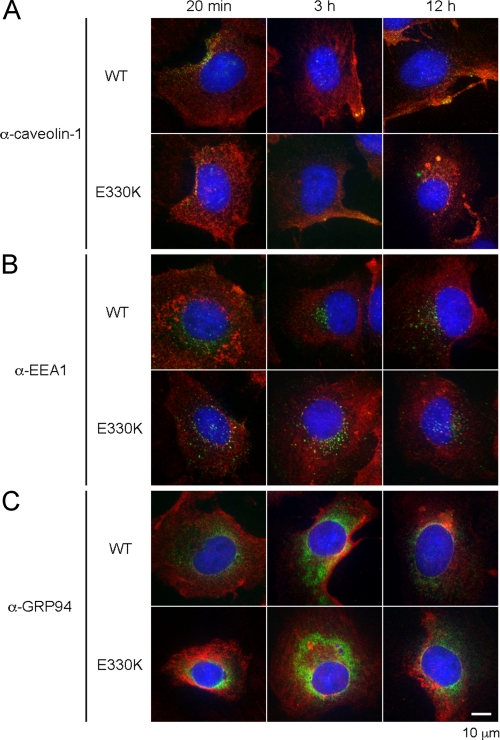
The calcium bridge between the pentamers of polyoma viruses maintains capsid metastability. It has been shown that viral infection is profoundly inhibited by the substitution of lysine for glutamate in one calcium-binding residue of the SV40 capsid protein, VP1. However, it is unclear how the calcium bridge affects SV40 infectivity. In this in vitro study, we analyzed the influence of host cell components on SV40 capsid stability. We used an SV40 mutant capsid (E330K) in which lysine had been substituted for glutamate 330 in protein VP1. The mutant capsid retained the ability to interact with the SV40 cellular receptor GM1, and the internalized mutant capsid accumulated in caveolin-1-mediated endocytic vesicles and was then translocated to the endoplasmic reticulum (ER) region. However, when placed in ER-rich microsome, the mutant capsid retained its spherical structure in contrast to the wild type, which disassembled. Structural analysis of the mutant capsid with cryo-electron microscopy and image reconstruction revealed altered pentamer coordination, possibly as a result of electrostatic interaction, although its overall structure resembled that of the wild type. These results indicate that the calcium ion serves as a trigger at the pentamer interface, which switches on capsid disassembly, and that the failure of the E330K mutant capsid to disassemble is attributable to an inadequate triggering system. Our data also indicate that calcium depletion-induced SV40 capsid disassembly may occur in the ER region and that this is essential for successful SV40 infection.


Figures

FIGURE 1.
FIGURE 1.
Cryo-electron micrograph and reconstruction of E330K capsid. A, cryo-electron micrograph of E330K capsid. The E330K VP1 assembled into spherical particles with diameters of 510 Å (black arrow), 360 Å (white arrow), and 260 Å (white arrowhead). Tubular assembly of E330K VP1 was also observed (black arrowhead). Bar, 100 nm. B, surface presentation of reconstructed E330K capsid. The positions of the icosahedral axes are marked with the corresponding numbers.
FIGURE 2.
FIGURE 2.
Attachment to gangliosides and cell uptake of the E330K capsid. A, a sucrose floatation assay was performed to determine whether capsids bind to a specific ganglioside. GM1 or GD1a containing phospholipids was incubated with wild type or E330K capsid, mixed with 73% sucrose, and placed at the bottom of the tube. A 10–45% sucrose gradient was layered over this mixture and ultracentrifuged for 10 h at 4 °C. Fractions were collected at the top of the tube and analyzed by silver staining. Lane 1, wild type with GM1; lane 2, wild type with GD1a; lane 3, wild type with phospholipids alone; lane 4, E330K with GM1; lane 5, E330K with GD1a; lane 6, E330K with phospholipids alone. B, CV-1 cells were incubated with wild type or E330K capsid for the time indicated at the top. Following incubation, the cells were collected by scraping or trypsin treatment. Collected cells were washed three times with PBS(−), resuspended with PBS(−), and sonicated to prepare the cell lysate. Wild type (WT; top) or E330K VP1 protein (bottom) was visualized by SDS-PAGE followed by Western blot analysis using anti-VP1 antibody. Lane 1, 1% of input wild type or E330K VP1 protein; lanes 2–5, 5% of CV-1 cell lysates obtained from cells incubated with wild type or E330K and collected by scraping; lanes 6–9, 5% of CV-1 cell lysates obtained from cells incubated with wild type or E330K and collected by trypsinization.
FIGURE 3.
FIGURE 3.
E330K VP1 localization in CV-1 cells. CV-1 cells were fixed after incubation with wild type or E330K capsid for 20 min, 3 h, or 12 h. In A–C, the localization of wild type (WT; top) and E330K VP1 (bottom) was visualized after staining with a rabbit anti-SV40 VP1 serum followed by Alexa Fluor-594-conjugated donkey anti-rabbit IgG antibody (red). A, caveolin-1-containing vesicle was visualized with a mouse anti-caveolin-1 antibody followed by Alexa Fluor-488-conjugated goat anti-mouse IgG antibody (green). B, endosomes were visualized with a goat anti-EEA1 antibody followed by Alexa Fluor-488-conjugated donkey anti-goat IgG antibody (green). C, the ER was visualized with a goat anti-GRP94 antibody followed by Alexa Fluor-488-conjugated donkey anti-goat IgG antibody (green). Co-localization of wild type or E330K VP1 with caveolin-1, EEA1, or GRP94 was visualized with yellow. Magnification was ×600. Scale bar, 10 μm.
FIGURE 4.
FIGURE 4.
Microsome effect on E330K capsid. A, wild type (WT; top) or E330K capsid (bottom) was incubated with microsomes at 37 °C for 3 h in the absence of (left) or in the presence of (right) 10 mm CaCl2 and observed under EM. The CaCl2 was added prior to microsome treatment. Scale bar, 100 nm. Discontinuous wild type capsids are indicated by black arrowheads. Continuous E330K capsids are indicated by arrows. In B and C, wild type capsid (B, top, lane 1), E330K capsid (B, bottom, lane 1), or wild type virion (C, lane 1) was incubated with microsomes in the absence of 10 mm CaCl2 for 1 h (lane 2) or 3 h (lane 3) or in the presence of 10 mm CaCl2 for 1 h (lane 4) or 3 h (lane 5) at 37 °C. The portion of these samples that contained 200 ng of capsids was loaded onto a 0.8% native agarose gel, electrophoresed, transferred to polyvinylidene difluoride membranes, and analyzed by immunoblotting using anti-VP1 antibody. To analyze disassembly in the presence of EGTA/DTT, wild type capsid, E330K capsid, or wild type virion was incubated with 5 mm EGTA and 5 mm DTT for 1 h at room temperature and analyzed with native agarose gel as described above (lane 6). White circle, capsid position; white arrowhead, the lower molecular weight position of microsome-treated VP1 protein.
FIGURE 5.
FIGURE 5.
E330K capsid stability in the presence of EGTA/DTT, at acidic or basic pH, high ionic strength, or high temperature. A, wild type (WT; top) or E330K capsid (bottom) was incubated with EGTA, DTT, and CaCl2 at the concentrations indicated at the top of each image for 1 h at room temperature and then observed by EM. CaCl2 (10 mm) was added before EGTA and DTT treatment. B, wild type (top) or E330K capsid (bottom) was incubated overnight at 4 °C in the buffer containing 20 mm Tris and 50 mm NaCl at pH 4 or at pH 10 or in buffer containing 20 mm Tris (pH 7.9) and 4 m NaCl as described at the top of each image and observed by EM. C, wild type (top) or E330K capsid (bottom) was incubated for 1 h at 45 or 50 °C in the presence or absence of 10 mm CaCl2 as described at the top of each image and observed by EM. Scale bar, 100 nm.
FIGURE 6.
FIGURE 6.
Docking of atomic structures into cryo-EM density reveals electrostatic interactions around the E330K mutation site. A, the fitting of SV40 coordinates (ribbon drawing) to the cryo-EM density map (gray mesh). B, the coordinates of six unique E330K VP1 monomers (blue) within an icosahedral asymmetric unit overlapped with the coordinates of wild type VP1 (yellow), which showed the position shifts of the E330K mutant pentamers. C, surface charge distribution of a hexavalent pentamer and the neighboring pentavalent monomer within an asymmetric unit shows the local environment around the E330K mutation. The docking structure prefers ion pairs (grouped by the arrow) between the positively charged residues Lys330 and Arg332 (blue) on the invading C-terminal tail and the negatively charged pocket formed by amino acids Glu216, Glu157, and Glu160 (red) on the surface of neighboring hexavalent pentamer and between the positively charged residue Lys214 on the pentamer and the negatively charged residue Glu329 on the invading C-terminal tail. D, surface charge distribution of wild type SV40 monomers in an asymmetric unit. The crystal structure indicates the calcium ion-mediated salt bridge between residue Glu330 on the invading C-terminal tail and residues Glu46, Glu48, and Glu216 at the core of the neighboring hexavalent pentamer. The figure was made with the program PyMOL.
FIGURE 7.
FIGURE 7.
Predicted cell entry pathway of the SV40 capsid. SV40 virion selectively interacts with the cellular receptor, GM1 ganglioside (1), is taken up into the cell (2), and is transported through the caveolae endocytosis pathway (3). Calcium is removed from the capsid in the vicinity of the ER region (4), and the interpentameric disulfide bonds are isomerized (5). The SV40 virion capsid is disassembled (partially or completely) in the ER prior to nuclear translocation (6). However, the E330K mutant was resistant to ER-induced calcium depletion and was thus blocked from proceeding to the next step.

Similar articles

Cited by

References

    1. Butel J. S., Lednicky J. A. (1999) J. Natl. Cancer Inst. 91, 119–134 - PubMed
    1. Hilleman M. R. (1998) Dev. Biol. Stand. 94, 183–190 - PubMed
    1. Sullivan C. S., Pipas J. M. (2002) Microbiol. Mol. Biol. Rev. 66, 179–202 - PMC - PubMed
    1. Baker T. S., Drak J., Bina M. (1989) Biophys. J. 55, 243–253 - PMC - PubMed
    1. Liddington R. C., Yan Y., Moulai J., Sahli R., Benjamin T. L., Harrison S. C. (1991) Nature 354, 278–284 - PubMed

Publication types

LinkOut - more resources