Review
. 2010 Jan 1;588(Pt 1):67-81.
doi: 10.1113/jphysiol.2009.178863. Epub 2009 Oct 12.

Ion-dependent gating of kainate receptors

Affiliations
Review

Ion-dependent gating of kainate receptors

Derek Bowie. J Physiol. .

Abstract

Ligand-gated ion channels are an important class of signalling protein that depend on small chemical neurotransmitters such as acetylcholine, l-glutamate, glycine and gamma-aminobutyrate for activation. Although numerous in number, neurotransmitter substances have always been thought to drive the receptor complex into the open state in much the same way and not rely substantially on other factors. However, recent work on kainate-type (KAR) ionotropic glutamate receptors (iGluRs) has identified an exception to this rule. Here, the activation process fails to occur unless external monovalent anions and cations are present. This absolute requirement of ions singles out KARs from all other ligand-gated ion channels, including closely related AMPA- and NMDA-type iGluR family members. The uniqueness of ion-dependent gating has earmarked this feature of KARs as a putative target for the development of selective ligands; a prospect all the more compelling with the recent elucidation of distinct anion and cation binding pockets. Despite these advances, much remains to be resolved. For example, it is still not clear how ion effects on KARs impacts glutamatergic transmission. I conclude by speculating that further analysis of ion-dependent gating may provide clues into how functionally diverse iGluRs families emerged by evolution. Consequently, ion-dependent gating of KARs looks set to continue to be a subject of topical inquiry well into the future.


Figures

Figure 1
Figure 1. N- and C-type inactivation of K+ channels shows first order kinetics
A, schematic diagrams showing that a single subunit and all four subunits undergo conformational change during N- and C-type inactivation, respectively. B, simulated data showing that recovery from inactivation with first order kinetics can be fitted by a single exponential function (red line).
Figure 2
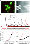
Figure 2. Kainate receptors recover from desensitization in multiple steps
A, photomicrographs showing the green fluorescent protein (GFP)-stained tsA201 cells and arrangement of the fast agonist perfusion system used to study KAR desensitization kinetics. B, typical experiment showing conditioning and test agonist pulses used to monitor rates into and out of desensitization. A conditioning and a test pulse are highlighted in red. C, summary plot of GluR6 KAR recovery from desensitization in its entirety (left) or in the early stages (right). The continuous red line in each denotes the relationship expected of first order kinetics. Adapted from Bowie & Lange (2002) with permission from the Society for Neuroscience.
Figure 3
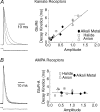
Figure 3. External anions and cations distinguish between AMPA and kainate receptors
A, macroscopic KAR desensitization is regulated by ion concentration and ion type. Left, plot showing typical membrane currents elicited by GluR6 KARs in symmetrical solutions of 55, 150 and 405 mm NaCl. Right, plot comparing the KAR response amplitude and desensitization kinetics in different external ions. Note that the data are fitted well by a linear regression analysis showing that ions have a concomitant effect on both response amplitude and decay kinetics. B, unlike KARs, AMPAR desensitization is not regulated by changes in external ion concentration (left) nor by the type of ion present (right). Adapted from Bowie & Lange (2002) with permission from the Society for Neuroscience and Bowie (2002).
Figure 4
Figure 4. KARs have an absolute requirement for external ions
A, a family of membrane currents evoked by 1 mm l-Glu in the absence of all external ions at a range of membrane potentials (−100 to +110 mV, 15 mV increments). Responses evoked at GluR1 AMPARs (middle) and GluR6M770K KARs (right) are outward currents due to the outflow of Na+ ions from the internal pipette solution. In contrast, in identical solutions, GluR6 KARs are unresponsive (left). B, sequence alignment of all iGluR subfamilies around the extracellular M2–M3 linker region. The methionine-770 residue is highlighted by the asterisk and is coloured red. Adapted from Wong et al. (2006) with permission from the Society for Neuroscience.
Figure 5
Figure 5. External ions regulate occupancy of a novel, high-affinity inactivated state
A, left, family of inhibition curves to l-Glu in 5 (filled triangle), 10 (open triangle), 150 (filled circle) and 405 mm (open circle) external NaCl. Continuous lines are fits to single- or double-binding site model. Right, inhibition curve observed in 5 mm external NaCl in more detail showing contribution of high- and low-affinity states. B, effect of external NaCl on the fractional occupancy of high- and low-affinity states. Dotted lines represent fit extrapolations. C, schematic diagram to illustrate a possible interpretation of data shown in panels A and B. At saturating levels of NaCl and agonist, all four subunits of the KAR tetramer are occupied at both the neurotransmitter- and ion-binding sites. In relative terms, this state has a low affinity for the agonist and is graphically represented as a tetramer containing four circles. However, as NaCl levels are lowered, fewer and fewer subunits contain bound Na+ ions, which has two effects. First, subunits that are unbound by ions (square symbol) exhibit a significantly higher agonist affinity. Second, ion-unbinding inactivates the KAR subunit and therefore the mature tetramer fails to gate. Adapted from Wong et al. (2006) with permission from the Society for Neuroscience.
Figure 6
Figure 6. Possible mechanisms for monovalent ion interactions at KARs
A, schematic diagram showing three distinct models to explain the effect of monovalent anions and cations on KARs. B, upper, crystal structure showing critical amino acid residues that constitute the proposed anion binding site for GluR5 KARs. Although only a single dimer is shown, the ion is also conjugated by the corresponding amino acids from the adjacent subunit. Lower, proposed anion binding pocket containing point mutations (R775K, D776E, and T779N) that interfere with both anion and cation modulation of KARs. C, summary plot showing that single point mutants which affect anion binding also have marked effects on cation modulation of KARs. Adapted from Wong et al. (2007) with permission from the Society for Neuroscience.
Figure 7
Figure 7. Variations in the cation binding pocket of iGluR families
Composite image showing the dimer interface of the δ-2 orphan-class iGluR, the GluR5 KAR and GluR2 AMPAR. In each case, amino acids have been labelled from the first Met residue in the N-terminal. Note that δ-2 subunits bind two Ca2+ ions (blue) (Naur et al. 2007) whereas the GluR5 subunit binds two Na+ (purple) and a single Cl (green) (Plested & Mayer, 2007). In contrast, the positively charged Lys residue acts as a tethered cation at GluR2 AMPARs. Figure is courtesy of Mark Aurousseau.
None

Similar articles

Cited by

References

    1. Akk G, Auerbach A. Inorganic, monovalent cations compete with agonists for the transmitter binding site of nicotinic acetylcholine receptors. Biophys J. 1996;70:2652–2658. - PMC - PubMed
    1. Aldrich RW, Corey DP, Stevens CF. A reinterpretation of mammalian sodium channel gating based on single channel recording. Nature. 1983;306:436–441. - PubMed
    1. Antonov SM, Gmiro VE, Johnson JW. Binding sites for permeant ions in the channel of NMDA receptors and their effects on channel block. Nat Neurosci. 1998;1:451–461. - PubMed
    1. Armstrong CM. Interaction of tetraethylammonium ion derivatives with the potassium channels of giant axons. J Gen Physiol. 1971;58:413–437. - PMC - PubMed
    1. Armstrong N, Gouaux E. Mechanisms for activation and antagonism of an AMPA-sensitive glutamate receptor: crystal structures of the GluR2 ligand binding core. Neuron. 2000;28:165–181. - PubMed

LinkOut - more resources