. 2009 Dec;29(24):6353-65.
doi: 10.1128/MCB.01051-09. Epub 2009 Oct 12.

Two fundamentally distinct PCNA interaction peptides contribute to chromatin assembly factor 1 function

Affiliations

Two fundamentally distinct PCNA interaction peptides contribute to chromatin assembly factor 1 function

Tom Rolef Ben-Shahar et al. Mol Cell Biol. 2009 Dec.

Abstract

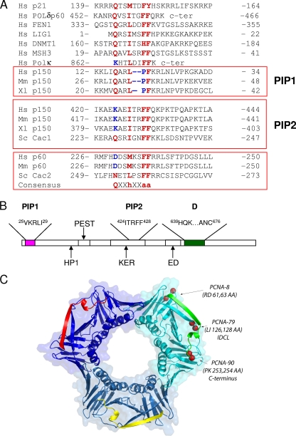
Chromatin assembly factor 1 (CAF-1) deposits histones H3 and H4 rapidly behind replication forks through an interaction with the proliferating cell nuclear antigen (PCNA), a DNA polymerase processivity factor that also binds to a number of replication enzymes and other proteins that act on nascent DNA. The mechanisms that enable CAF-1 and other PCNA-binding proteins to function harmoniously at the replication fork are poorly understood. Here we report that the large subunit of human CAF-1 (p150) contains two distinct PCNA interaction peptides (PIPs). The N-terminal PIP binds strongly to PCNA in vitro but, surprisingly, is dispensable for nucleosome assembly and only makes a modest contribution to targeting p150 to DNA replication foci in vivo. In contrast, the internal PIP (PIP2) lacks one of the highly conserved residues of canonical PIPs and binds weakly to PCNA. Surprisingly, PIP2 is essential for nucleosome assembly during DNA replication in vitro and plays a major role in targeting p150 to sites of DNA replication. Unlike canonical PIPs, such as that of p21, the two p150 PIPs are capable of preferentially inhibiting nucleosome assembly, rather than DNA synthesis, suggesting that intrinsic features of these peptides are part of the mechanism that enables CAF-1 to function behind replication forks without interfering with other PCNA-mediated processes.


Figures

FIG. 1.
FIG. 1.
CAF-1 p150 PIP and PCNA mutations. (A) Alignment of PIPs from human proteins to the motifs found in CAF-1 subunits derived from a number of distinct species: Hs, Homo sapiens; Mm, Mus musculus; Xl, Xenopus laevis; Sc, Saccharomyces cerevisiae. The conserved residues of canonical PIPs are highlighted in red, whereas nonconservative changes in the corresponding residues of CAF-1 p150 and DNA polymerase κ PIPs are shown in blue. (B) Location of PIPs in the primary structure of mouse CAF-1 p150 relative to previously described domains (25, 38, 44). (C) Front side view of a PCNA homotrimer (PDB 1AX8) (16) bound by three p21 PIPs (indicated in green, yellow, and red). The front side is the face of the PCNA ring that points in the direction of DNA synthesis. The mutations analyzed are present in the three PCNA monomers but, for clarity, are shown in the cyan subunit only.
FIG. 2.
FIG. 2.
Binding of CAF-1 p150 to the interdomain connector loop of PCNA is mediated by PIP1. (A) Wild-type human p150 and PIP mutants (∼9.5 pmol) were bound to p150 monoclonal antibody beads. Recombinant PCNA-His6 (∼100 pmol of trimer) was incubated with the p150 beads for 2 h at 4°C. After washes in buffer A, bound proteins were detected by immunoblotting. Results shown are for bound p150 (top panel) and bound PCNA (bottom panel). (B) Coomassie blue-stained gel of human p150 fragments purified from E. coli and bound to p150 monoclonal antibody beads. Recombinant CAF-1 purified from insect cells is also shown. (C) Only the PCNA-79 mutation in the interdomain connector loop affects PCNA binding via PIP1 in vitro. Wild-type and mutant forms of PCNA-His6 were incubated with beads coated with the p150 fragments shown in panel B for 1 h at 4°C. After washes in buffer A, bound PCNA-His6 was detected by Coomassie blue staining. Lane I, 40% of input PCNA; lane R, positive control with recombinant CAF-1 bound to beads; lane C, Negative control without p150 bound to beads.
FIG. 3.
FIG. 3.
Nucleosome assembly during SV40 DNA replication is dependent upon PIP2. (A) Wild-type and PIP mutants of human p150 were expressed in the rabbit reticulocyte lysate and detected by sodium dodecyl sulfate-polyacrylamide gel electrophoresis and autoradiography. (B) Nucleosome assembly reactions performed in the SV40 DNA replication system (without T antigen) in the presence of increasing amounts of wild-type p150 or PIP mutants. Each p150 protein was titrated over an eightfold range of protein concentrations in twofold increments. Replicated DNA was detected by incorporation of [α-32P]dAMP and autoradiography. Total DNA was stained with ethidium bromide. Lane 1, negative control reaction lacking p150.
FIG. 4.
FIG. 4.
PIP mutations result in mistargeting of mouse p150 to heterochromatin in early- and mid-S-phase cells. Mouse NIH 3T3 cells were transfected with constructs for cytomegalovirus promoter-driven expression of GFP-p150 PIP mutants and stained with a PCNA monoclonal antibody to detect DNA replication foci and Hoechst 33258 to reveal foci of pericentric heterochromatin. White arrows indicate examples of p150 PIP mutants that were mistargeted to heterochromatin in either early- or mid-S-phase cells. The images in panels A to F and J to O were obtained with the ΔPIP1 + 2 mutant, which rarely showed a full defect in GFP-p150 localization to DNA replication foci (Fig. 5). The images in panels G to I and P to R were derived from observations of the ΔD ΔPIP2 mutant, for which a complete lack of localization of GFP-p150 to DNA replication foci was very frequent (Fig. 5).
FIG. 5.
FIG. 5.
The two PIPs of mouse p150 function contribute to prevent mistargeting of CAF-1 p150 to heterochromatin. Severe defects in localization of p150 to DNA replication foci were observed in GFP-p150 mutants lacking both PIP2 and the dimerization region. Cells transfected with each PIP mutant were examined to assess the percentage of transfected cells showing mistargeting of GFP-p150 to heterochromatin in either early or mid-S phase. The fraction of cells with a partial defect in targeting (GFP-p150 observed in both replication foci and heterochromatin) is indicated by the stippled columns. The fraction of cells with a complete defect in targeting (GFP-p150 absent from replication foci and present in heterochromatin only) is represented by the black columns. ΔD, deletion of the dimerization motif.
FIG. 6.
FIG. 6.
Both PIPs of human p150 preferentially interfere with nucleosome assembly, rather than DNA synthesis, even though they exhibit striking differences in their respective abilities to bind to PCNA. (A) Synthetic peptides used in panels B to D. Residues characteristic of canonical PIPs are shown in bold. Wild-type and mutant peptides are denoted as w and m, respectively, and the mutated residues are underlined. (B) Biotinylated synthetic PIPs and PCNA-His6 were incubated in an equimolar ratio (ratio of peptide to PCNA monomer) in buffer A for 1 h at 4°C. The biotinylated peptides were isolated using streptavidin-coated magnetic beads and, after washes, bound PCNA-His6 was detected by immunoblotting. Lane I, 10% of input PCNA. (C) The p21 PIP has a higher affinity for PCNA than p150 PIP1. The same experiment as described for panel B was performed using S100 extract as a source of PCNA, either in the absence or presence of nonbiotinylated peptides that competed for PCNA binding to biotinylated PIPs. The molar ratio of biotinylated and competitor peptide in lanes 4 and 6 was 1:1. Lane I, 25% of input S100 extract. (D) T antigen-independent DNA synthesis reactions were performed in the S100 extract in the absence (negative control) or presence of 0.45 pmol of recombinant CAF-1 (rCAF-1; positive control), which was sufficient to promote supercoiling of all the replicated DNA (lanes 1 and 2 and 11 and 12). Increasing amounts of wild-type and mutant PIPs were titrated into reaction mixtures that contained fixed amounts of rCAF-1 to determine whether the peptides had any preference to inhibit nucleosome assembly rather than DNA synthesis. The amounts of peptide used were 0.2, 0.4, 0.8, and 1.6 nmol for the p21 PIPs (lanes 3 to 6 and 7 to 10) and 1.6, 3.2, and 6.4 nmol for all the p150 PIPs (lanes 13 to 24). Replicated DNA was detected by incorporation of [α-32P]dAMP and autoradiography. Total DNA was stained with ethidium bromide.
FIG. 7.
FIG. 7.
The noncanonical CAF-1 p150 PIP2 and DNA polymerase κ PIP bind directly to PCNA at low salt concentration based on isothermal titration calorimetry. Synthetic peptides were progressively titrated into a sample cell containing PCNA-His6. In each panel, the upper half shows the measured heat exchanges during each peptide injection. The lower half of each panel shows the enthalpic changes as a function of the molar ratio of peptide to PCNA monomer. The black squares correspond to the experimental data points. (A to C) Titrations performed in 10 mM sodium phosphate, pH 7.0, 10 mM NaCl. (D and E) Titrations performed in 20 mM HEPES-NaOH, pH 7.6, 100 mM NaCl.
FIG. 8.
FIG. 8.
Hydrophobic residues of CAF-1 p150 PIP2 are critical for its binding to PCNA-His6 as measured by STD NMR in 10 mM sodium phosphate, pH 7.0, 10 mM NaCl. 1H STD NMR spectra of wild-type PIP2 (A) and lysine-to-glutamine PIP2 (B) in the presence of PCNA-His6 (see Materials and Methods for details) are shown. Given their sequence similarities, many of the same peptide resonances were observed in the STD NMR spectra. In particular, the intense peaks originating from the ring protons of the Phe residues (∼7.25 ppm; labeled with an asterisk) and the methyl protons of Ile (∼0.85 ppm; labeled with an X) suggest they are in close proximity to PCNA-His6. (C) The 1H STD NMR spectrum of the the PIP2 FF-to-AA mutant in the presence of PCNA-His6 shows no signal, indicating that they do not interact under these conditions.

References

    1. Ayyagari, R., K. J. Impellizzeri, B. L. Yoder, S. L. Gary, and P. M. J. Burgers. 1995. A mutational analysis of the yeast proliferating cell nuclear antigen indicates distinct roles in DNA replication and DNA repair. Mol. Cell. Biol. 15:4420-4429. - PMC - PubMed
    1. Bi, X., L. R. Barkley, D. M. Slater, S. Tateishi, M. Yamaizumi, H. Ohmori, and C. Vaziri. 2006. Rad18 regulates DNA polymerase κ and is required for recovery from S-phase checkpoint-mediated arrest. Mol. Cell. Biol. 26:3527-3540. - PMC - PubMed
    1. Bienko, M., C. M. Green, N. Crosetto, F. Rudolf, G. Zapart, B. Coull, P. Kannouche, G. Wider, M. Peter, A. R. Lehmann, K. Hofmann, and I. Dikic. 2005. Ubiquiting-binding domains in Y-family polymerases regulate translesion synthesis. Science 310:1821-1824. - PubMed
    1. Bowman, G. D., M. O'Donnell, and J. Kuriyan. 2004. Structural analysis of a eukaryotic sliding DNA clamp-clamp loader complex. Nature 429:724-730. - PubMed
    1. Bruning, J. B., and Y. Shamoo. 2004. Structural and thermodynamic analysis of human PCNA with peptides derived from DNA polymerase δ p66 subunit and FLAP endonuclease 1. Structure 12:2209-2219. - PubMed

Publication types

MeSH terms

LinkOut - more resources