. 2009 Dec;29(24):6515-26.
doi: 10.1128/MCB.00489-09. Epub 2009 Oct 12.

Identification of a novel amino acid response pathway triggering ATF2 phosphorylation in mammals

Affiliations

Identification of a novel amino acid response pathway triggering ATF2 phosphorylation in mammals

Cédric Chaveroux et al. Mol Cell Biol. 2009 Dec.

Abstract

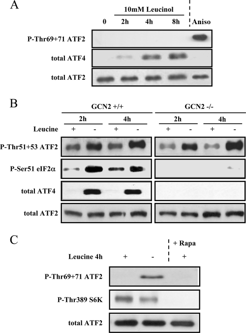
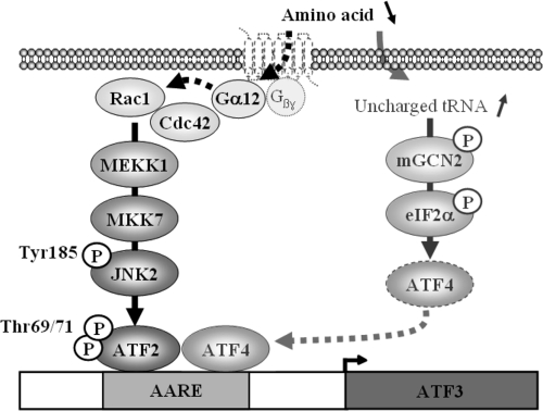
It has been well established that amino acid availability can control gene expression. Previous studies have shown that amino acid depletion induces transcription of the ATF3 (activation transcription factor 3) gene through an amino acid response element (AARE) located in its promoter. This event requires phosphorylation of activating transcription factor 2 (ATF2), a constitutive AARE-bound factor. To identify the signaling cascade leading to phosphorylation of ATF2 in response to amino acid starvation, we used an individual gene knockdown approach by small interfering RNA transfection. We identified the mitogen-activated protein kinase (MAPK) module MEKK1/MKK7/JNK2 as the pathway responsible for ATF2 phosphorylation on the threonine 69 (Thr69) and Thr71 residues. Then, we progressed backwards up the signal transduction pathway and showed that the GTPase Rac1/Cdc42 and the protein Galpha12 control the MAPK module, ATF2 phosphorylation, and AARE-dependent transcription. Taken together, our data reveal a new signaling pathway activated by amino acid starvation leading to ATF2 phosphorylation and subsequently positively affecting the transcription of amino acid-regulated genes.


Figures

FIG. 1.
FIG. 1.
Role of ATF2 in the transcriptional regulation of ATF3 in response to amino acid starvation. (A) ATF2−/−, ATF4−/−, and wild-type (WT) MEF were incubated for 4 h in either control or leucine-free (−Leu) medium and then harvested. Total RNA was extracted and analyzed by quantitative RT-PCR for ATF3 mRNA content as described in Materials and Methods. Each point represents the mean value of results from three independent experiments. We obtained the same results using the wild-type counterparts of the ATF2−/− and the ATF4−/− MEF (not shown). (B) ChIP analysis was performed using antibodies specific for ATF4, ATF2, and phosphorylated ATF2 (Thr71). Quantitative RT-PCR was performed with immunoprecipitated DNA and a dilution of input DNA samples, using primers to amplify the AARE region of the ATF3 promoter. Data were plotted as percentages of PCR product from immunoprecipitated DNA versus input DNA. Each point represents the mean value of results from three independent experiments, and the error bars represent the standard errors of the means (* indicates statistical significance at P values of <0.05). IgG, immunoglobulin G. (C) Cells were incubated in either control or leucine-free medium and harvested after the indicated incubation times. Western blot analysis of phospho-ATF2 (Thr71 and Thr69+Thr71) and total ATF2 was performed with nuclear extract as described in Materials and Methods. (D) Cells were incubated for 4 h either in control medium (Ctrl) or in a medium devoid of one individual amino acid (leucine, lysine, methionine, or glutamine), and then analysis of phospho-ATF2 (Thr69+Thr71), ATF2, and ATF4 was performed.
FIG. 2.
FIG. 2.
ATF2 phosphorylation upon leucine starvation does not involve the GCN2 and mTORC1 pathways. (A) Cells were incubated with 10 mM leucinol for the indicated times, and then ATF2 phosphorylation and ATF4 expression were analyzed as previously described. A 30-minute incubation with 20 μg/ml anisomycin (Aniso) was used as a positive control for the measurement of ATF2 phosphorylation. (B) GCN2+/+ or GCN2−/− MEF were incubated in either control or leucine-free medium for 2 and 4 h. Cells were then harvested for analysis of ATF4 expression and phosphorylation of ATF2 and eIF-2α. (C) Cells were leucine starved for 4 h or treated with 50 nM rapamycin (Rapa) for 4 h, and then the phosphorylations of ATF2 and S6K were measured.
FIG. 3.
FIG. 3.
Pharmacological inhibition of JNK prevents ATF2 phosphorylation by leucine starvation. Cells were incubated in either control or leucine-free medium for 4 hours in the presence of various MAPK inhibitors (20 μM SB203580 inhibits p38, 50 μM U0126 inhibits MEK1/2 and thus ERK, and 20 μM JI8 and 50 μM SP600125 inhibit JNK). The phosphorylation of ATF2 was then measured. For this experiment, cells were first preincubated for 1 h with MAPK inhibitors or vehicle (dimethyl sulfoxide [DMSO]).
FIG. 4.
FIG. 4.
Silencing of JNK2 by siRNA prevents ATF2 phosphorylation by leucine starvation. (A) Cells were incubated in either control or leucine-free medium and harvested after the indicated incubation times. The phosphorylated forms of JNK (Tyr185) and ATF2 were measured. Each blot was obtained from a representative experiment among three independent experiments. (B) HeLa cells were transfected with control siRNA, JNK1 siRNA, JNK2 siRNA, or siRNA targeting both JNK1 and JNK2, as described in Materials and Methods. At 72 h posttransfection, cells were incubated in either DMEM or leucine-free DMEM for 4 h and then harvested for analysis of ATF2, JNK1, JNK2, ATF4, and the phosphorylated form of ATF2. Each blot was obtained from a representative experiment among three independent experiments.
FIG. 5.
FIG. 5.
Knockdown of MKK7 or MEKK1 inhibits leucine starvation-induced ATF2 phosphorylation. (A) HeLa cells were transfected with either control siRNA, MKK4 siRNA, MKK7 siRNA, or both MKK4 siRNA and MKK7 siRNA. At 72 h posttransfection, cells were incubated in either DMEM or leucine-free DMEM for 4 h. Cells were then harvested for analysis of MKK4, MKK7, ATF4, and the phosphorylated form of ATF2 as previously described. (B) MEKK1, MEKK2, MEKK3, or all three proteins were silenced as previously described. Then, the expression levels of MEKK1, MEKK2, MEKK3, ATF4, and the phosphorylated form of ATF2 were analyzed. (C) Protein extracts from cells transfected with either control, MKK7, or MEKK1 siRNA were analyzed for JNK phosphorylation. Each blot was obtained from a representative experiment among three independent experiments.
FIG. 6.
FIG. 6.
Silencing of Cdc42 and Rac1 prevents ATF2 phosphorylation in response to leucine starvation. Rac1, Cdc42, or both were silenced as previously described. The phosphorylated form of ATF2 and the expression of Rac1, Cdc42, and ATF4 were measured. Each blot was obtained from a representative experiment among four independent experiments.
FIG. 7.
FIG. 7.
Involvement of the Gα12 protein in leucine starvation-induced ATF2 phosphorylation. HeLa cells were transfected with either control siRNA, Gα12 siRNA, Gα13 siRNA, or both Gα12 siRNA and Gα13 siRNA. At 72 h posttransfection, cells were incubated in either DMEM or leucine-free DMEM for 4 h. Cells were then harvested for analysis of Gα12, Gα13, ATF4, and the phosphorylated form of ATF2 as previously described. Each blot was obtained from a representative experiment among three independent experiments. (B) HEK293 cells were transfected with an activated mutant of Gα12 (Gα12 QL) or empty vector (pcDNA3). At 48 h posttransfection, cells were lysed, then protein was harvested for analysis of Gα12, and the phosphorylated forms of ATF2 and JNK were measured as previously described.
FIG. 8.
FIG. 8.
Role of JNK2 and Rac1/Cdc42 in the transcriptional regulation of ATF3 in response to amino acid starvation. (A) HeLa cells were transfected with control siRNA, JNK1 siRNA, or JNK2 siRNA and then incubated in a control medium or leucine-free medium (−Leu) for 4 h as described for Fig. 4A. Total RNA was then extracted and analyzed by real-time RT-PCR for ATF3 or ASNS mRNA content. (B) Cells were transfected with the appropriate siRNA, together with a reporter plasmid containing two copies of the ATF3 or CHOP AARE inserted 5′ to the thymidine kinase promoter driving the Luc gene (2xAARE-Tk-Luc). A plasmid encoding β-galactosidase, driven by a cytomegalovirus promoter, was cotransfected to normalize transfection (see Materials and Methods). At 48 h posttransfection, cells were incubated in either DMEM or leucine-free DMEM for 16 h and then harvested to measure Luc activity. (C) HeLa cells were transfected with control siRNA or with Rac1 siRNA and Cdc42 siRNA. Cells were then treated and analyzed as described for panel B. All values are means calculated from the results for at least three independent experiments performed in triplicate. Data are expressed as mean ± standard error of the mean. Statistical analyses were performed using a Student test. Asterisks indicate that −Leu/siRNA-treated cells had statistical significance at P values of <0.05 for comparison with the −Leu/control siRNA-treated cells.
FIG. 9.
FIG. 9.
Model of amino acid starvation-induced AARE-dependent transcription in human cells. In response to amino acid starvation, two pathways are activated. The first one is activated by uncharged tRNA and leads to ATF4 induction. The second one involves the Gα12 protein and leads to ATF2 phosphorylation.

References

    1. Almeida, E. A., D. Ilic, Q. Han, C. R. Hauck, F. Jin, H. Kawakatsu, D. D. Schlaepfer, and C. H. Damsky. 2000. Matrix survival signaling: from fibronectin via focal adhesion kinase to c-Jun NH(2)-terminal kinase. J. Cell Biol. 149:741-754. - PMC - PubMed
    1. Averous, J., A. Bruhat, C. Jousse, V. Carraro, G. Thiel, and P. Fafournoux. 2004. Induction of CHOP expression by amino acid limitation requires both ATF4 expression and ATF2 phosphorylation. J. Biol. Chem. 279:5288-5297. - PubMed
    1. Baan, B., H. van Dam, G. C. van der Zon, J. A. Maassen, and D. M. Ouwens. 2006. The role of c-Jun N-terminal kinase, p38, and extracellular signal-regulated kinase in insulin-induced Thr69 and Thr71 phosphorylation of activating transcription factor 2. Mol. Endocrinol. 20:1786-1795. - PubMed
    1. Baertl, J. M., R. P. Placko, and G. G. Graham. 1974. Serum proteins and plasma free amino acids in severe malnutrition. Am. J. Clin. Nutr. 27:733-742. - PubMed
    1. Barbosa-Tessmann, I. P., C. Chen, C. Zhong, F. Siu, S. M. Schuster, H. S. Nick, and M. S. Kilberg. 2000. Activation of the human asparagine synthetase gene by the amino acid response and the endoplasmic reticulum stress response pathways occurs by common genomic elements. J. Biol. Chem. 275:26976-26985. - PubMed

Publication types

MeSH terms

Substances