. 2009 Oct 19;187(2):201-17.
doi: 10.1083/jcb.200903024. Epub 2009 Oct 12.

Ubiquilin and p97/VCP bind erasin, forming a complex involved in ERAD

Affiliations

Ubiquilin and p97/VCP bind erasin, forming a complex involved in ERAD

Precious J Lim et al. J Cell Biol. .

Abstract

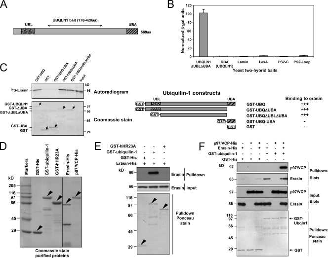
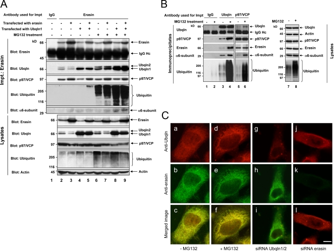
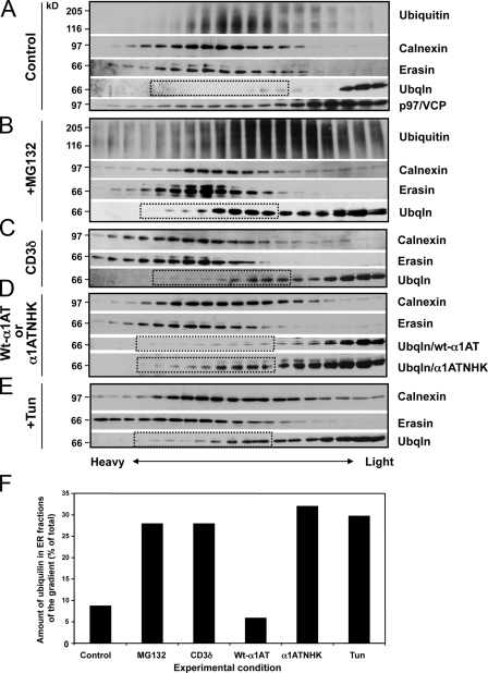
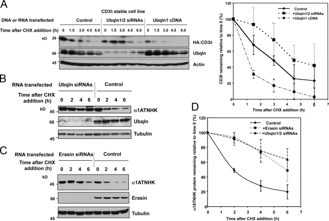
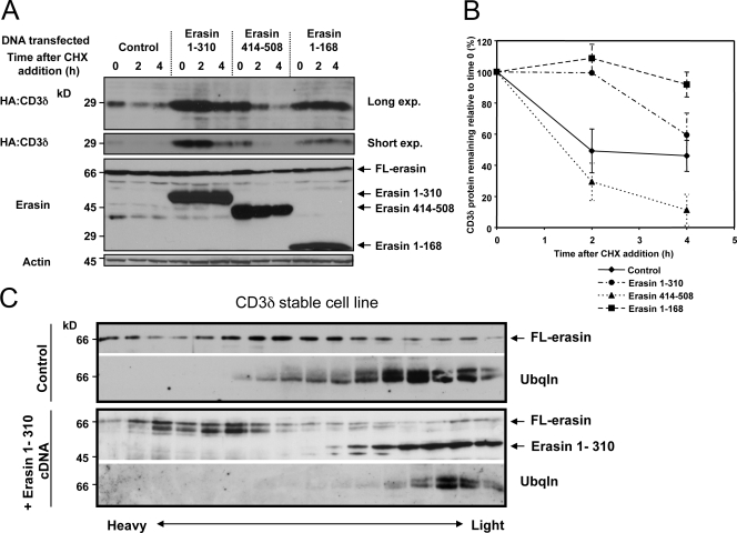
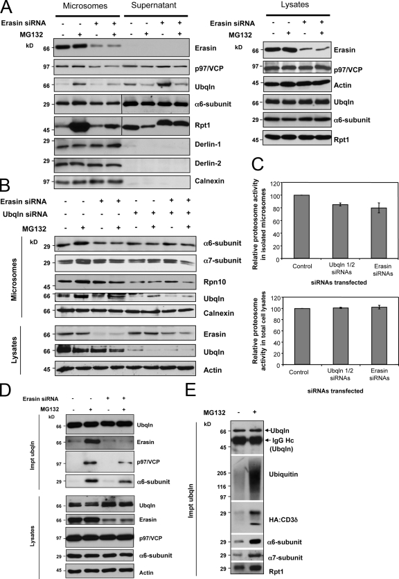
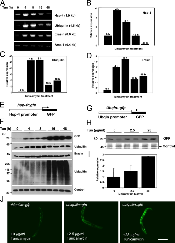
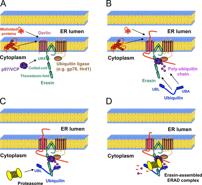
Unwanted proteins in the endoplasmic reticulum (ER) are exported into the cytoplasm and degraded by the proteasome through the ER-associated protein degradation pathway (ERAD). Disturbances in ERAD are linked to ER stress, which has been implicated in the pathogenesis of several human diseases. However, the composition and organization of ERAD complexes in human cells is still poorly understood. In this paper, we describe a trimeric complex that we propose functions in ERAD. Knockdown of erasin, a platform for p97/VCP and ubiquilin binding, or knockdown of ubiquilin in human cells slowed degradation of two classical ERAD substrates. In Caenorhabditis elegans, ubiquilin and erasin are ER stress-response genes that are regulated by the ire-1 branch of the unfolded protein response pathway. Loss of ubiquilin or erasin resulted in activation of ER stress, increased accumulation of polyubiquitinated proteins, and shortened lifespan in worms. Our results strongly support a role for this complex in ERAD and in the regulation of ER stress.


Figures

Figure 1.
Figure 1.
Erasin interaction with ubiquilin-1, p97/VCP, and hHR23A. (A) Schematic drawing of ubiquilin-1. Ubiquilin-1ΔUBLΔUBA (178–428 aa) was used as the bait in a Y2H screen. (B) Yeast interaction assay of an erasin prey clone with different baits measured in five independent colonies showing the mean and SD (error bars). (C) GST pull-down assays of ubiquilin-1 (UBQ) fusion proteins with 35S-radiolabeled erasin. (C, right) Schematic drawings of the GST constructs and a summary of their binding (+, binding; −, no binding). (C, left) Autoradiogram (top panel) and Coomassie-stained gel (bottom panel) of the pull-down experiment. (D) Coomassie-stained gel of purified proteins used for pull-down assays. The black line indicates that intervening lanes have been spliced out. (E) GST pull-down assays showing that erasin-His binds to GST-ubiquilin but not to GST or GST-hHR23A proteins. The input erasin, the recovered erasin, and GST proteins are shown. (F) GST pull-down assays showing that erasin, p97/VCP, and ubiquilin bind together in a trimeric complex. The input p97/VCP and erasin proteins and the recovered GST complexes (ponceau) are also shown. Arrowheads indicate the position of full-length GST-fusion proteins.
Figure 2.
Figure 2.
Erasin, ubiquilin, and p97/VCP coimmunoprecipitate with each other in cells. (A) HEK293 cells were either left untransfected (lanes 1, 2, and 6), transfected with either erasin (lanes 3 and 7) or ubiquilin-1 cDNA (lane 4 and 8), or cotransfected with both cDNAs (lane 5 and 9). After 20 h, MG132 was added for 4 h to one set of the cultures (lanes 6–9), whereas the other set was left untreated (lanes 1–5). Equivalent amounts of protein lysates were used for immunoprecipitation using either an erasin antibody (lanes 2–9) or control IgG antibody (lane 1). The immunoprecipitates as well as 1/10 the amount of each protein lysate were probed by immunoblotting for the indicated proteins. (B) Similar to A except that cells were not transfected and the antibodies used for immunoprecipitation were a different control antibody (lanes 1 and 2), a ubiquilin antibody (lanes 3 and 4), and a p97/VCP antibody (lanes 5 and 6). (lanes 7 and 8) Blots for proteins in equal amounts of the lysates used in the experiments. (C) Double immunofluorescence localization of ubiquilin and erasin proteins in HEK293 cells that were either not treated (−) or treated (+) with MG132, or transfected with siRNAs designed to knock down ubiquilin-1 and ubiquilin-2 proteins together, or erasin protein alone. Quantification of the colocalization of the proteins is provided in Fig. S3. Bar, 5 µm.
Figure 3.
Figure 3.
Proteasome inhibition or expression of ERAD substrates leads to a redistribution of ubiquilin to the ER. Immunoblot analysis of HEK293 cell homogenates separated on 0–25% iodixanol gradients. (A–E) The panels show the distribution of erasin, calnexin, and ubiquilin in fractions of the gradient (heavy, bottom; light, top) of cells that were either left untreated; treated with MG132 for 4 h; transfected with either CD3δ, α1ATNHK, or wild-type α1AT cDNAs; or treated with tunicamycin for 4 h (+Tun). Ubiquitin immunoreactivity is shown for the −MG132 and +MG132 and treatments. Because its profile did not change significantly, p97/VCP is only shown for the control. (F) The amount of ubiquilin in the ER fractions was estimated by dividing the intensity of the anti-ubiquilin–immunoreactive bands in nine fractions surrounding the calnexin peak (boxed with broken lines in A–E) by the sum of the bands in the whole gradient.
Figure 4.
Figure 4.
Knockdown of ubiquilin or erasin proteins impedes ERAD. (A) HEK293-CD3δ cells were transfected with either a ubiquilin-1 expression plasmid, the empty vector, or with UBQLN1 and UBQLN2 siRNAs. 48 h after transfection, cycloheximide was added to the cultures, and protein lysates were collected at intervals as indicated. Equal amounts of protein lysates were immunoblotted for CD3δ (HA-tag), ubiquilin, and actin. The graph shows the profile of CD3δ turnover determined from three independent experiments showing the mean and the SD (error bars). (B–D) Similar to A, except that turnover of α1ATNHK was analyzed after knockdown of either ubiquilin (B) or erasin (C). In these experiments, HEK293 cells were transfected with an α1ATNHK-HA expression construct 40 h after transfection of the siRNAs, with the turnover studies conducted 20 h later. Equal amounts of protein were immunoblotted for α1ATNHK (HA-tag), tubulin, and erasin or ubiquilin, depending on which of them was knocked down. (D) Quantification of α1ATNHK turnover after knockdown of either ubiquilin or erasin proteins, determined from three separate experiments showing the mean and SD (error bars).
Figure 5.
Figure 5.
Dominant-negative erasin fragments interfere with CD3δ degradation. (A) HEK293-CD3δ cells were either mock transfected (control) or transfected with erasin deletion constructs encoding portions of the erasin polypeptide from amino acids 1–310, 414–508, or 1–168. Protein turnover was analyzed similar to Fig 4. Both a long and short exposure of the CD3δ signal is shown. Please note that the erasin 414–508 runs aberrantly on SDS-PAGE. (B) Graphs of CD3δ turnover determined from three separate experiments showing the mean and SD (error bars). (C) Immunoblots showing ubiquilin and erasin distribution in fractions of HEK293-CD3δ cell homogenates separated on 0–25% iodixanol gradients.
Figure 6.
Figure 6.
Microsomes of cells depleted of erasin contain less ubiquilin, p97/VCP, and proteasomes. HEK293 cells were either mock transfected or transfected with siRNAs against erasin. 72 h after transfection, the cells were incubated with MG132 for an additional 4 h, whereas a duplicate set was left untreated. (A) Equal amounts of protein in microsome and supernatant fractions of the cells were separated by SDS-PAGE and probed for the proteins shown. Blots of equal amounts of proteins in total lysates of the cultures are also shown. (B) Similar to A, except that this time erasin or ubiquilin-1 and -2 were knocked down separately, or simultaneously. Additionally, only the microsome fractions were probed for proteasome subunits (α6, α7, and Rpn10), ubiquilin, and calnexin (to ensure equal protein loading). Also shown are the blots of the total lysates of the cultures. (C) Assays of proteasome activity in microsomes and total lysates of HEK293-CD3δ cells that were either mock transfected or transfected with siRNAs against erasin or ubiquilin-1 and -2 of three separate cultures showing the mean and SD (error bars). (D) A repeat of the experiment shown in A, but this time we immunoprecipitated ubiquilin from the cells and examined equal fractions of the precipitates by immunoblotting for the indicated proteins. Blots of the total lysates are also shown. (E) Ubiquilin was immunoprecipitated from HEK293-CD3δ cells that were either left untreated or treated with MG132 for 4 h. Equal portions of the immunoprecipitates were probed for the proteins shown.
Figure 7.
Figure 7.
Ubiquilin and erasin expression are induced by ER stress in C. elegans. (A) Mixed-stage zcls4 [hsp-4::GFP] worms were grown on plates with 28 µg/ml tunicamycin for 0, 4, 8, 16, and 48 h, and transcript levels of hsp-4, ubiquilin, erasin, and ama-I were measured by semiquantitative PCR and gel electrophoresis. Quantification of Hsp-4 (B), ubiquilin (C), and erasin (D) expression relative to the 0 time point. Similar trends were observed in two other experiments. (E) Schematic diagram of the hsp-4::GFP reporter construct. (F) Immunoblot analysis of equal amounts of protein lysate (two independent samples for each time point) prepared from worms treated with 28 µg/ml of tunicamycin for the periods indicated. An unknown protein whose expression was insensitive to ER stress was used as a loading control. (G) Schematic diagram of the C. elegans ubiquilin promoter fragment fused to GFP. (H) Immunoblot analysis of equal amounts of protein lysate of worms treated with 0, 2.5, or 28 µg/ml tunicamycin for 24 h. (I) Quantification of GFP expression determined by scanning of immunoblots as described in H showing the mean and SD (error bars) seen in three different experiments. (J) GFP fluorescence of worms containing the integrated ubiquilin::GFP fusion construct treated for 24 h with 0, 2.5, or 28 µg/ml tunicamycin. Bar, 0.2 mm.
Figure 8.
Figure 8.
The ire-1 branch of the UPR regulates induction of ubiquilin and erasin expression during ER stress. (A) Mixed-stage worms of the indicated mutant strains were grown on plates with 28 µg/ml tunicamycin for 0 or 6 h. Transcript levels of hsp-4 (regulated by ire-1; Shen et al., 2001), ubiquilin, erasin, and CBP were analyzed by PCR and agarose gel electrophoresis. (B–E) Quantification of the relative change in transcript expression determined from the experiment shown in A. Levels were normalized to the 0 h time point in each mutant. Similar results were observed in two other experiments.
Figure 9.
Figure 9.
Knockdown of ubiquilin or erasin proteins in worms results in the induction of the ER stress reporter hsp-4::GFP, accumulation of misfolded proteins, and a shorter lifespan. (A) Mixed-stage zcls4 [hsp-4::GFP] worms were either fed standard bacteria in the presence or absence of 28 µg/ml tunicamycin or bacteria to induce RNAi of the ubiquilin gene. Immunoblot analysis of equal amounts of protein lysate prepared after 48 h of treatment for GFP, ubiquilin, erasin, ubiquitin, and an unknown protein used as a loading control. (B) Identical to A, except that worms were fed bacteria to specifically knock down erasin. (C) Lifespan curves comparing N2 (wild-type) worms fed bacteria transformed with the empty RNAi vector or the vector containing cDNAs to induce RNAi of either ubiquilin or erasin genes. Results are cumulative from three independent experiments, with ≥60 animals per trial. Log-rank (Mantel-Cox) and Gehan-Breslow-Wilcoxon tests, P < 0.0001 versus control.
Figure 10.
Figure 10.
Model of the mammalian erasin-assembled ERAD complex. A schematic diagram of the putative erasin-containing ERAD complex (see Discussion).

References

    1. Alexandru G., Graumann J., Smith G.T., Kolawa N.J., Fang R., Deshaies R.J. 2008. UBXD7 binds multiple ubiquitin ligases and implicates p97 in HIF1alpha turnover.Cell. 134:804–816 doi: - DOI - PMC - PubMed
    1. Ballar P., Shen Y., Yang H., Fang S. 2006. The role of a novel p97/valosin-containing protein-interacting motif of gp78 in endoplasmic reticulum-associated degradation.J. Biol. Chem. 281:35359–35368 doi: - DOI - PubMed
    1. Ballar P., Zhong Y., Nagahama M., Tagaya M., Shen Y., Fang S. 2007. Identification of SVIP as an endogenous inhibitor of endoplasmic reticulum-associated degradation.J. Biol. Chem. 282:33908–33914 doi: - DOI - PubMed
    1. Brenner S. 1974. The genetics of Caenorhabditis elegans.Genetics. 77:71–94 - PMC - PubMed
    1. Brodsky J.L. 2007. The protective and destructive roles played by molecular chaperones during ERAD (endoplasmic-reticulum-associated degradation).Biochem. J. 404:353–363 doi: - DOI - PMC - PubMed

Publication types

MeSH terms