. 2009 Oct 19;187(2):165-73.
doi: 10.1083/jcb.200903096. Epub 2009 Oct 12.

Differential regulation of telomere and centromere cohesion by the Scc3 homologues SA1 and SA2, respectively, in human cells

Affiliations

Differential regulation of telomere and centromere cohesion by the Scc3 homologues SA1 and SA2, respectively, in human cells

Silvia Canudas et al. J Cell Biol. .

Abstract

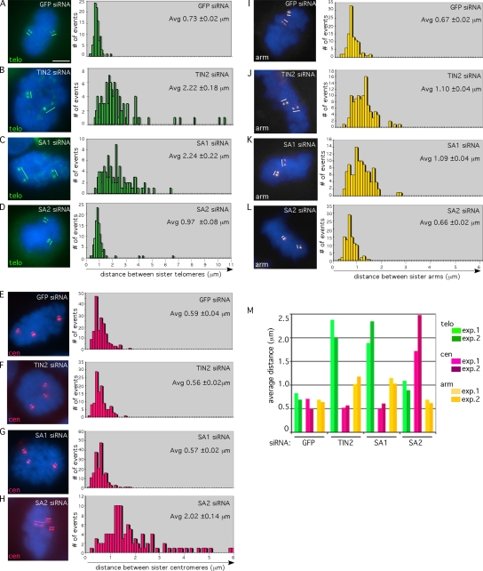
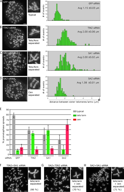
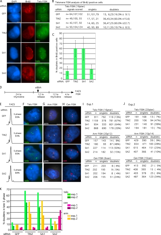
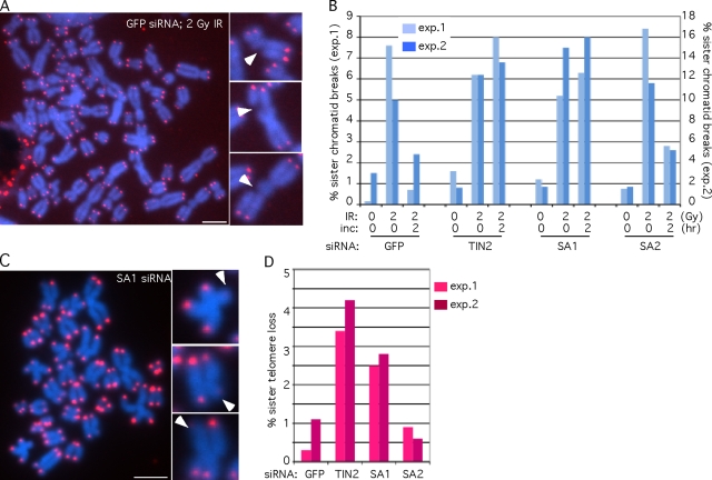
Replicated sister chromatids are held together until mitosis by cohesin, a conserved multisubunit complex comprised of Smc1, Smc3, Scc1, and Scc3, which in vertebrate cells exists as two closely related homologues (SA1 and SA2). Here, we show that cohesin(SA1) and cohesin(SA2) are differentially required for telomere and centromere cohesion, respectively. Cells deficient in SA1 are unable to establish or maintain cohesion between sister telomeres after DNA replication in S phase. The same phenotype is observed upon depletion of the telomeric protein TIN2. In contrast, in SA2-depleted cells telomere cohesion is normal, but centromere cohesion is prematurely lost. We demonstrate that loss of telomere cohesion has dramatic consequences on chromosome morphology and function. In the absence of sister telomere cohesion, cells are unable to repair chromatid breaks and suffer sister telomere loss. Our studies elucidate the functional distinction between the Scc3 homologues in human cells and further reveal an essential role for sister telomere cohesion in genomic integrity.


Figures

Figure 1.
Figure 1.
TIN2 and SA1 are required for telomere and arm cohesion, whereas SA2 is required for centromere cohesion. FISH analysis of siRNA-treated HeLaI.2.11 mitotic cells with chromosome-specific fluorescently labeled probes: (A–D) telomere 16pter (green), (E–H) centromere 6cen (red), and (I–L) arm 20p12 (white). The cen locus is trisomic. DNA was stained with DAPI (blue). Bar, 5 μm. Histograms (based on 100 measurements from two independent experiments [see Table S1]) showing the distance between FISH signals are on the right. Average (Avg) distance with SEs is indicated. (M) Graphical representation of the average distance from two independent experiments.
Figure 2.
Figure 2.
SA1 and TIN2, but not SA2, are required for sister chromatid cohesion along arms. (A–D) Chromosome spread analysis of siRNA-treated HeLaI.2.11 cells stained with antibodies to the condensin subunit Smc2. Histograms (based on ∼110 measurements from three spreads [see Table S2]) showing the distance between arms are on the right. Average distance (Avg) with SEs is indicated. (E) Graphical representation of the frequency of each type of chromosome morphology. The classification was assigned when five or more chromosomes in a spread displayed the indicated morphology. Bar graphs represent the average values from three independent experiments with SDs (see Table I). (F–H) Chromosome spread analysis from double siRNA–treated cells. 150–200 spreads were scored (see Table S3). The percentage indicates the frequency of the indicated morphology. Bars, 5 μm.
Figure 3.
Figure 3.
Sister telomere cohesion is lost prematurely (or not established) in S phase in SA1- and TIN2-depleted cells. (A–C) FISH analysis of BrdU-positive cells. (A) siRNA-treated HeLaI.2.11 cells were incubated with BrdU for 60 min before harvest, stained with anti-BrdU antibody (red), and hybridized with a telomere-specific fluorescently labeled probe 16pter (green). DNA was stained with DAPI (blue). (B) Table showing the number of FISH signals scored in BrdU-positive cells as singlet or doublets from three independent experiments with SDs. (C) Graphical representation of the frequency of doublets in BrdU-positive cells. Bar graphs represent the average values with SDs. (D–K) FISH analysis of late S phase synchronized cells. (D) Schematic representation of the experimental protocol to synchronize siRNA-treated cells. (E) FACS analysis and (F–H) FISH analysis of cells 4 h after release from the second thymidine arrest. Cells were hybridized with a telomere 16pter (F, green), arm 20p12 (G, white), or centromere 6cen (H, red) probe. Asterisks indicate a centromere that has lost cohesion. DNA was stained with DAPI (blue). Bars, 5 μm. (I) Tables showing the FISH signals scored as singlets or doublets from exp. 1 for the telomere (16pter), arm (20p12), or centromere (6cen) probe. (J) Tables showing the FISH signals scored as singlets or doublets from exp. 2 for the telomere (20qter), arm (10p14), or centromere (10cen) probe. (K) Graphical representation of the frequency of doublets from two independent experiments.
Figure 4.
Figure 4.
Sister telomere cohesion is required for double-strand break repair and telomere maintenance. (A and B) SA1- and TIN2-depleted cells are defective in sister chromatid repair after ionizing radiation. siRNA-treated HeLaI.2.11 cells were irradiated with 2 Gy of ionizing radiation (IR), and allowed to recover for 0 or 2 h. Prometaphase spreads were analyzed by hybridization to Cy3-conjugated telomere repeat (CCCTAAA)3 peptide nucleic acid (PNA) probe (red). DNA was stained with DAPI (blue). (A) Chromosome spread of GFP siRNA cells treated with 2 Gy IR. Three enlarged examples of sister chromatid breaks (indicated by arrowheads) are shown. (B) Graphical representation of the percentage of sister chromatid breaks. Approximately 500 chromosomes were scored for each sample from two independent experiments (exp.1, x axis on the left; exp. 2, x axis on the right; see Table II). (C and D) Sister telomeres are lost in SA1- and TIN2-depleted cells. siRNA-treated HeLaI.2.11 cells were processed for PNA-FISH as described above. (C) Chromosome spread of SA1 siRNA cells. Three enlarged examples of sister telomere loss (indicated by arrowheads) are shown. Bars, 5 μm. (D) Graphical representation of the percentage of sister telomere loss. Approximately 500 chromosomes were scored for each sample from two independent experiments (see Table II).

References

    1. Ancelin K., Brunori M., Bauwens S., Koering C.E., Brun C., Ricoul M., Pommier J.P., Sabatier L., Gilson E. 2002. Targeting assay to study the cis functions of human telomeric proteins: evidence for inhibition of telomerase by TRF1 and for activation of telomere degradation by TRF2.Mol. Cell. Biol. 22:3474–3487 doi:10.1128/MCB.22.10.3474-3487.2002 - DOI - PMC - PubMed
    1. Anderson D.E., Losada A., Erickson H.P., Hirano T. 2002. Condensin and cohesin display different arm conformations with characteristic hinge angles.J. Cell Biol. 156:419–424 doi:10.1083/jcb.200111002 - DOI - PMC - PubMed
    1. Blasco M.A. 2007. The epigenetic regulation of mammalian telomeres.Nat. Rev. Genet. 8:299–309 doi:10.1038/nrg2047 - DOI - PubMed
    1. Canudas S., Houghtaling B.R., Kim J.Y., Dynek J.N., Chang W.G., Smith S. 2007. Protein requirements for sister telomere association in human cells.EMBO J. 26:4867–4878 doi:10.1038/sj.emboj.7601903 - DOI - PMC - PubMed
    1. Cesare A.J., Reddel R.R. 2008. Telomere uncapping and alternative lengthening of telomeres.Mech. Ageing Dev. 129:99–108 doi:10.1016/j.mad.2007.11.006 - DOI - PubMed

Publication types