. 2009 Oct 19;187(2):233-46.
doi: 10.1083/jcb.200902117. Epub 2009 Oct 12.

Pex3 peroxisome biogenesis proteins function in peroxisome inheritance as class V myosin receptors

Affiliations

Pex3 peroxisome biogenesis proteins function in peroxisome inheritance as class V myosin receptors

Jinlan Chang et al. J Cell Biol. .

Abstract

In Saccharomyces cerevisiae, peroxisomal inheritance from mother cell to bud is conducted by the class V myosin motor, Myo2p. However, homologues of S. cerevisiae Myo2p peroxisomal receptor, Inp2p, are not readily identifiable outside the Saccharomycetaceae family. Here, we demonstrate an unexpected role for Pex3 proteins in peroxisome inheritance. Both Pex3p and Pex3Bp are peroxisomal integral membrane proteins that function as peroxisomal receptors for class V myosin through direct interaction with the myosin globular tail. In cells lacking Pex3Bp, peroxisomes are preferentially retained by the mother cell, whereas most peroxisomes gather and are transferred en masse to the bud in cells overexpressing Pex3Bp or Pex3p. Our results reveal an unprecedented role for members of the Pex3 protein family in peroxisome motility and inheritance in addition to their well-established role in peroxisome biogenesis at the endoplasmic reticulum. Our results point to a temporal link between peroxisome formation and inheritance and delineate a general mechanism of peroxisome inheritance in eukaryotic cells.


Figures

Figure 1.
Figure 1.
Sequence alignment of Pex3p with the hypothetical protein Pex3Bp encoded by the Y. lipolytica genome. Amino acid sequences were aligned with the use of the ClustalW program (EMBL-EBI; http://www.ebi.ac.uk/Tools/clustalw2). Identical residues (black) and similar residues (gray) in the two proteins are shaded. Similarity rules: G = A = S; A = V; V = I = L = M; I = L = M = F = Y = W; K = R = H; D = E = Q = N; and S = T = Q = N. Dashes represent gaps. Amino acid numbers are shown on the right.
Figure 2.
Figure 2.
Pex3Bp is a peroxisomal integral membrane protein. (A) Pex3Bp-mRFP colocalizes with the peroxisomal chimeric reporter Pot1p-GFP to punctate structures characteristic of peroxisomes by confocal microscopy. The right panel presents the merged image of the left and middle panels, with colocalization of Pex3Bp-mRFP and Pot1p-GFP shown in yellow. Bar, 5 µm. (B) Pex3Bp-mRFP localizes to the 20KgP subcellular fraction enriched for peroxisomes. Immunoblot analysis of equivalent portions of the PNS, 20KgP, and 20KgS subcellular fractions from cells expressing Pex3Bp-mRFP was performed with antibodies to mRFP and to the peroxisomal matrix enzyme thiolase (Pot1p). (C) Pex3Bp exhibits the characteristics of an integral membrane protein. The 20KgP fraction from cells expressing Pex3Bp-mRFP was treated with Ti8 buffer to lyse peroxisomes and then subjected to centrifugation to yield a supernatant fraction (Ti8S) enriched for matrix proteins and a pellet fraction (Ti8P) enriched for membrane proteins. The Ti8P fraction was further treated with alkali Na2CO3 and separated by centrifugation into a supernatant fraction (CO3S) enriched for peripheral membrane proteins and a pellet fraction (CO3P) enriched for integral membrane proteins. Equivalent portions of each fraction were analyzed by immunoblotting. Immunodetection of Pot1p and Pex2p marked the fractionation profiles of a peroxisomal matrix and integral membrane protein, respectively. (D) pex3BΔ cells exhibit slightly retarded growth on oleic acid medium. Cells of the wild-type strain E122 and of the deletion strains pex3BΔ and pex3Δ were grown to mid-log phase in liquid YPD, incubated in liquid YPBO for 1 d, spotted at dilutions of 10−1–10−4 on YPBO agar, and grown for 2 d at 30°C.
Figure 3.
Figure 3.
Deletion of the PEX3B gene affects peroxisome morphology. (A) Wild-type and pex3BΔ cells expressing genomically integrated POT1-GFP were grown in glucose-containing YPD for 16 h and then transferred to oleic acid–containing YPBO. Fluorescent images of cells at different times of incubation in YPBO were captured by confocal microscopy and deconvolved. Bar, 5 µm. (B) Cells lacking Pex3Bp contain elongated peroxisomes. An elongated peroxisome was functionally defined as being 2 µm or greater in length along its long axis. Graphic results are the means and SEM of three independent experiments. (C) Ultrastructure of wild-type E122 and pex3BΔ cells. Cells were cultured in YPD for 16 h, transferred to YPBO for 10 h, and then fixed and processed for EM. Arrowheads indicate Individual peroxisomes. Bar, 1 µm. (D) Tracings of individual peroxisomes in the electron micrographs of cells presented in C. Bar, 1 µm.
Figure 4.
Figure 4.
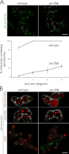
Deletion of the PEX3B gene affects peroxisome inheritance. (A) Wild-type and pex3BΔ cells expressing genomically integrated POT1-GFP were grown in YPD for 16 h and then transferred to YPBO for 2 h. Fluorescent images of randomly chosen fields of cells were acquired as a stack by confocal microcopy and deconvolved. Buds were sized according to four categories relative to the volume of the mother cell (see Materials and methods). The percentages of bud tips containing peroxisomes at each size category were plotted. Quantification was performed on at least 50 budded cells from each category. Graphic results are the means and SEM of three independent experiments. Bar, 5 µm. (B) Deletion of the PEX3B gene does not affect the actin structure of cells or the inheritance of vacuoles or mitochondria. Wild-type and pex3BΔ cells synthesizing Pot1p-GFP were grown in YPD. Mitochondria were stained with Mitotracker dye, vacuoles were stained with the fluorophore FM4-64, and actin was stained with rhodamine-phalloidin. Images were captured by confocal microscopy. Bar, 5 µm.
Figure 5.
Figure 5.
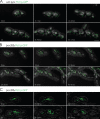
Peroxisome dynamics and morphogenesis in wild-type and pex3BΔ cells visualized by 4D in vivo video microscopy. Peroxisomes were fluorescently labeled with genomically encoded Pot1p-GFP. Cells were grown for 16 h in YPD, transferred to YPBO for 6 h, and visualized at 28°C (A and B) or 23°C (C) with an LSM 510 confocal microscope specifically modified for 4D in vivo microscopy (see Materials and methods). (A) Wild-type E122/POT1-GFP strain. Representative frames from Video 1 show the specific movements and division of peroxisomes through several cell divisions. The emergence of new buds at 1 h 1 min, 1 h 22 min, and 2 h 45 min is followed by the vectorial transfer of a portion of the mother cell's peroxisomes to the bud, where they associate with the bud tip. Bar, 5 µm. (B and C) pex3BΔ/POT1-GFP strain. (B) Representative frames from Video 2 display the specific movements and morphogenesis of peroxisomes in pex3B cells. At 0 min, both buds lack peroxisomes. By 12 min, several peroxisomes have entered the buds but have failed to associate with the bud tips. Subsequently, many peroxisomes undergo a morphogenic transition, becoming elongated and tubular-reticular in appearance. These peroxisomes often straddle the mother-bud neck (2 h 26 min). Also, peroxisome inheritance does not keep pace with cell division, as many buds are devoid of peroxisomes at later time points (4 h 5 min). Bar, 5 µm. (C) Representative frames from Video 3 display the inability of a tubular-reticular peroxisome to divide except through cytokinesis. A tubular reticular peroxisome is seen initially straddling the mother-bud neck (0 min). At 1 h 48 min, the peroxisome is cut in two by constriction of the septin ring, concluding cytokinesis. A second scission event occurs at 3 h with the conclusion of cytokinesis between the mother cell and the bud to her right. Subsequent buds fail to inherit peroxisomes (4 h 49 min). Bar, 5 µm.
Figure 6.
Figure 6.
Pex3Bp and Pex3p interact directly with the cargo-binding tail of Y. lipolytica class V myosin. (A) Peroxisome inheritance is reduced by overexpression of the Y. lipolytica class V myosin cargo-binding tail. Wild-type strain E122 expressing genomically encoded Pot1p-GFP to fluorescently label peroxisomes was transformed with the empty plasmid pUB4 or with pUB4 expressing the globular tail domain (amino acids 1,092–1,594) of Y. lipolytica class V myosin under the control of the oleic acid–inducible POT1 promoter. Cells were grown in YPD supplemented with hygromycin B and then transferred to and incubated in oleic acid–containing YPBO supplemented with hygromycin B for 6 h. Fluorescent images of randomly chosen fields of cells were acquired as a stack by confocal microscopy and then deconvolved. Buds were sized as “small” (0–39% of mother cell volume) or “large” (40–61% of mother cell volume; see Materials and methods). The percentages of buds containing peroxisomes in each size category are presented. Quantification was performed on at least 50 budded cells from each category. Graphic results are the means and SEM of three independent experiments. Bar, 5 µm. (B) Split-ubiquitin membrane yeast two-hybrid analysis. Cells of the S. cerevisiae strain DSY-1 synthesizing Cub protein fusions to Pex3Bp or Pex3p and NubG protein fusions to Pex3Bp, Pex3p, or the globular tail of the class V myosin of Y. lipolytica (amino acids 1,092–1,594) were tested for their ability to interact with each other by a β-galactosidase filter detection assay. A positive interaction was detected by the production of blue color. The color intensities of positive (+) and negative (−) controls are indicated. (C) Glutathione sepharose beads containing GST fused to the cargo-binding tail of the class V myosin of Y. lipolytica (GST-YlMyoV); the cargo-binding tail of the class V myosin, Myo2p, of S. cerevisiae (GST-ScMyoV); or GST alone were incubated with extracts of E. coli synthesizing MBP, MBP-Pex3p, MBP-Pex3Bp, MBP-YALI0E03124p, MBP-ScInp2p, or MBP-ScVam6p. Bound proteins, as well as 10% of input proteins, were analyzed by immunoblotting with anti-MBP antibodies. Total GST-YlMyoV, GST-ScMyoV, or GST protein levels were visualized by immunoblotting with anti-GST antibodies. Arrowheads indicate MBP or MBP fusion proteins. (D) Overexpression of PEX3B delivers peroxisomes preferentially to buds. pex3BΔ cells containing peroxisomes labeled with Pot1p-GFP and the plasmid pUB4 expressing PEX3B under the control of the oleic acid–inducible POT1 promoter were grown for 16 h in YPD supplemented with hygromycin B, then transferred to oleic acid–containing YPBO supplemented with hygromycin B for 6 h, and visualized at 23°C with an LSM 510 confocal microscope specifically modified for 4D in vivo microscopy (see Materials and methods). Representative frames from Video 4 show the specific movements of peroxisomes and their inheritance from mother cell to bud. At 0 min, two large peroxisome clusters are initially located next to the mother-bud neck. By 1 h 49 min, these peroxisomes have been transferred to their respective buds, and by 4 h 5 min, the cycle is repeated, with the peroxisomes now residing in the granddaughters of the original mother cells. De novo synthesis of peroxisomes can also be detected by the reappearance of fluorescent punctae in mother cells that had transferred their original peroxisome complement to their buds. These de novo formed peroxisomes are also vectorially transferred to newly formed buds (6 h 20 min). The formation of peroxisomes and subsequent transfer to buds continued (6 h 56 min). Bar, 5 µm.
Figure 7.
Figure 7.
A candidate Y. lipolytica Inp2p orthologue, YALI0E03124p, does not localize to peroxisomes. The chimeric protein YALI0E03124p-mRFP, whose expression is under the control of the oleic acid–inducible promoter POT1, was imaged in the wild-type strain E122 expressing genomically integrated Pot1p-GFP to fluorescently label peroxisomes. YALI0E03124p-mRFP did not localize to punctate peroxisomes, and when cells were incubated in oleic acid–containing medium, YALI0E03124p-mRFP exhibited a pattern typical of protein localization to the ER and secretory system. The top panels show representative images of cells grown in medium containing acetate, whereas the bottom panels show representative images of cells grown in medium containing oleic acid. Bar, 5 µm.
Figure 8.
Figure 8.
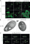
Pex3p can substitute for Pex3Bp in peroxisome inheritance. (A) pex3BΔ and pex3Δ cells expressing genomically integrated Pot1p-GFP were transformed with empty plasmid pUB4 or pUB4 containing PEX3B or PEX3 for overexpression in oleic acid–containing medium. Cells were grown in YPD supplemented with hygromycin B and then transferred and incubated for 6 h in oleic acid–containing YPBO supplemented with hygromycin B. Fluorescent images of cells were captured by confocal microscopy and deconvolved. Bar, 5 µm. (B) Ultrastructure of pex3BΔ cells overexpressing PEX3B or PEX3. Cells were cultured as in A and then fixed and processed for EM. Arrowheads indicate peroxisomes. Bar, 1 µm. (C) Overexpression of PEX3 can rescue the pex3BΔ phenotype. pex3BΔ cells containing peroxisomes labeled with Pot1p-GFP and the plasmid pUB4 expressing PEX3 under the control of the oleic acid–inducible POT1 promoter were grown and imaged as in Fig. 6 D (see Materials and methods). Representative frames from Video 5 show the specific movements of peroxisomes and their inheritance from mother cell to bud. At 0 min, one large peroxisome cluster is initially located near the mother-bud neck. By 28 min, the peroxisome cluster is split in two by cytokinesis. As new buds emerge, these peroxisome clusters are transferred to the new buds. Several single peroxisomes can be seen at the bud tip by 1 h 36 min. As the buds continue to grow, the peroxisome clusters also move to the bud tips. Bar, 5 µm.

References

    1. Altschul S.F., Madden T.L., Schäffer A.A., Zhang J., Zhang Z., Miller W., Lipman D.J. 1997. Gapped BLAST and PSI-BLAST: a new generation of protein database search programs.Nucleic Acids Res. 25:3389–3402 doi:10.1093/nar/25.17.3389 - DOI - PMC - PubMed
    1. Bartholomew C.R., Hardy C.F.J. 2009. p21-activated kinases Cla4 and Ste20 regulate vacuole inheritance in Saccharomyces cerevisiae.Eukaryot. Cell. 8:560–572 - PMC - PubMed
    1. Bascom R.A., Chan H., Rachubinski R.A. 2003. Peroxisome biogenesis occurs in an unsynchronized manner in close association with the endoplasmic reticulum in temperature-sensitive Yarrowia lipolytica Pex3p mutants.Mol. Biol. Cell. 14:939–957 doi:10.1091/mbc.E02-10-0633 - DOI - PMC - PubMed
    1. Chang J., Fagarasanu A., Rachubinski R.A. 2007. Peroxisomal peripheral membrane protein YlInp1p is required for peroxisome inheritance and influences the dimorphic transition in the yeast Yarrowia lipolytica.Eukaryot. Cell. 6:1528–1537 - PMC - PubMed
    1. Davidson R.C., Blankenship J.R., Kraus P.R., de Jesus Berrios M., Hull C.M., D'Souza C., Wang P., Heitman J. 2002. A PCR-based strategy to generate integrative targeting alleles with large regions of homology.Microbiology. 148:2607–2615 - PubMed

Publication types