. 2009 Oct 27;106(43):18177-82.
doi: 10.1073/pnas.0905558106. Epub 2009 Oct 12.

Solution structure of a paradigm ArsR family zinc sensor in the DNA-bound state

Affiliations

Solution structure of a paradigm ArsR family zinc sensor in the DNA-bound state

Alphonse I Arunkumar et al. Proc Natl Acad Sci U S A. .

Abstract

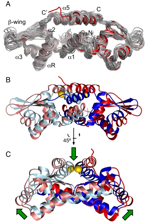
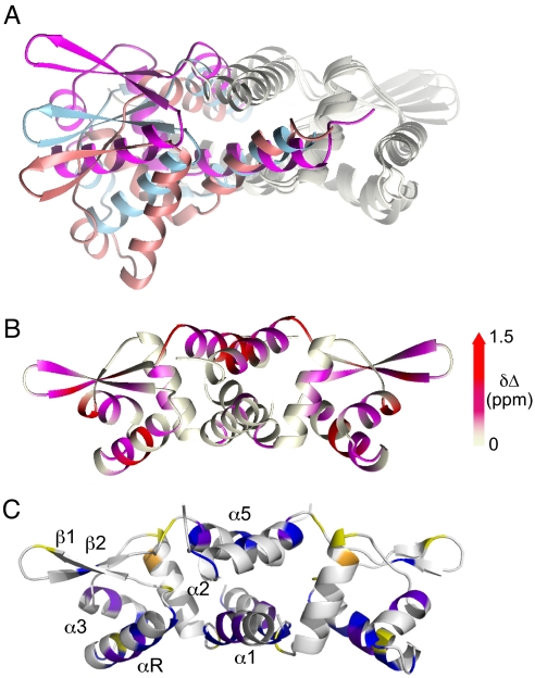
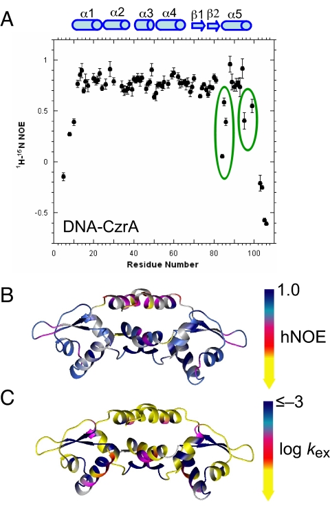
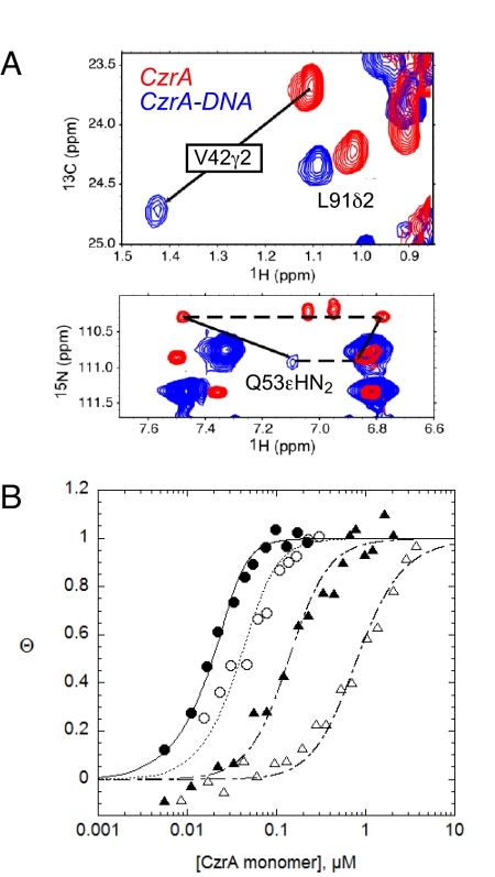
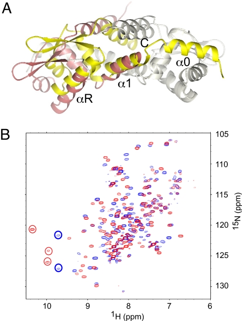
Staphylococcus aureus CzrA is a zinc-dependent transcriptional repressor from the ubiquitous ArsR family of metal sensor proteins. Zn(II) binds to a pair of intersubunit C-terminal alpha5-sensing sites, some 15 A distant from the DNA-binding interface, and allosterically inhibits DNA binding. This regulation is characterized by a large allosteric coupling free energy (DeltaGc) of approximately +6 kcal mol(-1), the molecular origin of which is poorly understood. Here, we report the solution quaternary structure of homodimeric CzrA bound to a palindromic 28-bp czr operator, a structure that provides an opportunity to compare the two allosteric "end" states of an ArsR family sensor. Zn(II) binding drives a quaternary structural switch from a "closed" DNA-binding state to a low affinity "open" conformation as a result of a dramatic change in the relative orientations of the winged helical DNA binding domains within the dimer. Zn(II) binding also effectively quenches both rapid and intermediate timescale internal motions of apo-CzrA while stabilizing the native state ensemble. In contrast, DNA binding significantly enhances protein motions in the allosteric sites and reduces the stability of the alpha5 helices as measured by H-D solvent exchange. This study reveals how changes in the global structure and dynamics drive a long-range allosteric response in a large subfamily of bacterial metal sensor proteins, and provides insights on how other structural classes of ArsR sensor proteins may be regulated by metal binding.


Conflict of interest statement

The authors declare no conflict of interest.

Figures

Fig. 1.
Fig. 1.
Solution structure of CzrO DNA-bound CzrA. (A) Backbone heavy atom (N, Cα, and C') overlay of 20 lowest-energy structures of DNA bound CzrA (see Table S2 for structure statistics; unstructured residues 100–103 of the bundle are not shown in this view for clarity). The ribbon diagram in the overlay represents the mean structure of the ensemble and the two subunits are shaded salmon and red. (B) Ribbon diagram representation of the global overlay of DNA bound CzrA and the crystal structure of Zn(II) CzrA (8). The subunits of DNA bound CzrA are colored as in A and the two subunits of Zn(II) CzrA are colored slate and blue. Zn(II) ions are colored yellow. (C) Another view of the same overlay as in B (rotated 45°) and the green arrows represent the direction of the quaternary structural change.
Fig. 2.
Fig. 2.
Distinct conformational states of α5 metal sensor proteins. (A) Overlay of the apo SmtB (magenta ribbon), Zn(II)-bound CzrA (light blue ribbon) and DNA-bound CzrA (salmon ribbon). The “right” subunit is overlaid to better show the quaternary structural differences. (B) Effect of binding DNA on apo CzrA structure is mapped using a 1H HN chemical shift perturbation experiment. Colors on the ribbon are ramped according to Δδ ppm as follows: gray, Δδ<0.2 ppm; magenta, 0.2<Δδ<0.8 and red; 0.8<Δδ<1.5 ppm. (C) A comparison of the short time scale 15N relaxation dynamics of Zn2 CzrA vs. apo-CzrA (see Fig. S2 for primary data). Blue, increased S2 by ≥ 0.02 on Zn(II) binding; yellow, decreased S2 by ≤ –0.02 on Zn(II) binding; purple, residues in apo-CzrA that exhibit measurable Rex ≥ 1 s−1 that is completely dampened upon Zn(II) binding; orange, the single residue in Zn2 CzrA for which there is measurable Rex ≥ 1 s−1 (L35).
Fig. 3.
Fig. 3.
Fast motion dynamics and hydrogen-deuterium (H-D) exchange rates for DNA-bound CzrA. (A) The 1H-15N steady-state heteronuclear NOE (hNOE) of DNA-CzrA complex. The region where there are pronounced internal motions are highlighted with solid circles. (B) Mapping of the hNOE values on ribbon diagram of DNA-bound CzrA. The colors are ramped as follows: dark blue, hNOE ≥ 0.8, light blue, 0.7 ≤ hNOE <0.8; magenta, 0.6 ≤ hNOE <0.7; red, 0.4 ≤ hNOE <0.6; yellow, NOE <0.4. Gray shading, no information due to spectral overlap. (C) Mapping of the H-D exchange rates (as log kex, h–1) on a ribbon diagram of DNA-bound CzrA. The colors are ramped as follows and correspond to roughly linear free energy increments: yellow, –0.2 ≤ log kex < 0.75; red, –0.9 ≤ log kex < –0.2; magenta, –1.6 ≤ log kex < –0.9; light blue, –2.3 ≤ log kex < –1.6; dark blue, log kex < –2.3.
Fig. 4.
Fig. 4.
CzrO binding isotherms for wild-type and mutant CzrAs. (A) Overlay of the same selected region of 1H-13C HMQC spectra (Upper) the 1H-15N TROSY spectra (Lower) acquired for apo-CzrA (red cross peaks) and the CzrA-CzrO complex (blue cross peaks) illustrating the large shifts in the side chains of Val-42 and Gln-53 upon binding DNA. (B) Representative CzrO binding isotherms obtained for wild-type (filled circles), Q53A (open circles), V42A (filled triangles) and Q53E (Δ) CzrAs. The continuous line through each data set represents a nonlinear least squares fit to a dissociable dimer model with the parameters compiled in Table 1.
Fig. 5.
Fig. 5.
Distinct mechanisms for allosteric control in α3N and α5 sensors. (A) A right protomer (shaded gray) superposition of the apo C11G S. aureus pI258 CadC dimer (35) and the DNA-bound form of apo-CzrA (this work). The subunits of apo-CadC are colored gray and yellow, while that for the DNA-bound CzrA are shaded gray and salmon. Note the N-terminal α0 helix in CadC is not present in CzrA; the N-terminal residue on this protomer is Gly-11, with the N-terminal 10 residues disordered. (B) Overlay of 1H-15N TROSY spectra acquired for apo C10G L. monocytogenes CadC (blue cross peaks) with the CadC-CadO complex (red cross peaks). See text for details.

References

    1. Outten CE, O'Halloran TV. Femtomolar sensitivity of metalloregulatory proteins controlling zinc homeostasis. Science. 2001;292:2488–2492. - PubMed
    1. Ma Z, Jacobsen FE, Giedroc DP. Metal transporters and metal sensors: How coordination chemistry controls bacterial metal homeostasis. Chem Rev. 2009;109 10.1021/cr900077w. - DOI - PMC - PubMed
    1. Giedroc DP, Arunkumar AI. Metal sensor proteins: Nature's metalloregulated allosteric switches. Dalton Trans. 2007:3107–3120. - PubMed
    1. Lu M, Fu D. Structure of the zinc transporter YiiP. Science. 2007;317:1746–1748. - PubMed
    1. Cherezov V, et al. Insights into the mode of action of a putative zinc transporter CzrB in Thermus thermophilus. Structure. 2008;16:1378–1388. - PMC - PubMed

Publication types

MeSH terms

LinkOut - more resources