

Prediction of functional loss in glaucoma from progressive optic disc damage
- PMID: 19822839
- PMCID: PMC2828324
- DOI: 10.1001/archophthalmol.2009.276
Prediction of functional loss in glaucoma from progressive optic disc damage
Abstract
Objective: To evaluate the ability of progressive optic disc damage detected by assessment of longitudinal stereophotographs to predict future development of functional loss in those with suspected glaucoma.
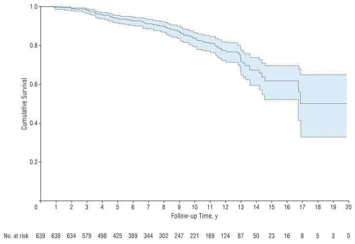
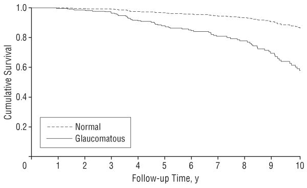
Methods: The study included 639 eyes of 407 patients with suspected glaucoma followed up for an average of 8.0 years with annual standard automated perimetry visual field and optic disc stereophotographs. All patients had normal and reliable standard automated perimetry results at baseline. Conversion to glaucoma was defined as development of 3 consecutive abnormal visual fields during follow-up. Presence of progressive optic disc damage was evaluated by grading longitudinally acquired simultaneous stereophotographs. Other predictive factors included age, intraocular pressure, central corneal thickness, pattern standard deviation, and baseline stereophotograph grading. Hazard ratios for predicting visual field loss were obtained by extended Cox models, with optic disc progression as a time-dependent covariate. Predictive accuracy was evaluated using a modified R(2) index.
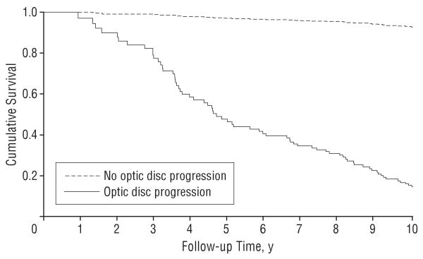
Results: Progressive optic disc damage had a hazard ratio of 25.8 (95% confidence interval, 16.0-41.7) and was the most important risk factor for development of visual field loss with an R(2) of 79%. The R(2)s for other predictive factors ranged from 6% to 26%.
Conclusions: Presence of progressive optic disc damage on stereophotographs was a highly predictive factor for future development of functional loss in glaucoma. These findings suggest the importance of careful monitoring of the optic disc appearance and a potential role for longitudinal assessment of the optic disc as an end point in clinical trials and as a reference for evaluation of diagnostic tests in glaucoma.
Figures
Comment in
-
Progressive optic disc change: implications for clinical practice and trial design.Arch Ophthalmol. 2009 Oct;127(10):1382-3. doi: 10.1001/archophthalmol.2009.281. Arch Ophthalmol. 2009. PMID: 19822858 No abstract available.
References
-
- Weinreb RN, Khaw PT. Primary open-angle glaucoma. Lancet. 2004;363(9422):1711–1720. - PubMed
-
- Jonas JB, Budde WM, Panda-Jonas S. Ophthalmoscopic evaluation of the optic nerve head. Surv Ophthalmol. 1999;43(4):293–320. - PubMed
-
- Gonzalez-Hernandez M, Pablo LE, Armas-Domingue K, Rodriguez de la Vega R, Ferreras A, Gonzalez de la Rosa M. Structure-function relationship depends on glaucoma severity. Br J Ophthalmol. 2009 - PubMed
-
- Racette L, Medeiros FA, Bowd C, Zangwill LM, Weinreb RN, Sample PA. The impact of the perimetric measurement scale, sample composition, and statistical method on the structure-function relationship in glaucoma. J Glaucoma. 2007;16(8):676–684. - PubMed
-
- Johnson CA, Cioffi GA, Liebmann JR, Sample PA, Zangwill LM, Weinreb RN. The relationship between structural and functional alterations in glaucoma: a review. Semin Ophthalmol. 2000;15(4):221–233. - PubMed
Publication types
MeSH terms
Grants and funding
LinkOut - more resources
Full Text Sources
Medical
