Review
. 2009 Nov 1;8(21):3527-36.
doi: 10.4161/cc.8.21.9888. Epub 2009 Nov 24.

Bioelectric controls of cell proliferation: ion channels, membrane voltage and the cell cycle

Affiliations
Review

Bioelectric controls of cell proliferation: ion channels, membrane voltage and the cell cycle

Douglas J Blackiston et al. Cell Cycle. .

Abstract

All cells possess long-term, steady-state voltage gradients across the plasma membrane. These transmembrane potentials arise from the combined activity of numerous ion channels, pumps and gap junction complexes. Increasing data from molecular physiology now reveal that the role of changes in membrane voltage controls, and is in turn controlled by, progression through the cell cycle. We review recent functional data on the regulation of mitosis by bioelectric signals, and the function of membrane voltage and specific potassium, sodium and chloride ion channels in the proliferation of embryonic, somatic and neoplastic cells. Its unique properties place this powerful, well-conserved, but still poorly-understood signaling system at the center of the coordinated cellular interactions required for complex pattern formation. Moreover, disregulation of ion channel expression and function is increasingly observed to be not only a useful marker but likely a functional element in oncogenesis. New advances in genomics and the development of in vivo biophysical techniques suggest exciting opportunities for molecular medicine, bioengineering and regenerative approaches to human health.


Figures

Figure 1
Figure 1
Linkage between bioelectric signals and cell cycle control via Vmem changes. (A) Schematic illustrating the linkage of membrane potential modulation to ion channel dynamics during the cell cycle. During G1/S transition, the membrane potential becomes hyperpolarized relative to the normal resting potential. Potassium channels from the ATP-sensitive, voltage gated and Ca2+-activated families become active allowing for potassium efflux from the cell; sodium channels also become activated. During the G2/S transition the membrane becomes depolarized, and there is a decrease in potassium channel activity. In addition, G2/M is characterized by the activation of chloride channels and a subsequent efflux of chloride. While the role of potassium channels are the most well-studied in relation to the cell cycle, a number other ion gradients are involved, each contributing to the net membrane potential as described by Goldman-Hodgkin-Katz equation. (B) Membrane voltage in a cell can be altered by a variety of factors, including channel/pump activity in it’s own membrane (cell-autonomous effects), gap junctional communication to neighboring cells of different potential, or nearby electric fields and ion flows from wounded and intact epithelia (the latter two being non-cell-autonomous control mechanisms). In turn, changes in ion flow can be transduced into alterations of the mitotic program by voltage-gated calcium channels and calcium-dependent second-messenger pathways, changes in cell volume, and alterations of transport of mitogens such as serotonin. These can arrive in cells by two voltage-dependent mechanisms: electrophoresis through gap junctions, or changes in the activity of transporters like SERT that are powered by transmembrane potential.
Figure 2
Figure 2
Sample of recursive feedback among physiological parameters and ion channel/pump activity. Bioelectric controls of cell functions are inherently non-linear because channels and pumps produce effects on voltage and pH that in turn regulate those same channels and pumps. Here is shown one example taken from a circuit used in vertebrate left-right patterning. The V-ATPase creates both a pH gradient and contributes to membrane hyperpolarization. At the same time, the H,K-ATPase functions together with a K+ channel to regulate Vmem; however, both of these components are themselves voltage- and pH-sensitive. Quantitative models of such networks, which take into account both the molecular biology of components expressed in relevant cells and the time-dependent physiology of the resulting circuit.
Figure 3
Figure 3
Perturbation of growth in Xenopus embryos caused by manipulation of ion channels and electroporation. A variety of mRNAs encoding wild-type and mutant channels were microinjected into frog embryo blastomeres to screen for bioelectrical signals with roles in growth and pattern control. The VSOP proton channel (kindly provided by Yasushi Okamura) (A), the Cx32 gap junction subunit (plasmid kindly provided by Dan Goodenough) (B and C), and the HERG K+ channel (plasmid kindly provided by Annarosa Arcangeli) (D) result in ectopic growth and abnormal duplication of body structures such as eyes, sometimes forming extensive fin-like protrusions that are clearly associated with increases in cell growth. (E–E”’) Electroporation of embryos at stage 33, with no DNA, (95 msec interval, 5 msec pulse, 10X repeated) results in significant areas of ectopic growth 24 hours later. Red arrows indicate hyperproliferation.
Figure 4
Figure 4
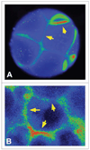
Membrane voltage levels are not homogenous around the cell surface. Using the voltage-sensitive fluorescent dye DiBAC, early frog embryo blastomeres (A) as well as COS cells in monolayer culture (B) exhibit significant variations of membrane voltage level around the cell surface, indicating that a single Vmem number for a given cell drastically under-estimates the amount of information that can be encoded in the plasma membrane’s physiological state and potentially communicated to neighboring cells. Images courtesy of Dany S. Adams.

References

    1. Vermeulen K, Van Bockstaele DR, Berneman ZN. The cell cycle: a review of regulation, deregulation and therapeutic targets in cancer. Cell Prolif. 2003;36:131–149. - PMC - PubMed
    1. Sundelacruz S, Levin M, Kaplan DL. Role of membrane potential in the regulation of cell proliferation and differentiation. Stem Cell Rev Rep. 2009;5:231–246. - PMC - PubMed
    1. Binggeli R, Weinstein R. Membrane potentials and sodium channels: hypotheses for growth regulation and cancer formation based on changes in sodium channels and gap junctions. J Theor Biol. 1986;123:377–401. - PubMed
    1. Balitsky KP, Shuba EP. Resting potential of malignant tumour cells. Acta Unio Int Contra Cancrum. 1964;20:1391–1393. - PubMed
    1. Johnstone BM. Micro-electrode penetration of ascites tumour cells. Nature. 1959;183:411. - PubMed

Publication types

LinkOut - more resources