. 2009 Nov 15;8(22):3729-41.
doi: 10.4161/cc.8.22.10033. Epub 2009 Nov 10.

microRNAs regulate human embryonic stem cell division

Affiliations

microRNAs regulate human embryonic stem cell division

Junlin Qi et al. Cell Cycle. .

Abstract

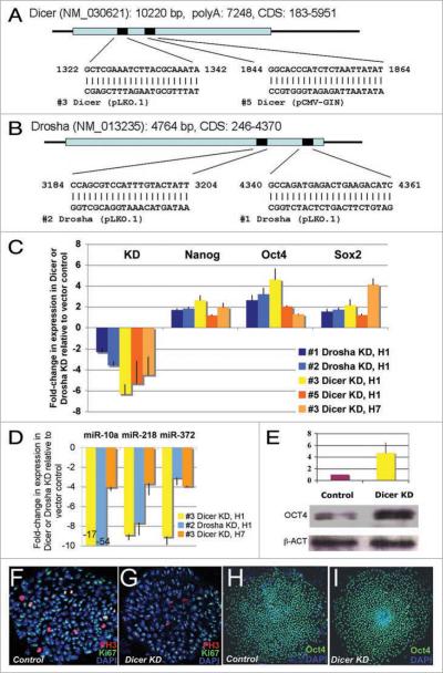
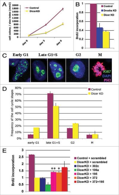
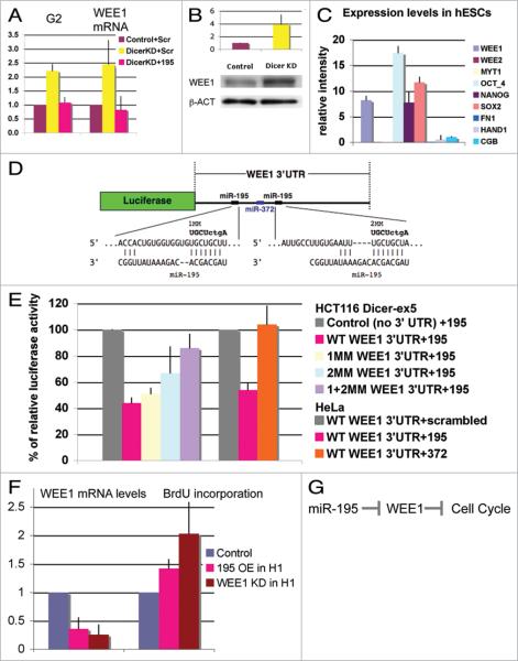
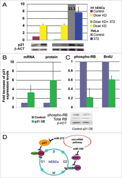
microRNAs (miRNAs) regulate numerous physiological processes such as cell division and differentiation in many tissue types including stem cells. To probe the role that miRNAs play in regulating processes relevant to embryonic stem cell biology, we used RNA interference to silence DICER and DROSHA, the two main miRNA processing enzymes. Consistent with a role for miRNAs in maintaining normal stem cell division and renewal, we found that perturbation of miRNA pathway function in human embryonic stem cells (hESCs) attenuates cell proliferation. Normal cell growth can be partially restored by introduction of the mature miRNAs miR-195 and miR-372. These miRNAs regulate two tumor suppressor genes, respectively: WEE1, which encodes a negative G2/M kinase modulator of the CycB/CDK complex and CDKN1A, which encodes p21, a CycE/CDK cyclin dependent kinase inhibitor that regulates the G1/S transition. We show that in wild-type hESCs, WEE 1 levels control the rate of hESC division, whereas p21 levels must be maintained at a low level for hESC division to proceed. These data support a model for hESC cell cycle control in which miRNAs regulate negative cell cycle modulators at two phases of the cell cycle to ensure proper replenishment of the stem cell population.


Figures

Figure 1
Figure 1
Dicer and Drosha knockdown H1 hESCs. (A and B) two different lentiviral short hairpin RNA (shRNA) constructs per gene were used in hESCs to knock down Dicer (A) or Drosha (B). (C) the silencing levels for Drosha and Dicer in the H1 lines and Dicer in the H7 line are shown as fold changes in expression compared with cells treated with the empty vector control lines. The mean (±SD) was calculated from at least three independent experiments. Stem cell regulators, Nanog, Oct4 and Sox2 were upregulated in these knockdown lines. Standard deviations were calculated from 6 independent experiments for Nanog and Oct4 and two experiments for Sox2 in H1 and H7 cells. (D) the levels of mature miRNAs 10a, 218 and 372 were reduced in the Drosha- or Dicer-knockdown hESCs. The mean (±SD) was calculated from three independent experiments. (E) OCt4 protein expression was increased four-fold in the Dicer knockdown H1 compared with the control line. The mean (±range) was calculated from two independent experiments. (F and G) Morphologically the Dicer knockdown H1 hESC colonies are similar to control H1 hESC colonies. Similar colony structures were observed for Drosha knockdown lines #1 and #2, Dicer knockdown lines #3 and #5 and control lines #4 and #7. Immunofluorescence staining for DAPI, Ki67 and phospho-Histone 3 were examined by confocal microscopy and are represented in blue, green and red respectively. (H and I) Dicer-knockdown H1 hESCs and the control cells both express the stem cell marker OCT4. OCT4 is green and DAPI is blue.
Figure 2
Figure 2
Dicer and Drosha knockdown in H1 hESCs results in a cell division defect. (A) The Dicer-knockdown hESC line H1(#3) grows significantly slower than the control line (#4) based on a colony count assay. (B) Both Drosha- (#1) and Dicer- (#3) knockdown lines show significant reduction in BrdU incorporation compared with the control line (#4). (C and D) Based on Ki67 staining patterns (C), cells were categorized into different cell cycle phases. (D) Dicer-knockdown H1 hESCs (yellow bars) show an increase in frequency of G1 and G2 stages and a decrease in S-phase frequency compared to the control lines (brown bars). (E) miR-195 and miR-372 partially rescue the cell cycle defect in Dicer-knockdown hESCs as determined by BrdU incorporation assay. The standard deviations or ranges are calculated from two independent experiments for miR-302c and 372 + 195, three experiments for miR-106a and four for miR-195 and 372. The miRNA overexpression was verified by qPCR analysis.
Figure 3
Figure 3
miR-195 regulates normal hESC self-renewing division through the cell cycle regulator WEE1 kinase. (A) The percentage of cells in the G2 phase (Ki67 analysis) increases and WEE1 mRNA levels (qPCR analysis) are upregulated in the Dicer-knockdown line. Both of these defects can be rescued by overexpression of miR-195 in Dicer-knockdown cells by use of miR-195 duplexes. (B) Western blot analysis shows a four-fold upregulation of WEE1 protein in Dicer-knockdown H1 line #3 compared with control cell line #4. (A and B) Mean ± range was calculated from two independent experiments. (C) On the basis of microarray analysis, WEE1 is expressed at similar levels as the stem cell markers Nanog, Oct4 and Sox2, whereas WEE2 and MYT1 as well as differentiation markers are very low or undetectable. Mean ± SD was calculated from nine different hESC lines. Similar results were obtained with two different mRNA microarray platforms (Affymetrix and Agilent). (D) Diagram of the WEE1 3'UTR luciferase reporter construct. The entire WEE1 3'UTR was inserted after the luciferase open reading frame. Two miR-195 sites and one miR-372 site predicted by TargetScan are present in the WEE1 3'UTR. miR-195 and its complementary sequences in the WEE1 3'UTR as well as the mismatches (MM) made in miR-195 seed sequences are represented. (E) Cotransfection of this construct with miR-195 into HCT116 Dicer-ex5 cells results in downregulation of luciferase activity. The data are normalized to luciferase activity after cotransfection with miR-149, which has no predicted target site in WEE1 3'UTR, and, therefore, serves as a negative control. Mismatches of the miR-195 site(s) in WEE1-3'UTR release the repression of luciferase activity caused by miR-195 overexpression. The control plasmid used is SGC (SwitchGear Genomics), an empty vector with a luciferase cassette, but no inserted 3' UTR. Cotransfection of WT WEE1 3' UTR reporter construct with miR-195 in HeLa cells shows similar downregulation of luciferase activity whereas cotransfection with miR-372 has no effect. (F) The level of WEE1 mRNA is reduced upon miR-195 overexpression, in comparison with the overexpression of scrambled oligonucleotides or a miR-195 oligonucleotide bearing seed region mismatch mutations. BrdU incorporation is increased when miR-195 is overexpressed in wild-type H1 hESCs. WEE1 knockdown by siRNAs shows a similar effect, BrdU incorporation is increased 2-fold. Mean ± SD or range was calculated from three independent experiments for miR-195 OE and two independent experiments for WEE1 KD. (G) Model of miR-195 function in hESC cell cycle.
Figure 4
Figure 4
miR-372 regulates the cell cycle regulator p21 to control hESC division. (A) Western blot analysis shows a four-fold upregulation of p21 protein in Dicer-knockdown H1 line #3 compared to control cell line #4. HeLa cells show 23-fold higher expression of p21 than the control hESCs. Overexpression of miR-372 results in reduction of p21 protein levels in HeLa cells (US2-372 OE transient transfection) and in Dicer-knockdown hESCs (Lenti-372 OE stable cell line). Mean ± range is shown from two independent experiments after quantification and normalization to β-ACTIN expression level. Overexpression of p21 in wild-type H1 hESCs (B) reduced phospho-RB levels and resulted in reduced BrdU incorporation (C). Mean ± range is shown from two independent experiments. (D) Model for miRNA function in hESC cell cycle regulation. The data suggest that miRNAs are important for proper cell cycle control of both G1 and G2 checkpoints in hESCs. miR-195 is required for proper hESC division. miR-195 inhibits the CycB/CDK1 inhibitor, WEE1, and, therefore, indirectly controls the G2/M transition. miR-372 suppresses the CDK inhibitor p21 and, therefore, indirectly controls the G1/S transition.

References

    1. Cheng T. Cell cycle inhibitors in normal and tumor stem cells. Oncogene. 2004;23:7256–66. - PubMed
    1. Bucher N, Britten CD. G2 checkpoint abrogation and checkpoint kinase-1 targeting in the treatment of cancer. Br J Cancer. 2008;98:523–8. - PMC - PubMed
    1. Fluckiger AC, Marcy G, Marchand M, Negre D, Cosset FL, Mitalipov S, et al. Cell cycle features of primate embryonic stem cells. Stem Cells. 2006;24:547–56. - PMC - PubMed
    1. Shcherbata HR, Hatfield S, Ward EJ, Reynolds S, Fischer KA, Ruohola-Baker H. The MicroRNA pathway plays a regulatory role in stem cell division. Cell Cycle. 2006;5:172–5. - PubMed
    1. Becker KA, Ghule PN, Therrien JA, Lian JB, Stein JL, van Wijnen AJ, et al. Self-renewal of human embryonic stem cells is supported by a shortened G1 cell cycle phase. J Cell Physiol. 2006;209:883–93. - PubMed

Publication types

MeSH terms

LinkOut - more resources