. 2009 Oct 13;3(10):e529.
doi: 10.1371/journal.pntd.0000529.

Mosquito infection responses to developing filarial worms

Affiliations

Mosquito infection responses to developing filarial worms

Sara M Erickson et al. PLoS Negl Trop Dis. .

Abstract

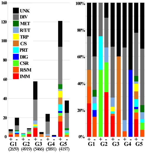
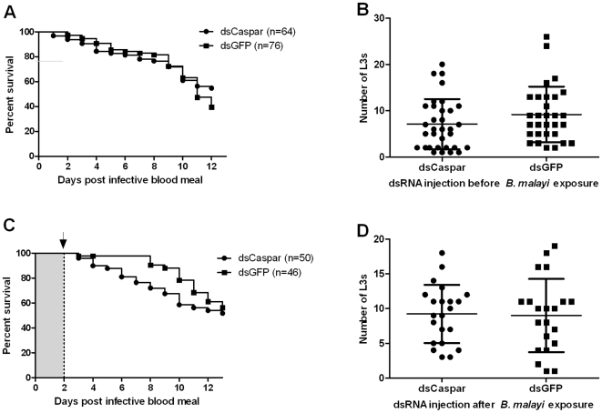
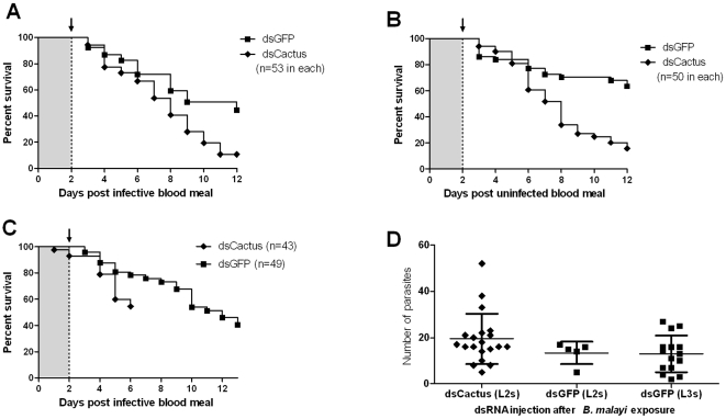
Human lymphatic filariasis is a mosquito-vectored disease caused by the nematode parasites Wuchereria bancrofti, Brugia malayi and Brugia timori. These are relatively large roundworms that can cause considerable damage in compatible mosquito vectors. In order to assess how mosquitoes respond to infection in compatible mosquito-filarial worm associations, microarray analysis was used to evaluate transcriptome changes in Aedes aegypti at various times during B. malayi development. Changes in transcript abundance in response to the different stages of B. malayi infection were diverse. At the early stages of midgut and thoracic muscle cell penetration, a greater number of genes were repressed compared to those that were induced (20 vs. 8). The non-feeding, intracellular first-stage larvae elicited few differences, with 4 transcripts showing an increased and 9 a decreased abundance relative to controls. Several cecropin transcripts increased in abundance after parasites molted to second-stage larvae. However, the greatest number of transcripts changed in abundance after larvae molted to third-stage larvae and migrated to the head and proboscis (120 induced, 38 repressed), including a large number of putative, immunity-related genes (approximately 13% of genes with predicted functions). To test whether the innate immune system of mosquitoes was capable of modulating permissiveness to the parasite, we activated the Toll and Imd pathway controlled rel family transcription factors Rel1 and Rel2 (by RNA interference knockdown of the pathway's negative regulators Cactus and Caspar) during the early stages of infection with B. malayi. The activation of either of these immune signaling pathways, or knockdown of the Toll pathway, did not affect B. malayi in Ae. aegypti. The possibility of LF parasites evading mosquito immune responses during successful development is discussed.


Conflict of interest statement

The authors have declared that no competing interests exist.

Figures

Figure 1
Figure 1. Relative sizes of Brugia malayi developmental stages that occur within compatible mosquito hosts: An example of cyclo-developmental transmission.
A. microfilariae are ingested during blood feeding; B. parasites differentiate into non-feeding, first-stage larvae within mosquito indirect flight muscle cells; C. following the first molt, second-stage larvae remain intracellular parasites which ingest cellular material into their newly developed digestive tract. D. third-stage larvae leave the muscle cells and migrate to the mosquito's head and proboscis where they will exit through the mosquito cuticle during blood feeding.
Figure 2
Figure 2. Global transcriptional analysis of the response of a compatible mosquito infected with successfully developing Brugia malayi.
A. Regulated (differentially expressed above a 1.7-fold threshold) genes for each experimental group. (+) indicates up-regulated in infected mosquitoes and (−) indicates down-regulated. Assayable genes (those that gave signal intensities above a standard cutoff threshold) are indicated below the graph. Functional group distributions are the same to those used in Nene et al., 2007 and Xi et al., 2008: IMM: immunity, R/S/M: redox, stress, mitochondrial, CSR: chemosensory reception, DIG: blood digestive, PRT: proteolysis, C/S: cytoskeletal, structural, TRP: transport, R/T/T: replication, transcription, translation, MET: metabolism, DIV: diverse, UKN: unknown. B. Percent distribution of functional group for each up- and down-regulated experimental group. Transcription data are presented in Table S5.
Figure 3
Figure 3. Gene knockdown of Ae. aegypti Caspar does not affect B. malayi development.
A, B. Gene knockdown of Ae. aegypti Caspar at the time of parasite ingestion did not affect mosquito mortality (A; P = 0.23) or B. malayi development (B; P = 0.10), in comparison to control mosquitoes. C, D. Gene knockdown of Ae. aegypti Caspar at the time of the parasite's first molt (L1 to L2) did not affect mosquito mortality (C; P = 0.32) or B. malayi development (D; P = 0.72). Bars indicate the mean intensity (total number of L3s recovered per infected individual) and standard deviation (B and D). Log-rank and Mann-Whitney tests were used to compare mosquito mortality curves and parasite mean intensities, respectively. Arrow: dsRNA was injected intrathoracically two days following infective blood meal.
Figure 4
Figure 4. Gene knockdown of Ae. aegypti MyD88 does not affect B. malayi development.
A, B. Gene knockdown of Ae. aegypti MyD88 during parasite ingestion did not affect mosquito mortality (A; P = 0.94) or B. malayi development (B; P = 0.14), compared to control mosquitoes. C, D. Knockdown of Ae. aegypti MyD88 when parasites first molt (L1 to L2) did not affect mosquito mortality (C; P = 0.30) or B. malayi development (D; P = 0.77). Bars indicate the mean intensity (total number of L3s recovered per infected individual) and standard deviation (B and D). Log-rank and Mann-Whitney tests were used to compare mosquito mortality curves and parasite mean intensities, respectively. Arrow: dsRNA was injected intrathoracically two days following infective blood meal.
Figure 5
Figure 5. Gene knockdown of Ae. aegypti Cactus decreases mosquito survival but does not affect B. malayi development (until death of the host).
A. Cactus-silenced mosquitoes showed a greater mortality compared to the GFP dsRNA treated controls (P<0.001). B. Mortality of Cactus-silenced mosquitoes fed on uninfected blood displayed a similar mortality rate to those that ingested parasite infected blood (4A). C. Cactus-silenced mosquitoes were dissected at 6 d post blood meal to observe parasite development. D. Gene silencing of Ae. aegypti Cactus did not affect the development of B. malayi (no difference in L2 mean intensities, P = 0.16). Bars indicate the mean intensity (total number of L3s recovered per infected individual) and standard deviation (D). Log-rank and Mann-Whitney tests were used to compare mosquito mortality curves and parasite mean intensities, respectively. Arrows indicate dsRNA was injected intrathoracically two days following the infective blood meal.

References

    1. WHO. Global Programme to Eliminate Lymphatic Filariasis: Annual Report on Lymphatic Filariasis 2001 . Geneva: World Health Organization.; 2002. 76p
    1. WHO. Global programme to eliminate lymphatic filariasis. Wkly Epidemiol Rec. 2007;82:361–380. - PubMed
    1. Christensen BM, Sutherland DR. Brugia pahangi: Exsheathment and midgut penetration in Aedes aegypti. Transactions of the American Microscopical Society. 1984;103:423–433.
    1. Murthy PK, Sen AB. Sequential development changes in microfilariae of subperiodic Brugia malayi to infective larvae in susceptible strain of Aedes aegypti (Macdonald). J Commun Dis. 1981;13:102–109. - PubMed
    1. Perrone JB, Spielman A. Microfilarial perforation of the midgut of a mosquito. J Parasitol. 1986;72:723–727. - PubMed

Publication types