. 2009 Sep;7(9):e1000188.
doi: 10.1371/journal.pbio.1000188. Epub 2009 Sep 8.

Dual regulation by pairs of cyclin-dependent protein kinases and histone deacetylases controls G1 transcription in budding yeast

Affiliations

Dual regulation by pairs of cyclin-dependent protein kinases and histone deacetylases controls G1 transcription in budding yeast

Dongqing Huang et al. PLoS Biol. 2009 Sep.

Abstract

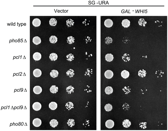
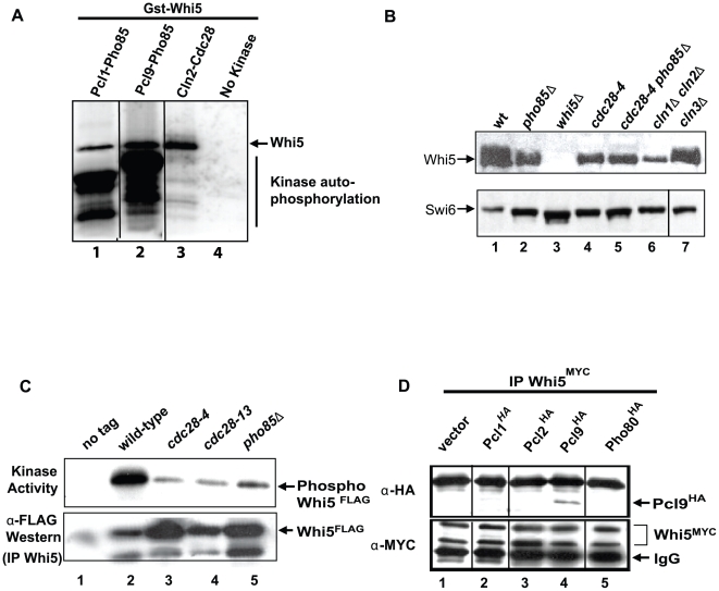
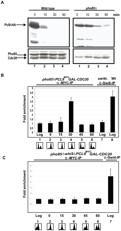
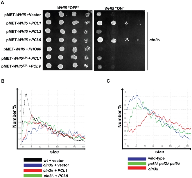
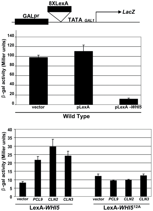
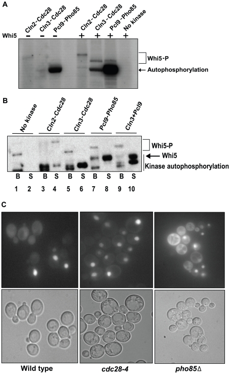
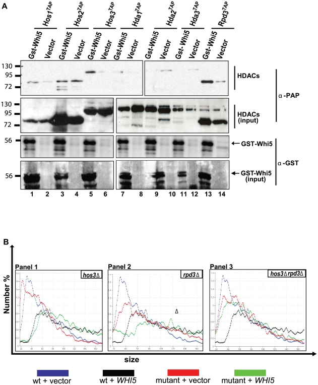
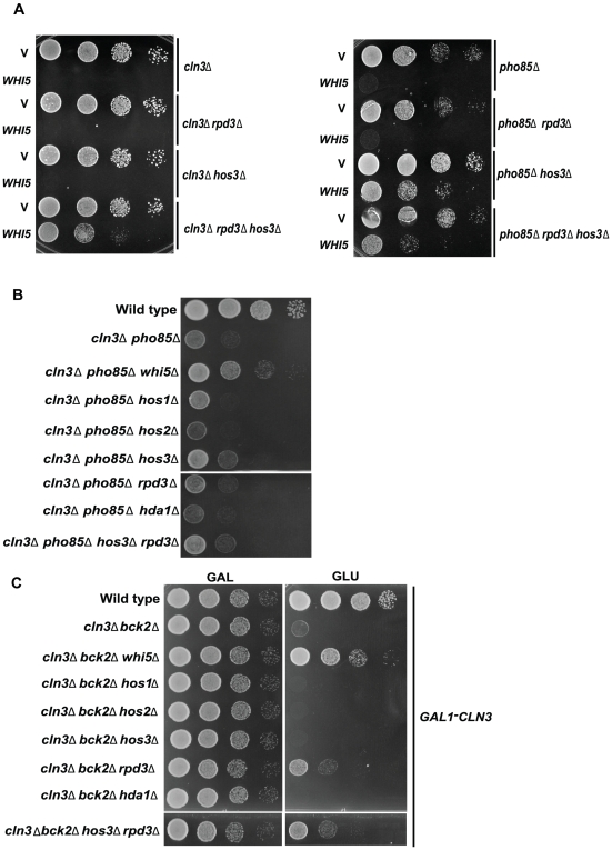
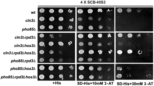
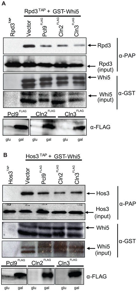
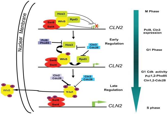
START-dependent transcription in Saccharomyces cerevisiae is regulated by two transcription factors SBF and MBF, whose activity is controlled by the binding of the repressor Whi5. Phosphorylation and removal of Whi5 by the cyclin-dependent kinase (CDK) Cln3-Cdc28 alleviates the Whi5-dependent repression on SBF and MBF, initiating entry into a new cell cycle. This Whi5-SBF/MBF transcriptional circuit is analogous to the regulatory pathway in mammalian cells that features the E2F family of G1 transcription factors and the retinoblastoma tumor suppressor protein (Rb). Here we describe genetic and biochemical evidence for the involvement of another CDK, Pcl-Pho85, in regulating G1 transcription, via phosphorylation and inhibition of Whi5. We show that a strain deleted for both PHO85 and CLN3 has a slow growth phenotype, a G1 delay, and is severely compromised for SBF-dependent reporter gene expression, yet all of these defects are alleviated by deletion of WHI5. Our biochemical and genetic tests suggest Whi5 mediates repression in part through interaction with two histone deacetylases (HDACs), Hos3 and Rpd3. In a manner analogous to cyclin D/CDK4/6, which phosphorylates Rb in mammalian cells disrupting its association with HDACs, phosphorylation by the early G1 CDKs Cln3-Cdc28 and Pcl9-Pho85 inhibits association of Whi5 with the HDACs. Contributions from multiple CDKs may provide the precision and accuracy necessary to activate G1 transcription when both internal and external cues are optimal.


Conflict of interest statement

The authors have declared that no competing interests exist.

Figures

Figure 1
Figure 1. WHI5 overexpression is toxic to strains compromised for Pho85 CDK activity.
Isogenic wt (BY263), pho85Δ (BY391), pcl1Δ (BY628), pcl2Δ (BY451), pcl9Δ (BY694), pcl1Δ pcl9Δ (BY760), and pho80Δ (BY490) strains bearing either GAL1-WHI5 (pBA1973) or empty vector control (pEG-H) were spotted in serial 10-fold dilutions on galactose media and incubated for 72 h at 30°C.
Figure 2
Figure 2. Whi5 is a substrate for Pcl9-Pho85 CDK-dependent phosphorylation.
(A) In vitro phosphorylation of Whi5 by Pho85 kinase. Purified Whi5GST and γ-32P-ATP were incubated alone (lane 4) or in the presence of recombinant Pcl1-Pho85 (lane 1), Pcl9-Pho85 (lane 2), or Cln2-Cdc28 (lane 3) kinases. Phosphorylated Whi5 protein was resolved by SDS-PAGE and autoradiography. (B) Slower-migrating forms of Whi5 are dependent on Cdc28 and Pho85. Cell extracts were prepared from wt (BY2507, lane 1), pho85Δ (BY4152, lane 2), cdc28-4 (BY4153, lane 4), cdc28-4 pho85Δ (BY4154, lane 5), cln1Δ cln2Δ (BY4289, lane 6), and cln3Δ (BY4288, lane 7) strains expressing WHI5MYC along with a whi5Δ control strain (BY4454, lane 3). Cells were grown at 30°C (semipermissive temperature for cdc28-4 strains) to log phase (Optical Density [OD] = 0.6) before harvesting. cdc28-4 cells were placed at 37°C for 2 h to inactivate Cdc28 before harvesting. Whi5MYC mobility was assessed by immunoblotting. (C) Whi5 associates with Pho85-dependent kinase activity. Wt (BY263) (lane 2), cdc28-4 (BY465) (lane 3), cdc28-13 (BY462) (lane 4), or pho85Δ (BY867) (lane 5) strains bearing a GAL-WHI5FLAG plasmid (pMT3586) or control vector control (pMT3164) were grown at 30°C (semipermissive temperature for cdc28-4 and cdc28-13 strains) in galactose media for 3 h. Whi5 complexes were recovered on anti-FLAG resin, incubated in kinase buffer with γ-32P-ATP at 30°C and resolved by SDS-PAGE. Capture of Whi5 protein was detected with anti-FLAG antibody. (D) Whi5 interacts with the Pcl, Pcl9. Anti-MYC immune precipitates of WHI5MYC strain lysates (BY2507) bearing either PCL1HA (pBA1820, lane 2), PCL2HA (pBA1821, lane 3), PCL9HA (pBA1822, lane 4), PHO80HA (pBA1823, lane 5), or a vector control (pBA330v, lane 1) were probed with 9E10 anti-MYC and 12CA5 anti-HA antibodies. The black lines in (A, B, and D) indicate empty lanes that were removed from the original blot.
Figure 3
Figure 3. Pcl9 localizes to G1-specific promoters in a cell cycle-dependent manner.
(A) Pho85 regulates Pcl9 protein stability. Wt (BY263) and pho85Δ strains (BY391) harboring a GAL1-PCL9HA plasmid (pBA2112) were grown to exponential phase in galactose media (lane 1). PCL9 expression was repressed by addition of glucose to final concentration of 2% and cells were harvested 10 (lane 2), 30 (lane 3), and 90 (lane 4) min after addition of glucose. Pcl9 abundance was assessed by immunoblotting using 12CA5 anti-HA antibodies. (B) Pcl9 localizes to SBF-dependent promoters. An exponentially growing GAL1-CDC20 pho85ΔPCL9MYCstrain (BY4148, lane 1) was arrested at M/G1 phase in glucose-containing medium (lane 2). Cultures were harvested 15 (lane 3), 30 (lane 4), 45 (lane 5), and 60 (lane 6) min after release from CDC20-induced arrest in galactose medium. Cell cycle progression was monitored by FACS analysis. Anti-MYC and anti-Swi6 ChIPs from the indicated strains were analyzed for CLN2 promoter sequences by quantitative RT-PCR. (C) In a strain lacking Whi5, GAL1-CDC20 pho85Δ whi5ΔPCL9MYC, Pcl9 no longer localizes to the CLN2 promoter. Anti-Swi4 ChIPs are shown as a positive control.
Figure 4
Figure 4. PHO85 affects growth and cell size defects associated with cln3Δ.
(A) Ectopic PCL1 and PCL9 expression alleviates WHI5 toxicity in a cln3Δ strain. A cln3Δ strain (BY653) bearing a methionine-repressible WHI5GST (pBA1975) or WHI512A-GST low-copy plasmid (pBA2249) along with an additional vector control (pBA330v), PCL1HA (pBA1820), PCL2HA (pBA1821, PCL9HA, pBA1822), or PHO80HA (pBA1823) construct were spotted in serial 10-fold dilutions on media supplemented with or lacking methionine (WHI5 “OFF”, WHI5 “ON,” respectively) and incubated for 72 h at 30°C. (B) PCL1 and PCL9 cyclins modulate cell size. Cell size distributions were analyzed for wt (BY263) and cln3Δ strains (BY653) bearing vector control (pBA330v), PCL9HA (pBA1822), or PCL1HA (pBA1820) plasmids. The median cell volume based on three replicates was: 42.33 fl±1.13 (wt+vector control); 71.78 fl±1.43 (cln3Δ+vector control); 55.67 fl±1.66 (cln3Δ+PCL1); 54.25 fl±1.21 (cln3Δ+PCL9). (C) Cells lacking PHO85 G1 cyclins exhibit an enlarged cell size. Cell size distributions were analyzed for wt (BY263), pcl1Δ pcl2Δ pcl9Δ (BY764), and cln3Δ strains (BY653). The median cell volume based on three replicates was: 46.73 fl±0.63 (wt); 53.96 fl±0.75 (pcl1Δ pcl2Δ pcl9Δ); 72.72 fl±1.22 (cln3Δ).
Figure 5
Figure 5. PHO85 regulates G1 transcription via WHI5.
(A) The G1 delay phenotype associated with a cln3Δ pho85Δ strain is dependent on WHI5. Wt (BY263), cln3Δ (BY653), pho85Δ (BY391), cln3Δ pho85Δ (BY4291), and cln3Δ pho85Δ whi5Δ (BY4292) strains were spotted in serial 10-fold dilutions on rich media (YPED) and incubated for 24 h at 30°C. DNA content of exponentially growing cultures was determined by FACS analysis. Liquid growth assays were also performed for these strains and growth rate is reported relative to wt as shown in the bar graph. Graphical representations of growth rates are shown above the bar graph as line plots, where the upper red line represents the growth of WT and the black line shows the growth of each mutant. (B) A cln3Δ pho85Δ strain exhibits defects in SCB-driven gene expression. Wt (BY4302), cln3Δ (BY4303), pho85Δ (BY4304), cln3Δ pho85Δ (BY4305), cln3Δ pho85Δ whi5Δ (BY4306), pho85Δwhi5Δ (BY4308), and cln3Δwhi5Δ (BY4307) strains harboring an integrated SCB-HIS3 reporter were spotted in serial 10-fold dilutions on histidine-containing medium or media lacking histidine and supplemented with 10 or 30 mM 3-AT. Plates were incubated at 30°C for 48 h. We note that the synthetic growth defect of a cln3 pho85 mutant is most pronounced on rich medium (A), and is not as evident when strains are grown on minimal medium.
Figure 6
Figure 6. Whi5-mediated transcriptional repression is antagonized by PHO85 and CDC28.
A reporter gene consisting of eight LexA binding sites flanked by the GAL1 promoter and the LacZ coding sequence was constructed (pBA1976). β-galactosidase activity (upper histogram) was measured in a wt strain (BY263) bearing the LacZ reporter along with one of the following: a vector control (pBA230v); a LexA expressing plasmid (pLexA; pBA1977); or a construct expressing a LexA-Whi5 fusion protein (pLexA-WHI5; pBA1978). β-galactosidase activities were also assayed (lower histogram) in a wt strain harboring the LacZ reporter construct alone (vector control; pBA1976) or overexpressing the G1 cyclins, PCL9 (pBA1974), CLN2 (pBA2247), or CLN3 (pBA2248) in the presence of LexA-Whi5 (pBA1978) or LexA-Whi512A (pBA1979) fusion proteins.
Figure 7
Figure 7. Pho85 does not affect known Whi5 regulatory mechanisms.
(A) Determination of relative Cdc28 and Pho85 kinase activity. In vitro kinase assays using varying amounts of recombinant Cln2-Cdc28, Cln3-Cdc28, and Pcl9-Pho85 in the absence (lane 1–3) or presence of purified Whi5 (lanes 5–8) were conducted and the degree of Whi5 phosphorylation was determined by SDS-PAGE and autoradiography. Purified Whi5 and γ-32P-ATP were incubated in the absence of kinase in lane 8, and lane 4 is empty. A 3 µM final concentration of Cln3-Cdc28 and Pcl9-Pho85 and a 60 nM final concentration of Cln2-Cdc28 give similar amounts of 32P-incorporation in Whi5, although phosphorylation by Cln2-Cdc28 caused Whi5 to migrate more slowly than Whi5 phosphorylated by Cln3-Cdc28 or Pcl9-Pho85. The concentration of kinase used in (B) was based on these experiments. (B) Cln3-Cdc28 and Pcl9-Pho85 do not influence Whi5-SBF complex stability. A preassembled recombinant Whi5-Swi4FLAG-Swi6 complex bound to anti-FLAG resin was incubated with Cln2-Cdc28, Cln3-Cdc28, Pcl9-Pho85, or both Cln3-Cdc28 and Pcl9-Pho85 in the presence of radiolabeled ATP. After washing, proteins in the bound and supernatant fractions were identified by autoradiography. (C) Subcellular localization of Whi5 in cdk mutant strains. Wt (BY263), pho85Δ (BY391), and cdc28-4 strains (BY465) expressing WHI5GFP from a methionine-repressible promoter (pBA1981) were examined for Whi5GFP fluorescence. Representative fields are shown.
Figure 8
Figure 8. Whi5 function is dependent on HDAC activity.
(A) Whi5 associates with Hos3 and Rpd3. Lysates prepared from the indicated epitope-tagged HDAC strains (BY4309–4315) harboring a vector control (pEG-H) or construct expressing WHI5GST (pBA1973) were incubated with glutathione sepharose beads. Whi5GST -HDAC interactions were detected by immunoblot using α-GST and α-PAP antibodies. (B) Hos3 and Rpd3 modulate Whi5 cell size effects. A plasmid expressing WHI5 (pBA1980) or vector control (pBA230v) were introduced into wt (BY263), hos3Δ (BY4293), rpd3Δ (BY4294), and hos3Δ rpd3Δ (BY4295) strains, and cell size distributions were measured. Each panel corresponds to a specific mutant and wt distributions are superimposed in each panel. The median cell volume based on three replicates was: 42.06 fl±1.09 (wt+vector control, blue); 73.12 fl±1.16 (wt+WHI5, black); 30.57 fl±1.23 (hos3Δ+vector control, panel 1, red); 71.35 fl±1.59 (hos3Δ+WHI5, panel 1, green); 51.20 fl±1.73 (rpd3Δ+vector control, panel 2, red); 69.75 fl±2.79 (rpd3Δ+WHI5, panel 2, green); 45.62 fl±1.22 (hos3Δ rpd3Δ+vector control; panel 3, red); 50.26 fl±1.14 (hos3Δ rpd3Δ+WHI5, panel 3, green).
Figure 9
Figure 9. WHI5 toxicity is dependent on HOS3 and RPD3.
(A) cln3Δ (BY4290), cln3Δ rpd3Δ (BY4297), cln3Δ hos3Δ (BY4296), and cln3Δ rpd3Δ hos3Δ (BY4298) strains harboring a methionine-repressible WHI5 construct (pBA1975) or vector control (pBA228v) were spotted in serial 10-fold dilutions on medium lacking methionine. In a similar experiment, pho85Δ (BY391), pho85Δ rpd3Δ (BY4300), pho85Δ hos3Δ (BY4299), and pho85Δ rpd3Δ hos3Δ (BY4301) strains bearing a galactose-inducible WHI5 plasmid (pBA1973) or appropriate vector control (pEG-H) were spotted in serial 10-fold dilutions on galactose-containing medium. Plates were incubated at 30°C for 48 h. (B) Deletion of HOS3 partially restores growth of a cln3Δ pho85Δ strain. The indicated strains (BY263; BY4291, BY4292, BY4455–4461) were spotted in serial 10-fold dilutions on rich medium (YPED) and incubated at 30°C for 48 h. (C) Deletion of RPD3 and HOS3 partially restore viability of a cln3Δ bck2Δ strain. The indicated strains (BY4741; BY2948, BY4462–4468) were spotted in serial 10-fold dilutions on glucose-containing medium (YPED) to repress CLN3 expression. Strains were also spotted on medium containing galactose as a control. Plates were incubated at 30°C for 72 h.
Figure 10
Figure 10. Repression of gene expression by Whi5 is dependent on HOS3 and RPD3.
The growth defects of cln3Δ and pho85Δ strains can be rescued by removing RPD3 and HOS3 in SCB-driven gene expression. Wt (BY4302), cln3Δ (BY4303), pho85Δ (BY4304), cln3Δ rpd3Δ (BY4297), cln3Δ hos3Δ (BY4296), cln3Δrpd3Δhos3Δ (BY4298), pho85Δrpd3Δ (BY4300), pho85Δhos3Δ (BY4299), and pho85Δrpd3Δhos3Δ (BY4301) strains harboring an integrated SCB-HIS3 reporter were spotted in serial 10-fold dilutions on histidine-containing medium or media lacking histidine and supplemented with 10 or 30 mM 3-AT. Plates were incubated at 30°C for 48 h.
Figure 11
Figure 11. CDK activity antagonizes Whi5-HDAC interactions.
(A) Pho85 and Cdc28 activity inhibits interaction between Whi5 and Rpd3. PCL9FLAG (pBA1974), CLN2FLAG (pBA2247), CLN3FLAG (pBA2248), or a vector control (pMT3164) were introduced into a strain harboring RPD3TAP at the chromosomal locus (BY4315) and a WHI5GST plasmid (pBA1973). Cyclin expression was confirmed by immunoblot using anti-FLAG antibodies. Lysates were incubated with glutathione sepharose beads. Whi5GST-Rpd3TAP interactions were detected by immunoblot using α-GST and α-PAP antibodies, (B) Pho85 and Cdc28 activity inhibits interaction of Whi5 and Hos3. Experiments were conducted as described in (A) but using a strain bearing HOS3TAP at the chromosomal locus.
Figure 12
Figure 12. Model for CDK-dependent regulation of Whi5 activity and G1/S-specific transcription.
Shown is a schematic of the disruption of interactions between Whi5 and the HDACs, Hos3 and Rpd3, by Cln3-Cdc28 and Pcl9-Pho85-dependent phosphorylation, leading to transcription of G1 genes, including the CLN1 and CLN2 cyclins. Whi5 is then further phosphorylated by Cln1- and Cln2-Cdc28 complexes leading to complete disassembly of the Whi5-SBF complex, Whi5 nuclear export and a burst in gene expression necessary for the G1/S phase transition.

References

    1. Bloom J, Cross F. R. Multiple levels of cyclin specificity in cell-cycle control. Nat Rev Mol Cell Biol. 2007;8:149–160. - PubMed
    1. Pardee A. B. G1 events and regulation of cell proliferation. Science. 1989;246:603–608. - PubMed
    1. Cross F. R. Starting the cell cycle: what's the point? Curr Opin Cell Biol. 1995;7:790–797. - PubMed
    1. Bahler J. Cell-cycle control of gene expression in budding and fission yeast. Annu Rev Genet. 2005;39:69–94. - PubMed
    1. Wittenberg C, Reed S. I. Cell cycle-dependent transcription in yeast: promoters, transcription factors, and transcriptomes. Oncogene. 2005;24:2746–2755. - PubMed

Publication types

MeSH terms