

TgICMAP1 is a novel microtubule binding protein in Toxoplasma gondii
- PMID: 19823689
- PMCID: PMC2758671
- DOI: 10.1371/journal.pone.0007406
TgICMAP1 is a novel microtubule binding protein in Toxoplasma gondii
Abstract
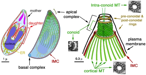
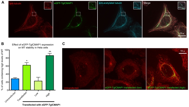
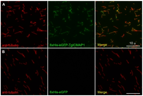
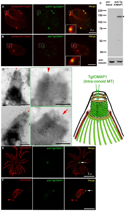
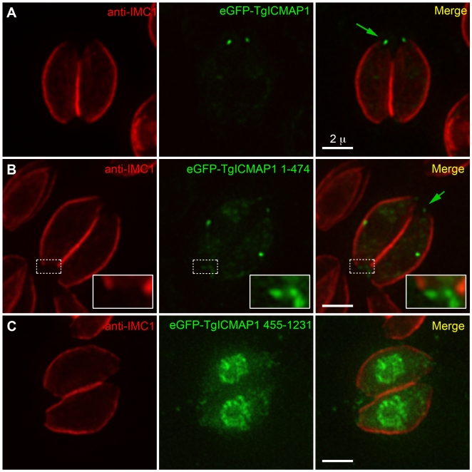
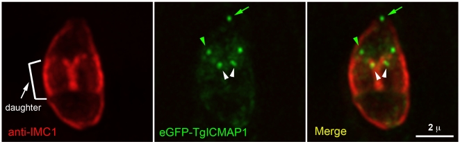
The microtubule cytoskeleton provides essential structural support for all eukaryotic cells and can be assembled into various higher order structures that perform drastically different functions. Understanding how microtubule-containing assemblies are built in a spatially and temporally controlled manner is therefore fundamental to understanding cell physiology. Toxoplasma gondii, a protozoan parasite, contains at least five distinct tubulin-containing structures, the spindle pole, centrioles, cortical microtubules, the conoid, and the intra-conoid microtubules. How these five structurally and functionally distinct sets of tubulin containing structures are constructed and maintained in the same cell is an intriguing problem. Previously, we performed a proteomic analysis of the T. gondii apical complex, a cytoskeletal complex located at the apical end of the parasite that is composed of the conoid, three ring-like structures, and the two short intra-conoid microtubules. Here we report the characterization of one of the proteins identified in that analysis, TgICMAP1. We show that TgICMAP1 is a novel microtubule binding protein that can directly bind to microtubules in vitro and stabilizes microtubules when ectopically expressed in mammalian cells. Interestingly, in T. gondii, TgICMAP1 preferentially binds to the intra-conoid microtubules, providing us the first molecular tool to investigate the intra-conoid microtubule assembly process during daughter construction.
Conflict of interest statement
Figures
Similar articles
-
Novel thioredoxin-like proteins are components of a protein complex coating the cortical microtubules of Toxoplasma gondii.Eukaryot Cell. 2013 Dec;12(12):1588-99. doi: 10.1128/EC.00082-13. Epub 2013 Jul 19. Eukaryot Cell. 2013. PMID: 23873863 Free PMC article.
-
Roles of the tubulin-based cytoskeleton in the Toxoplasma gondii apical complex.Trends Parasitol. 2024 May;40(5):401-415. doi: 10.1016/j.pt.2024.02.010. Epub 2024 Mar 25. Trends Parasitol. 2024. PMID: 38531711 Review.
-
Targeting Toxoplasma tubules: tubulin, microtubules, and associated proteins in a human pathogen.Eukaryot Cell. 2015 Jan;14(1):2-12. doi: 10.1128/EC.00225-14. Epub 2014 Nov 7. Eukaryot Cell. 2015. PMID: 25380753 Free PMC article. Review.
-
A doublecortin-domain protein of Toxoplasma and its orthologues bind to and modify the structure and organization of tubulin polymers.BMC Mol Cell Biol. 2020 Feb 28;21(1):8. doi: 10.1186/s12860-020-0249-5. BMC Mol Cell Biol. 2020. PMID: 32111164 Free PMC article.
-
Cryo-ET of Toxoplasma parasites gives subnanometer insight into tubulin-based structures.Proc Natl Acad Sci U S A. 2022 Feb 8;119(6):e2111661119. doi: 10.1073/pnas.2111661119. Proc Natl Acad Sci U S A. 2022. PMID: 35121661 Free PMC article.
Cited by
-
Essential function of the alveolin network in the subpellicular microtubules and conoid assembly in Toxoplasma gondii.Elife. 2020 May 7;9:e56635. doi: 10.7554/eLife.56635. Elife. 2020. PMID: 32379047 Free PMC article.
-
An evolutionarily conserved SSNA1/DIP13 homologue is a component of both basal and apical complexes of Toxoplasma gondii.Sci Rep. 2016 Jun 21;6:27809. doi: 10.1038/srep27809. Sci Rep. 2016. PMID: 27324377 Free PMC article.
-
Cell division in apicomplexan parasites.Nat Rev Microbiol. 2014 Feb;12(2):125-36. doi: 10.1038/nrmicro3184. Epub 2014 Jan 2. Nat Rev Microbiol. 2014. PMID: 24384598 Review.
-
Architecture of the Toxoplasma gondii apical polar ring and its role in gliding motility and invasion.Proc Natl Acad Sci U S A. 2024 Nov 12;121(46):e2416602121. doi: 10.1073/pnas.2416602121. Epub 2024 Nov 8. Proc Natl Acad Sci U S A. 2024. PMID: 39514309 Free PMC article.
-
SAS6-like protein in Plasmodium indicates that conoid-associated apical complex proteins persist in invasive stages within the mosquito vector.Sci Rep. 2016 Jun 24;6:28604. doi: 10.1038/srep28604. Sci Rep. 2016. PMID: 27339728 Free PMC article.
References
-
- Morrissette NS, Murray JM, Roos DS. Subpellicular microtubules associate with an intramembranous particle lattice in the protozoan parasite Toxoplasma gondii. J Cell Sci. 1997;110 (Pt 1):35–42. - PubMed
-
- Shaw MK, Compton HL, Roos DS, Tilney LG. Microtubules, but not actin filaments, drive daughter cell budding and cell division in Toxoplasma gondii. J Cell Sci. 2000;113 (Pt 7):1241–1254. - PubMed
-
- Nichols BA, Chiappino ML. Cytoskeleton of Toxoplasma gondii. J Protozool. 1987;34:217–226. - PubMed
-
- Morrissette NS, Sibley LD. Disruption of microtubules uncouples budding and nuclear division in Toxoplasma gondii. J Cell Sci. 2002;115:1017–1025. - PubMed
Publication types
MeSH terms
Substances
LinkOut - more resources
Full Text Sources
