. 2009 Dec;66(24):3979-91.
doi: 10.1007/s00018-009-0158-4. Epub 2009 Oct 13.

Inhibition of cholesterol recycling impairs cellular PrP(Sc) propagation

Affiliations

Inhibition of cholesterol recycling impairs cellular PrP(Sc) propagation

Sabine Gilch et al. Cell Mol Life Sci. 2009 Dec.

Abstract

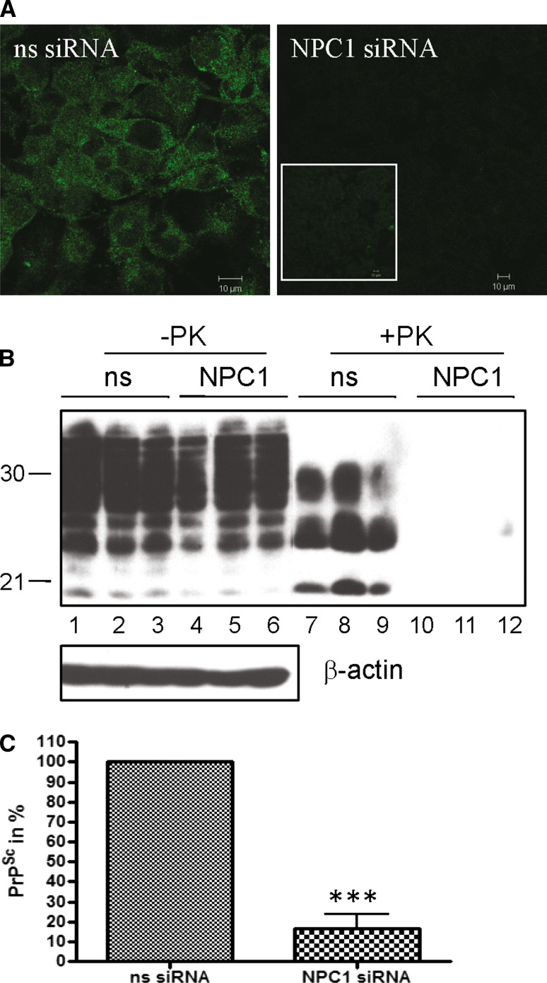
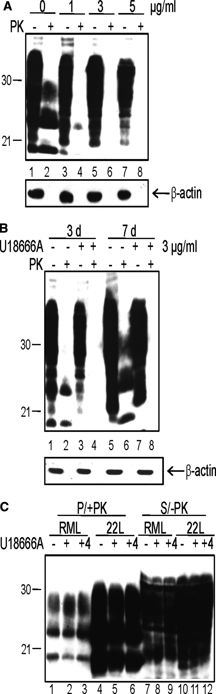
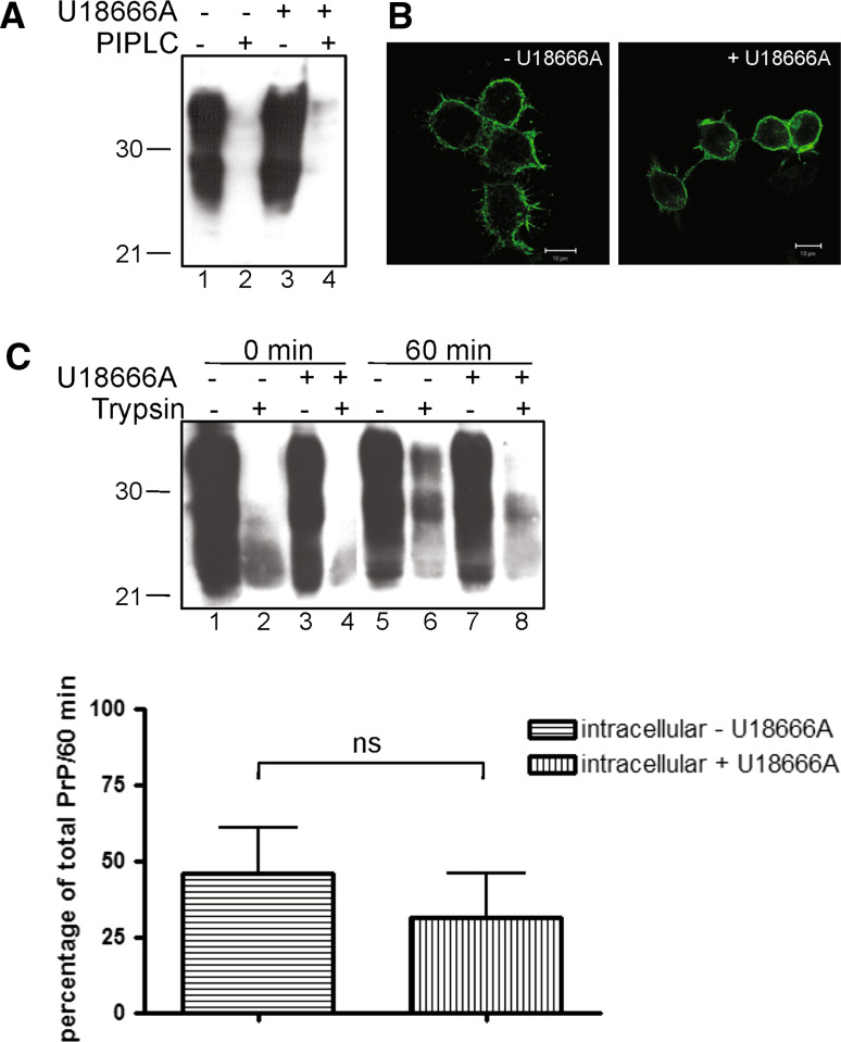
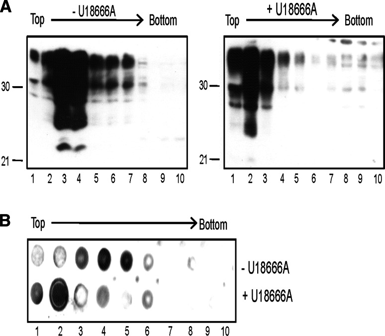
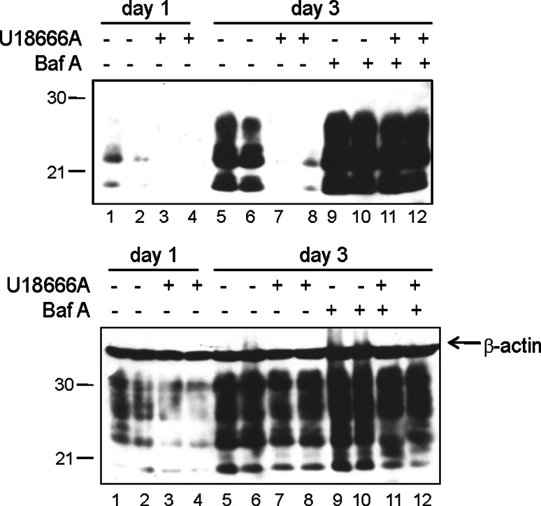
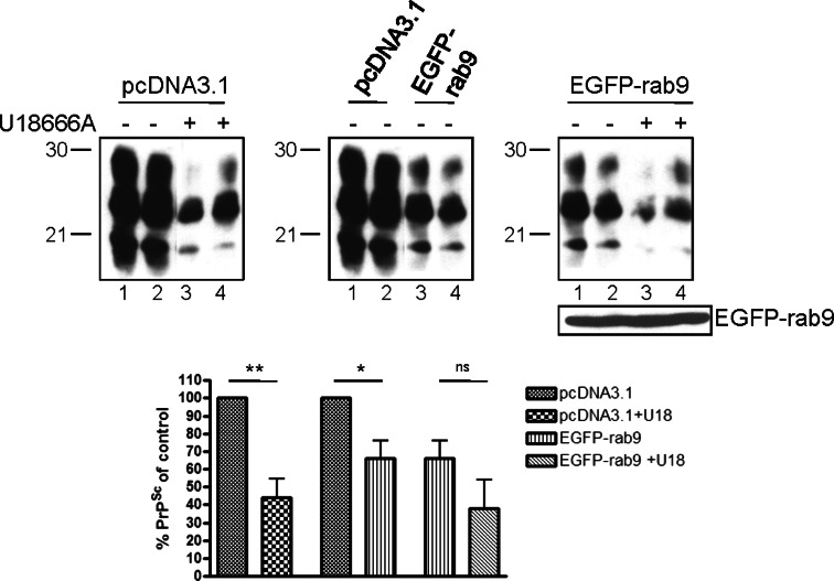
The infectious agent in prion diseases consists of an aberrantly folded isoform of the cellular prion protein (PrP(c)), termed PrP(Sc), which accumulates in brains of affected individuals. Studies on prion-infected cultured cells indicate that cellular cholesterol homeostasis influences PrP(Sc) propagation. Here, we demonstrate that the cellular PrP(Sc) content decreases upon accumulation of cholesterol in late endosomes, as induced by NPC-1 knock-down or treatment with U18666A. PrP(c) trafficking, lipid raft association, and membrane turnover are not significantly altered by such treatments. Cellular PrP(Sc) formation is not impaired, suggesting that PrP(Sc) degradation is increased by intracellular cholesterol accumulation. Interestingly, PrP(Sc) propagation in U18666A-treated cells was partially restored by overexpression of rab 9, which causes redistribution of cholesterol and possibly of PrP(Sc) to the trans-Golgi network. Surprisingly, rab 9 overexpression itself reduced cellular PrP(Sc) content, indicating that PrP(Sc) production is highly sensitive to alterations in dynamics of vesicle trafficking.


Figures

Fig. 1
Fig. 1
Knock-down of NPC-1 drastically reduces PrPSc. a ScN2a cells were transiently transfected with non-silencing (ns) siRNA or siRNA targeting NPC-1 mRNA. Two days post transfection, cells were stained with anti-NPC-1 antibody and Cy2-conjugated anti-rabbit IgG. Staining was visualized by confocal laser scanning microscopy, using identical immunofluorescence settings. The inset shows the image of cells transfected with NPC-1 siRNA excited with higher voltage to demonstrate presence of cells. b Transient transfections of ScN2a cells were performed in triplicate with ns siRNA or NPC-1 siRNA. Two days after transfection, cells were lysed and lysates were treated with (+PK) or without (−PK) PK. SDS-PAGE and immunoblot were performed, and for detection of PrP, monoclonal antibody 4H11 was used. To confirm equal loading, the immunoblot was reprobed with an antibody against β-actin (lower panel; -PK). c Quantification and statistical analysis of the effect of NPC-1 knock-down on PrPSc propagation. ScN2a cells transfected transiently with ns or NPC-1 siRNA were lysed 2 days post transfection. The amount of PrPSc upon PK digestion detected by immunoblot was densitometrically evaluated. The value for PrPSc in ns siRNA transfected cells was set to 100%; the amount in NPC-1 siRNA transfected cells was expressed as a percentage thereof. For statistical analysis, results of four independent experiments were used. Two out of four experiments were performed in duplicate and triplicate, respectively, and of these, the average percentage of PrPSc in relation to the control (ns siRNA) was used for statistical analysis (*** P <0.005; t-test)
Fig. 2
Fig. 2
Dose- and time-dependent reduction of PrPSc in persistently infected cells, but no inhibition of primary infection upon chemical induction of lysosomal cholesterol accumulation. a ScN2a cells were treated for 3 days with various concentrations (0, 1, 3, or 5 μg/ml) of U18666A as indicated. Cells were lysed, digested with PK (+PK) or not (−PK), and proteins were analyzed by immunoblot. Mab 4H11 was used for detection of PrP-specific bands. The immunoblot was dehybridized and incubated with an anti-β-actin mAb to control for equal loading (lower panel). b Treatment of ScN2a cells with U18666A (3 μg/ml) was performed for 3 and 7 days in parallel to mock treatment. At the different time points, U18666A-treated and mock-treated cells were lysed. Lysates were digested with PK (+PK) or not (−PK) and were subjected to immunoblot analysis using mAb 4H11. Equal loading of lysates of the different time points was confirmed by reprobing the membrane with anti-β-actin mAb (lower panel). c 3F4-N2a cells were left untreated or were pretreated for 4 h with U18666A (3 μg/ml) prior to prion infection. For prion infection, brain homogenates (1%) from RML- or 22L-infected terminally sick mice were added for 24 h to the culture medium. Cells that had not been pretreated were infected either in the presence (+) or absence (−) of U18666A for 24 h. For pretreated cells, the U18666A treatment was continued for the time period of infection (+4; 28 h treatment in total). After 24 h, brain homogenates and U18666A were removed, cells were washed and passaged three times 1:10. Passage 3 was lysed, lysates were treated with PK (+PK) or left untreated (−PK) and were then subjected to a solubility assay. Pellet fractions of samples + PK (P/+PK) and supernatant fractions of samples −PK (S/−PK) were analyzed by immunoblot with mAb 3F4, which selectively detects newly generated 3F4-PrPSc in the pellet fractions (P/+PK) and 3F4-PrPc in the supernatant (S/−PK)
Fig. 3
Fig. 3
No changes in membrane localization and turn-over of PrPc in U18666A treated N2a cells. a N2a cells were treated for 3 days with U18666A (3 μg/ml) or left untreated as indicated. For release of PrPc by PIPLC (left panel), cells were incubated for 4 h with or without the enzyme. Treatment with U18666A was continued during PIPLC digestion. Then cells were lysed and lysates were subjected to immunoblot analysis using mAb 4H11for detection of PrP specific bands. b N2a cells with or without U18666A treatment for 3 days were fixed, permeabilized, and stained in indirect immunofluorescence with anti-PrP mAb 4H11 and Cy2-conjugated anti-mouse IgG. c Biotinylation of N2a cells treated for 2 days with or without U18666A was performed and cultures were chased at 37°C in culture medium −/+U18666A for 60 min or were processed immediately. After 0 or 60 min chase, one culture dish each of treated or untreated cells was incubated with trypsin. PrP from cell lysates was immunoprecipitated using pAb A7, subjected to immunoblot, and detected by incubation with horseradish peroxidase (HRP)-conjugated streptavidin. A representative immunoblot of three independent experiments is shown. PrPc signals of the 60 min time point with (internalized PrPc) or without (total PrPc) trypsin digestion were densitometrically analyzed. The amount of internalized PrPc was expressed as a percentage of total PrPc. The lower panel depicts statistical evaluation of the difference between internalized PrPc in mock-treated or U18666A-treated cells using Student’s t-test (ns not significant; P-value >0.05; bars indicate standard error)
Fig. 4
Fig. 4
Lipid raft association of PrPc is not altered in U18666A-treated cells. a N2a cells were treated for 3 days with U18666A (right panel) or left untreated (left panel) and were subjected to a flotation assay. Ten fractions from the top to the bottom of the centrifuge tube were collected and analyzed by immunoblot with mAb 4H11. b Aliquots of gradient fractions were spotted on a nitrocellulose membrane and GM1 was detected by incubation of the membrane with HRP-conjugated cholera toxin subunit B (CtxB)
Fig. 5
Fig. 5
U18666A treatment increases PrPSc degradation. Upper panel ScN2a cells were treated for 1 day with U18666A (3 μg/ml) or were left untreated. Cells were either lysed after 1 day and lysates were treated with PK (lanes 1–4) or were incubated for further 2 days (day 3; lanes 5–12) with or without U18666A in presence or absence of bafilomycin A (10 nM). Then cells were lysed and subjected to PK digestion. All samples were separated by SDS-PAGE followed by immunoblot. For detection of PrP, mAb 4H11 was used. The experiment was performed in duplicate. Lower panel Samples without PK digestion were analyzed by immunoblot for total PrP using mAb 4H11 and for β-actin levels (arrow) for control of equal loading
Fig. 6
Fig. 6
Rab 9 overexpression impairs PrPSc propagation but partially rescues the interference induced by cholesterol accumulation. ScN2a cells were transiently transfected with pEGFP-rab 9 or with pcDNA3.1 as a control. Following transfection, treatment with U18666A (3 μg/ml) was performed for 2 days. Then cells were lysed, aliquots were digested with PK, and samples were analyzed by immunoblot using mAb 4H11. A representative duplicate experiment is shown (upper panel). To enable better comparison, samples analyzed by one immunoblot were opposed to each other. Expression of EGFP-rab 9 was detected using a polyclonal anti-GFP antibody. PrPSc signals of five independent experiments were evaluated densitometrically (ImageQuant TL), and statistical analysis was performed (lower panel, ns not significant; *P <0.05, **P <0.005; bars indicate standard error). The signal intensity of untreated mock-transfected controls was set as 100%, and all other values are expressed as a percentage of this value

References

    1. Prusiner SB. Prions. Proc Natl Acad Sci USA. 1998;95:13363–13383. doi: 10.1073/pnas.95.23.13363. - DOI - PMC - PubMed
    1. Weissmann C, Enari M, Klohn PC, Rossi D, Flechsig E. Transmission of prions. J Infect Dis. 2002;186(Suppl 2):S157–S165. doi: 10.1086/344575. - DOI - PubMed
    1. Aguzzi A, Polymenidou M. Mammalian prion biology: one century of evolving concepts. Cell. 2004;116:313–327. doi: 10.1016/S0092-8674(03)01031-6. - DOI - PubMed
    1. Kretzschmar HA, Prusiner SB, Stowring LE, Dearmond SJ. Scrapie prion proteins are synthesized in neurons. Am J Pathol. 1986;122:1–5. - PMC - PubMed
    1. Nunziante M, Gilch S, Schatzl HM. Prion diseases: from molecular biology to intervention strategies. Chembiochem. 2003;4:1268–1284. doi: 10.1002/cbic.200300704. - DOI - PubMed

Publication types

MeSH terms