. 2010 Jan;129(1):28-40.
doi: 10.1111/j.1365-2567.2009.03155.x. Epub 2009 Oct 12.

Efficiency of T-cell costimulation by CD80 and CD86 cross-linking correlates with calcium entry

Affiliations

Efficiency of T-cell costimulation by CD80 and CD86 cross-linking correlates with calcium entry

Markus Thiel et al. Immunology. 2010 Jan.

Abstract

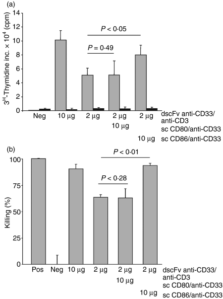
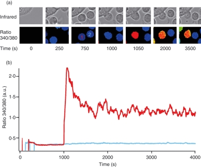
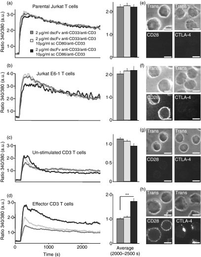
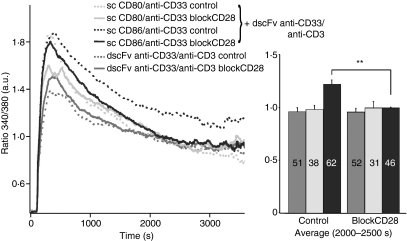
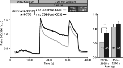
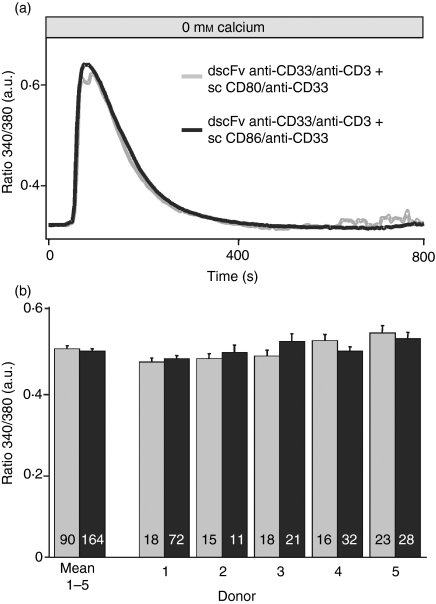
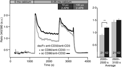
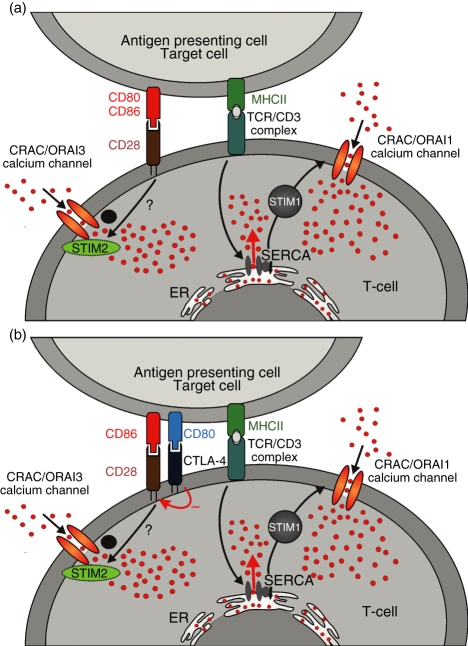
Costimulation is a fundamental principle of T-cell activation. In addition to T-cell receptor engagement, the interaction between CD80 and/or CD86 with CD28 and/or cytotoxic T-lymphocyte antigen 4 (CTLA-4) receptors is required to regulate T-cell activation and tolerance. While the importance of costimulation is clearly established, the exact molecular mechanism is unknown. We demonstrate that T-cell proliferation and the ability of CD8(+) T-effector cells to kill were enhanced slightly by CD80 but dramatically by CD86 costimulation. To further analyse the cellular process of costimulation, we developed a single-cell assay to analyse Ca(2+) signals following costimulation with bi-specific antibodies. We found that this stimulation method worked in every human T-cell that was analysed, making it one of the most efficient T-cell activation methods to date for primary human T cells. The enhanced proliferation and killing by costimulation was paralleled by an increase of Ca(2+) influx following CD86 costimulation and it was dependent on CD28/CTLA-4 expression. The enhanced Ca(2+) influx following CD86 costimulation was abrogated by an antibody that interfered with CD28 function. The differences in Ca(2+) influx between CD80 and CD86 costimulation were not dependent on the depletion of Ca(2+) stores but were eliminated by the application of 10 mum 2-aminoethyldiphenyl borate which has recently been shown to enhance stromal interaction molecule 2 (STIM2)-dependent Ca(2+) entry while reducing STIM1-dependent Ca(2+) entry. Our data indicate that differences in the efficiency of costimulation are linked to differences in Ca(2+) entry.


Figures

Figure 2
Figure 2
CD86 but not CD80 increased T-cell proliferation and cytotoxicity. (a) Suboptimal doses of double single-chain fragment variable (dscFv) anti-CD33/anti-CD3 (2 μg/ml) and 10 μg/ml of costimulatory molecules, either sc CD80/anti-CD33 or sc CD86/anti-CD33, were used to initiate the proliferation of the cytotoxic activity of naïve T cells in the presence of immobilized CD33 antigen (grey column) or phosphate-buffered saline (PBS; black column). The dscFv anti-CD19/anti-CD3 served as negative control (Neg). c.p.m., counts per minute. (b) Same constructs as in (a) were used. P-values for the difference between absence and presence of costimulatory molecules are indicated. Negative controls (Neg, dscFv anti-CD19/anti-CD3) and positive controls (Pos, 1% Triton X-100) were included in every experiment. Results are representative of three independent experiments.
Figure 1
Figure 1
Double single-chain fragment variable antibodies (dscFv) anti-CD33/anti-CD3 induced proliferation of T cells in a dose-dependent manner. Antigen-immobilized dscFv anti-CD33/anti-CD3 at various concentrations or control antibody fusion proteins single-chain fragment variable (scFv) anti-CD33, sc CD80/anti-CD33 and sc CD86/anti-CD33 (10 μg/ml) were used to stimulate T cells for 3 days. Cells were then pulsed for 16 hr before harvesting. Mean values of triplicate cultures are shown. Results are representative of three independent experiments. c.p.m., counts per minute.
Figure 3
Figure 3
Chinese hamster ovary (CHO) cells loaded with double single-chain fragment variable (dscFv) anti-CD33/anti-CD3 increase Ca2+ concentration in CD4+ T-cells. (a) Infrared and [Ca2+]i (as ratio 340/380) images. T cells are visible by the fura-2/AM loading in the ratio 340/380 pictures and in the infrared channel whereas CHO cells are only visible in the infrared channel. T cell 1 makes contact with a CHO cell, red Ca2+ trace in (b), whereas T-cell 2 makes no contact with a CHO cell during the entire experiment, blue Ca2+ trace in (b). (b) [Ca2+]i kinetics from typical fura-2/AM-loaded CD4+ T-cells either stimulated (T-cell 1, red trace) or not stimulated (T-cell 2, blue trace) by CHO cells loaded with 2 μg/ml dscFv anti-CD33/anti-CD3 in 0·25 mm Ca2+ solution. [Ca2+]i and infrared images were recorded in parallel every 5 seconds.
Figure 4
Figure 4
CD86 costimulation increases net Ca2+ entry in effector T cells. (a–d) Kinetics of intracellular [Ca2+] (as ratio 340/380) of parental Jurkat T cells, E6-1 Jurkat T cells, naïve, unstimulated primary CD4+ T cells or effector T cells. Effector T cells were prestimulated for 4 days with phytohaemagglutinin and interleukin-2. Cells were stimulated by Chinese hamster ovary (CHO) cells loaded with 2 μg/ml of dscFv anti-CD33/anti-CD3 alone or together with 10 μg/ml of sc CD80/anti-CD33 or sc CD86/anti-CD33. (e–h) CD28 and a cytotoxic T-lymphocyte antigen 4 (CTLA-4) staining of the corresponding cell type are shown. Scale bars represent 5 μm (**P < 0·01).
Figure 5
Figure 5
An inhibitory anti-CD28 antibody abrogated the differences between single-chain (sc) CD80/anti-CD33 or sc CD86/anti-CD33 costimulation. Kinetics and quantification of intracellular [Ca2+] (as ratio 340/380) of CD4+ T cells stimulated by CHO cells loaded with 2 μg/ml double single-chain fragment variable (dscFv) anti-CD33/anti-CD3 alone or together with 10 μg/ml sc CD80/anti-CD33 or sc CD86/anti-CD33. CD4+ T-cells were prestimulated with anti-CD3/anti-CD28-coated beads for 4 days. Cells were incubated for 10 min with the inhibitory anti-CD28 antibody in a 1 : 20 solution before measurement. The numbers of cells are given in the bars (**P < 0·01).
Figure 7
Figure 7
The differences in Ca2+ influx were eliminated by the application of 10 μm 2-aminoethyldiphenyl borate (2-APB). Kinetics of intracellular [Ca2+] (as ratio 340/380) of CD4+ T cells stimulated by Chinese hamster ovary (CHO) cells loaded with 2 μg/ml double single-chain fragment variable (dscFv) anti-CD33/anti-CD3 together with 10 μg/ml sc CD80/anti-CD33 or sc CD86/anti-CD33. CD4+ T cells were pre-stimulated with anti-CD3/anti-CD28-coated beads for 5 days. After 1000 seconds, cells were treated with thapsigargin (TG) in the absence of extracellular Ca2+ to completely deplete Ca2+ stores. A Ca2+ concentration of 0.25 mm was added to analyse Ca2+ entry. Finally, 10 μm 2-APB and 100 μm 2-APB were added. Numbers of analysed cells are given in the bars. (**P < 0·01).
Figure 6
Figure 6
Ca2+ release was identical for CD80 and CD86 costimulation. (a) Intracellular [Ca2+] (as ratio 340/380) of CD4+ T cells stimulated by Chinese hamster ovary (CHO) cells loaded with 2 μg/ml double single-chain fragment variable (dscFv) anti-CD33/anti-CD3 together with 10 μg/ml sc CD80/anti-CD33 or sc CD86/anti-CD33 following store depletion in the absence of extracellular Ca2+. The time-point zero was defined for every cell as 100 seconds before the release started. (b) Bars show the average of five different donors and all five donors individually (cell numbers are given in the bars).
Figure 8
Figure 8
The differences in Ca2+ influx were eliminated by the application of 10 μm 2-aminoethyldiphenyl borate. Same as Fig. 7, only that thapsigargin was omitted. Numbers of analysed cells are given in the bars (**P < 0·01).
Figure 9
Figure 9
A model for the activation of store-independent Ca2+ entry by costimulation is shown. Details are discussed in the text.

References

    1. Friedl P, den Boer AT, Gunzer M. Tuning immune responses: diversity and adaptation of the immunological synapse. Nat Rev Immunol. 2005;5:532. - PubMed
    1. Jacobelli J, Andres PG, Boisvert J, Krummel MF. New views of the immunological synapse: variations in assembly and function. Curr Opin Immunol. 2004;16:345. - PubMed
    1. Samelson LE. Signal transduction mediated by the T cell antigen receptor: the role of adapter proteins. Annu Rev Immunol. 2002;20:371. - PubMed
    1. Lanzavecchia A, Sallusto F. Antigen decoding by T lymphocytes: from synapses to fate determination. Nat Immunol. 2001;2:487. - PubMed
    1. Crabtree GR, Olson EN. NFAT signaling: choreographing the social lives of cells. Cell. 2002;109(Suppl):S67. - PubMed

Publication types

MeSH terms