. 2010 Jan 1;19(1):135-46.
doi: 10.1093/hmg/ddp474.

MDA5 and PTPN2, two candidate genes for type 1 diabetes, modify pancreatic beta-cell responses to the viral by-product double-stranded RNA

Affiliations

MDA5 and PTPN2, two candidate genes for type 1 diabetes, modify pancreatic beta-cell responses to the viral by-product double-stranded RNA

Maikel L Colli et al. Hum Mol Genet. .

Abstract

beta-Cell destruction in type 1 diabetes (T1D) is at least in part consequence of a 'dialog' between beta-cells and immune system. This dialog may be affected by the individual's genetic background. We presently evaluated whether modulation of MDA5 and PTPN2, two candidate genes for T1D, affects beta-cell responses to double-stranded RNA (dsRNA), a by-product of viral replication. These genes were selected following comparison between known candidate genes for T1D and genes expressed in pancreatic beta-cells, as identified in previous array analysis. INS-1E cells and primary fluorescence-activated cell sorting-purified rat beta-cells were transfected with small interference RNAs (siRNAs) targeting MDA5 or PTPN2 and subsequently exposed to intracellular synthetic dsRNA (polyinosinic-polycitidilic acid-PIC). Real-time RT-PCR, western blot and viability assays were performed to characterize gene/protein expression and viability. PIC increased MDA5 and PTPN2 mRNA expression, which was inhibited by the specific siRNAs. PIC triggered apoptosis in INS-1E and primary beta-cells and this was augmented by PTPN2 knockdown (KD), although inhibition of MDA5 did not modify PIC-induced apoptosis. In contrast, MDA5 silencing decreased PIC-induced cytokine and chemokine expression, although inhibition of PTPN2 induced minor or no changes in these inflammatory mediators. These findings indicate that changes in MDA5 and PTPN2 expression modify beta-cell responses to dsRNA. MDA5 regulates inflammatory signals, whereas PTPN2 may function as a defence mechanism against pro-apoptotic signals generated by dsRNA. These two candidate genes for T1D may thus modulate beta-cell apoptosis and/or local release of inflammatory mediators in the course of a viral infection by acting, at least in part, at the pancreatic beta-cell level.


Figures

Figure 1.
Figure 1.
siMDA5 and siRIG-I prevent PIC-induced activation of interferon-β promoter and NF-κB reporter, but do not modify INS-1E cell death. INS-1E cells were transfected with either an siControl (black bars), or siMDA5 (grey bars) or siRIG-I (striped bars). After 24 h of recovery, cells were left untreated or transfected with PIC1 (<2000 bp, used for siRIG-I experiments) or PIC2 (>2000 bp, siMDA5 experiments) as described in Materials and Methods. (A and B) MDA5 and RIG-I mRNA expression was assayed by real-time RT–PCR and corrected for the housekeeping gene GAPDH. Results are mean ± SEM of three to four independent experiments; *P < 0.01 versus siControl; **P < 0.01 versus siControl+PIC; ANOVA. (C and D) Twenty-four hours after transfection with siMDA5, cells were transfected with IFN-β (C) or NF-κB (D) reporters plus a pRL-CMV plasmid (used as internal control); cells were then exposed to internal PIC for 24 h and luciferase activity was assayed. The values were corrected for the activity of the internal control, pRL-CMV, and are presented as fold induction in relation to siControl. Results are mean ± SEM of 5–11 independent experiments; *P < 0.05 versus siControl, **P < 0.05 versus siControl+PIC, paired t-test. (E and F) INS-1E cells were transfected with siMDA5 or siRIG-I and then PIC as described earlier. Apoptosis was evaluated using HO/PI staining. Results are mean ± SEM of five independent experiments; *P < 0.01 versus siControl; ANOVA.
Figure 2.
Figure 2.
MDA5 or RIG-I KD partially prevents PIC-induced upregulation of mRNA expression of cytokines and chemokines in INS-1E cells. INS-1E cells were transfected with siControl (black bars), siMDA5 (grey bars) or siRIG-I (striped bars), and after recovery left untreated or exposed to different internal PICs (PIC1 for siRIG-I experiments and PIC2 for siMDA5 experiments) for 24 h. Poly(A)+ mRNA was extracted, assayed by real-time RT–PCR for rat IFN-β, IL15, CCL2, CCL5 and CXCL10 mRNAs and corrected for the housekeeping gene GAPDH. Results are mean ± SEM of three to four independent experiments; *P < 0.05 versus siControl, **P < 0.01 versus siControl, ***P < 0.01 versus siControl+PIC, ANOVA.
Figure 3.
Figure 3.
MDA5 but not RIG-I KD prevents PIC-induced CCL5 secretion in INS-1E cells. INS-1E cells were transfected with siControl (black bars), siMDA5 (A, grey bars) or siRIG-I (B, striped bars) and then treated as described in Figure 2. CCL5 secretion was evaluated in the supernatant by ELISA 24 h after internal PIC exposure (PIC1 for siRIG-I experiments and PIC2 for siMDA5 experiments), as described in Materials and Methods. Results are mean ± SEM of three to four independent experiments; *P < 0.01 versus siControl, **P < 0.01 versus siControl+PIC, ANOVA.
Figure 4.
Figure 4.
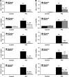
siMDA5 or siRIG-I prevent PIC-induced upregulation of cytokines and chemokines, but not apoptosis, in primary rat β-cells. FACS-purified rat β-cells were transfected with siControl (black bars), siMDA5 (grey bars) or siRIG-I (striped bars) as described in Materials and Methods. After 48 h of recovery, cells were left untreated or transfected with different PICs (PIC1 for siRIG-I experiments and PIC2 for siMDA5 experiments) for 24 h. (A and B) MDA5 and RIG-I mRNA expression was assayed by real-time RT–PCR and corrected for the housekeeping gene GAPDH. Results are mean ± SEM of four to six independent experiments; *P < 0.01 versus siControl; **P < 0.01 versus siControl+PIC, ANOVA. (C and D) Apoptosis was evaluated 24 h after PIC exposure using HO/PI staining. Results are mean ± SEM of three to five independent experiments; *P < 0.01 versus siControl, ANOVA. (E and F) INF-β, IL15, CCL2, CCL5 and CXCL10 mRNA expression were assayed by real-time RT–PCR and corrected for the housekeeping gene GAPDH. Results are mean ± SEM of four to six independent experiments; *P < 0.01 versus siControl; **P < 0.01 versus siControl+PIC, ANOVA.
Figure 5.
Figure 5.
Inhibition of JNK activation partially prevents PIC-induced JunB degradation and apoptosis in INS-1E cells. INS-1E cells were transfected with PIC1 for the indicated time points. (A and B) Expression of phospho-JNK, JunB and α-tubulin were evaluated by western blot. Pictures are representative of three independent experiments. (C) INS-1E cells were pre-treated with 10 µm of the JNK inhibitor SP600125 for 4 h and then transfected with PIC before being retrieved for protein extraction at the indicated time points; SP600125 was maintained in the medium following PIC treatment. Phospho-JNK, JunB and α-tubulin protein were evaluated by western blot. Pictures are representative of three independent experiments. (D and E) INS-1E cells were treated with the JNK chemical inhibitor SP600125 (D) or the JNK peptide inhibitor (E) for 4 h, and then transfected with PIC plus SP600125 or JNK peptide inhibitor. Apoptosis was evaluated 24 h after PIC exposure using HO/PI staining. Results are mean ± SEM of four independent experiments; *P < 0.05 versus siControl+PIC, paired t-test.
Figure 6.
Figure 6.
JunB KD augments PIC-induced β-cell death. INS-1E cells (AC) and primary rat β-cells (D) were transfected with siControl, siJunB 1 or siJunB 2 and then treated as described in Figures 2 and 4. (A) JunB protein expression was evaluated after transfection with two specific siRNAs. Pictures are representative of three independent experiments. (B) PIC-induced JunB mRNA expression was blocked by siRNA against JunB. Results are mean ± SEM of three to four independent experiments; *P < 0.01 versus siControl, **P < 0.01 versus siControl+PIC, ANOVA. (C and D) Apoptosis was evaluated in INS-1E cells (C) or FACS-purified rat β-cells (D) 24 h after PIC exposure using HO/PI staining. Results are mean ± SEM of four to six independent experiments; *P < 0.01 versus siControl, **P < 0.01 versus siControl+PIC, ANOVA.
Figure 7.
Figure 7.
KD of PTPN2 sensitizes INS-1E cells and primary β-cells to PIC-induced apoptosis. INS-1E cells (A) or FACS-purified rat β-cells (B) were transfected with siControl (black bars) or siPTPN2 (grey bars) and 48 h after recovery exposed to PIC1 for 24 h. (A and B) PTPN2 mRNA expression was evaluated by real-time RT–PCR and corrected for the housekeeping gene GAPDH. Results are mean ± SEM of three to five independent experiments; *P < 0.01 versus siControl, **P < 0.01 versus siControl+PIC, ANOVA. (C and D) INS-1E cells (C) or primary β-cells (D) were transfected with siControl or siPTPN2 and 48 h after recovery exposed to internal PIC1 for 24 h. Apoptosis was evaluated using HO/PI staining. Results are mean ± SEM of six independent experiments; *P < 0.01 versus siControl, **P < 0.05 versus siControl+PIC, ***P < 0.01 versus siControl+PIC, ANOVA. (E) PTPN2 and α-tubulin protein expression was assessed by western blot in FACS-purified rat β-cells exposed to internal PIC1 for 24 h; the picture is representative of two independent experiments. (F) INS-1E cells were transfected with control siRNA, or with an siRNA targeting PTPN2 as described in Materials and Methods, and 48 h after recovery exposed to internal PIC1 for 24 h. Cleavage of caspase-3 was observed by western blot. The picture is representative of two independent experiments.

References

    1. Eizirik D.L., Colli M.L., Ortis F. The role of inflammation in insulitis and β-cell loss in type 1 diabetes. Nat. Rev. Endocrinol. 2009;5:219–226. - PubMed
    1. Filippi C.M., von Herrath M.G. Viral trigger for type 1 diabetes: pros and cons. Diabetes. 2008;57:2863–2871. - PMC - PubMed
    1. Harrison L.C., Honeyman M.C., Morahan G., Wentworth J.M., Elkassaby S., Colman P.G., Fourlanos S. Type 1 diabetes: lessons for other autoimmune diseases? J. Autoimmun. 2008;31:306–310. - PubMed
    1. Peng H., Hagopian W. Environmental factors in the development of type 1 diabetes. Rev. Endocr. Metab. Disord. 2006;7:149–162. - PubMed
    1. Hardy J., Singleton A. Genomewide association studies and human disease. N. Engl. J. Med. 2009;360:1759–1768. - PMC - PubMed

Publication types

MeSH terms