. 2009 Nov 1;122(Pt 21):3954-65.
doi: 10.1242/jcs.044537. Epub 2009 Oct 13.

Differential VASP phosphorylation controls remodeling of the actin cytoskeleton

Affiliations

Differential VASP phosphorylation controls remodeling of the actin cytoskeleton

Peter M Benz et al. J Cell Sci. .

Abstract

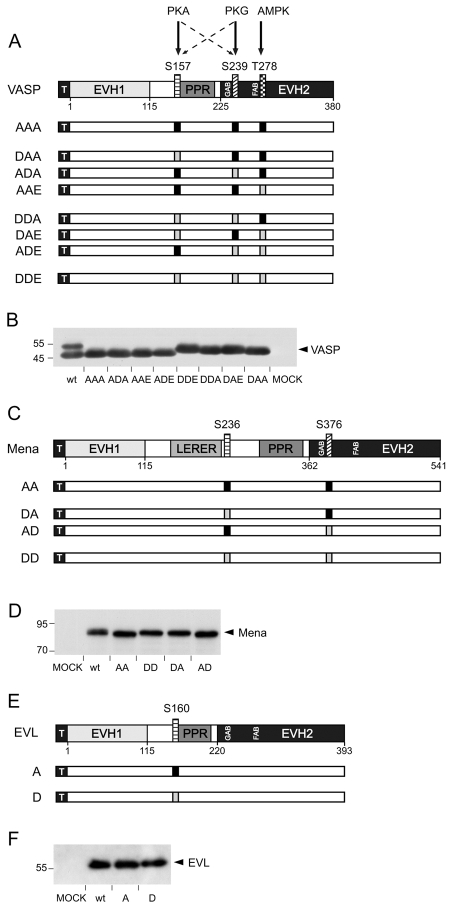
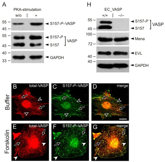
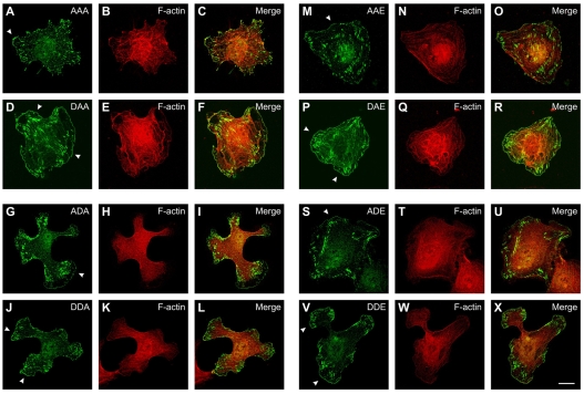
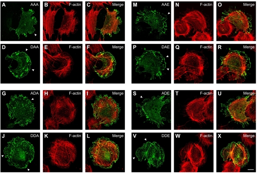
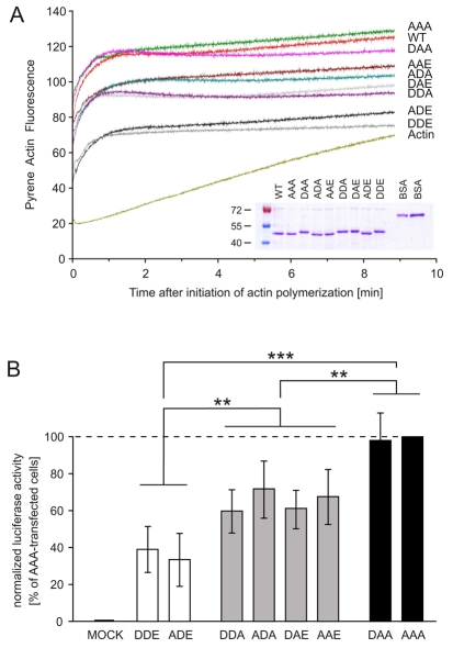
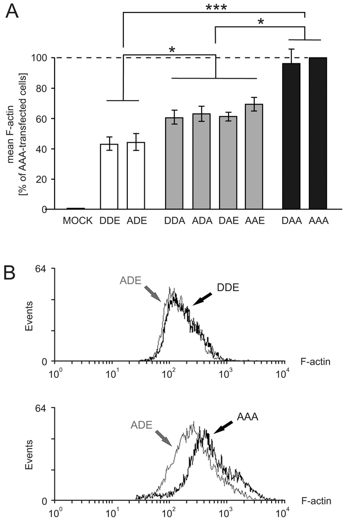
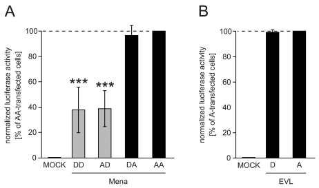
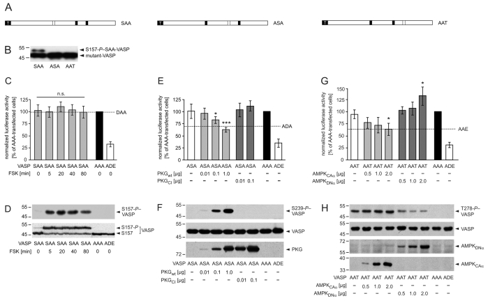
Proteins of the Enabled/vasodilator-stimulated phosphoprotein (Ena/VASP) family link signal transduction pathways to actin cytoskeleton dynamics. VASP is substrate of cAMP-dependent, cGMP-dependent and AMP-activated protein kinases that primarily phosphorylate the sites S157, S239 and T278, respectively. Here, we systematically analyzed functions of VASP phosphorylation patterns for actin assembly and subcellular targeting in vivo and compared the phosphorylation effects of Ena/VASP family members. Methods used were the reconstitution of VASP-null cells with ;locked' phosphomimetic VASP mutants, actin polymerization of VASP mutants in vitro and in living cells, site-specific kinase-mediated VASP phosphorylation, and analysis of the endogenous protein with phosphorylation-status-specific antibodies. Phosphorylation at S157 influenced VASP localization, but had a minor impact on F-actin assembly. Phosphorylation of the S157-equivalent site in the Ena/VASP family members Mena and EVL had no effect on the ratio of cellular F-actin to G-actin. By contrast, VASP phosphorylation at S239 (and the equivalent site in Mena) or T278 impaired VASP-driven actin filament formation. The data show that VASP functions are precisely regulated by differential phosphorylation and provide new insights into cytoskeletal control by serine/threonine kinase-dependent signaling pathways.


Figures

Fig. 1.
Fig. 1.
Arrested phosphomimetic Ena/VASP mutants. (A) Domain organization and phosphorylation sites of N-terminally 6×His-tagged human wild-type VASP (wt) and phosphomimetic VASP mutants. VASP is composed of an N-terminal EVH1 domain, a PPR and a C-terminal EVH2 domain. The black square with white `T' indicates the tag. PKA phosphorylates S157 (box shaded with horizontal lines) and S239 in order; S239 (box shaded with diagonal lines) is the primary target for PKG, which might also phosphorylate S157. T278 (box shaded with squares) is exclusively phosphorylated by AMPK. Phosphorylation sites were substituted with alanines (black boxes) or acidic residues (light boxes). Sites for G-actin binding (GAB, residues 234-237) and F-actin binding (FAB, residues 259-276), are located upstream of the preferred PKG and AMPK phosphorylation sites, respectively. The preferred PKA phosphorylation site is located upstream of the PPR. (B) Western blot with anti-6×His antibodies to confirm expression in EC_VASP–/– cells transiently transfected with cDNAs coding for VASP, arrested phosphomimetic mutants or vector without insert (MOCK). (C) Domain organization and phosphorylation sites of N-terminally 6×His-tagged murine wild-type Mena and phosphomimetic Mena mutants. LERER indicates the low complexity region harboring LERER repeats. Mena S236 and S376 correspond to VASP S157 and S239, respectively. Shading as for VASP. (D) Western blot with anti-6×His antibodies shows expression of wild-type and mutant Mena. (E) Domain organization and phosphorylation sites of N-terminally 6×His-tagged human wild-type EVL and phosphomimetic EVL mutants. EVL phosphorylation site S160 corresponds to VASP S157. (D) Western blot with anti-6×His antibodies confirms expression of wild-type EVL and arrested phosphomimetic mutants A and D. Note that (pseudo)phosphorylation at the first phosphorylation site leads to a mobility shift in SDS-PAGE of all mammalian Ena/VASP proteins.
Fig. 2.
Fig. 2.
VASP translocation to the cell periphery depends on S157 phosphorylation. Wild-type endothelial cells (EC_VASP+/+) were incubated with forskolin (5 μM) or buffer and analyzed using antibodies against S157-P–VASP and total-VASP. (A) Western blots of cell lysates normalized for GAPDH. (B-G) Immunofluorescence images of fixed and permeabilized cells. The merge of total-VASP (red; B,E) and S157-P–VASP (green; C,F) is given in yellow (D,G). Black arrowheads indicate stress fibers, black arrows indicate focal adhesions, and white arrowheads indicate plasma membranes. Images were taken with a 100× objective and are representative of a series of eight experiments. Scale bar: 15 μm. (H) Comparison of VASP, Mena and EVL expression in EC_VASP–/– and EC_VASP+/+ cells by western blotting. The cell lysates are normalized for GAPDH.
Fig. 3.
Fig. 3.
Subcellular localization of phosphomimetic VASP mutants in EC_VASP–/– cells. Confocal immunofluorescence images of EC_VASP–/– transiently transfected with phosphomimetic VASP mutants AAA (A-C), DAA (D-F), ADA (G-I), DDA (J-L), AAE (M-O), DAE (P-R), ADE (S-U), and DDE (V-X). At 24 hours after transfection, cells were trypsinized and replated on gelatinized chamberslides for 3 hours before fixation and staining. Reconstituted VASP mutants were immunolocalized with anti-VASP antibodies (green) and F-actin with fluorescent phalloidin (red). Arrowheads indicate the leading edge of lamellipodia. Scale bar: 15 μm.
Fig. 4.
Fig. 4.
Subcellular localization of phosphomimetic VASP mutants in MVD7 cells. Confocal immunofluorescence images of MVD7 cells transiently transfected with phosphomimetic VASP mutants AAA (A-C), DAA (D-F), ADA (G-I), DDA (J-L), AAE (M-O), DAE (P-R), ADE (S-U), and DDE (V-X). At 24 hours after transfection, cells were trypsinized and replated on fibronectin-coated chamber slides for 2 hours before fixation and staining. VASP mutants were immunolocalized with anti-VASP antibodies (green) and F-actin with fluorescent phalloidin (red). Arrowheads indicate the leading edge of lamellipodia. Scale bar: 15 μm.
Fig. 5.
Fig. 5.
VASP pseudophosphorylation at S239 and T278 but not at S157 impairs VASP-driven actin polymerization in vitro and in living cells. (A) In vitro actin polymerization driven by phosphomimetic VASP mutants. Pyrene-labeled G-actin (1 μM) was mixed with 0.25 μM purified recombinant wild-type (WT) or mutant VASP and the increase in fluorescence followed for 9 minutes. The curve labeled `Actin' represents a reaction without VASP. A representative experiment of a series of five is shown. The inset shows a Coomassie-stained gel of purified wild-type and mutant VASP. A molecular mass standard is given on the left; 1 and 3 μg BSA per lane serves to calibrate the protein load. Acidic substitution at position 157 leads to a mobility shift of the constructs in SDS-PAGE. (B) SRE assay of HEK293 cells overexpressing phosphomimetic VASP mutants. Normalized luciferase activity in cells transfected with AAA or empty vector (MOCK) were assigned values of 100% and 0%, respectively. The normalized luciferase activity of the phosphomimetic mutants was grouped into three classes: black, grey and white bars corresponding to 0, 1 and 2 negative charges at either the second (S239) or third (T278) phosphorylation site, respectively. Bars represent the mean ± s.d. (n=9; ANOVA, **P<0.01, ***P<0.001).
Fig. 6.
Fig. 6.
F-actin determination in cells expressing phosphomimetic VASP mutant by FACS analysis. HEK293 cells were transiently transfected with cDNAs coding for the indicated VASP pseudophosphorylation mutants or empty vector. F-actin was stained with Alexa-Fluor-647-phalloidin and quantified using FACS-analysis. (A) F-actin content of cells transfected with AAA or empty vector (MOCK) was set to 100% and 0%, respectively. Bars represent mean ± s.d. (n=6; ANOVA, *P<0.05, ***P<0.001). (B) Comparison of F-actin signals for ADE- (grey) versus DDE- (black, upper histogram) and ADE- (grey) versus AAA-transfected (black, lower histogram) HEK293 cells.
Fig. 7.
Fig. 7.
Effect of Mena and EVL pseudophosphorylations for actin assembly in cells. (A) SRE assay of CHO-S cells overexpressing phosphomimetic Mena mutants. Normalized luciferase activity in cells transfected with AA or empty vector (MOCK) were assigned values of 100% and 0%, respectively. Mean values ± s.d. are given (n=13; ANOVA, *** P<0.001). (B) SRE assay of HEK293 cells transfected with EVL mutants D and A. Normalized luciferase activity in cells transfected with A or empty vector were assigned values of 100% and 0%, respectively. Mean ± s.d. (n=8; ANOVA, P>0.05 D versus A).
Fig. 8.
Fig. 8.
Phosphorylation at S239 and T278 but not at S157 impairs VASP-driven actin polymerization in living cells. (A) Schematic representation of partially arrested VASP mutants. To analyze defined VASP phosphorylation patterns, we substituted two of the three phosphorylation sites with alanine residues, retaining one to allow kinase-mediated phosphorylation specifically at this residue. Black and open boxes indicate alanine and an accessible phosphorylation site, respectively. White `T' indicates the tag. (B) Western blot with anti-VASP antibodies to confirm expression of mutants SAA, ASA or AAT in transfected EC_VASP–/– cells. (C-D) PKA-mediated phosphorylation of S157-VASP does not affect global cellular F-actin content. Transfected HEK293 cells were incubated with forskolin (FSK, 5 μM) for 5, 20, 40 and 80 minutes. (C) SRE assay. The luciferase activities in cells transfected with AAA or empty vector (MOCK) were set to 100% and 0%, respectively. For comparison, the luciferase activity of unstimulated cells expressing DAA is given (dashed line, n=6; ANOVA, P>0.05 stimulated versus unstimulated SAA-transfected cells). The transfection with ADE, which maximally blocked VASP-driven actin assembly (Fig. 5B), served as a positive control. (D) The expression of VASP mutants and phosphorylation of SAA at S157 was confirmed by immunoblotting using antibodies against S157-P (upper panel) and total-VASP (lower panel). (E-F) PKG-mediated VASP phosphorylation at S239 reduces global actin polymerization. HEK239 cells were co-transfected with VASP mutant ASA and the indicated amounts of PKGwt or PKGCI. (E) For comparison, the signal of unstimulated cells expressing ADA is given (dashed line). Normalized luciferase activities are plotted relative to the activity in cells transfected with AAA or empty vector (MOCK) (set to 100% and 0%, respectively; n=8; ANOVA, *P<0.05, ***P<0.001). ADE served as a positive control. (F) Anti-S239-P antibodies indicated the phosphorylation of S239-ASA (upper panel) in western blots. The expression of VASP mutants in transfected cells was confirmed by immunoblotting with anti-VASP antibodies (middle panel). The expression of PKG variants was probed using antibodies against PKG (lower panel). (G-H) AMPK-meditated phosphorylation of VASP at T278 interferes with F-actin assembly. HEK239 cells were transiently co-transfected with VASP-AAT and AMPKCAα or AMPKDNα. (G) For comparison, the activity of AAE-transfected cells is indicated (dashed line). Normalized luciferase activities are plotted relative to the activity in cells transfected with AAA or empty vector (MOCK) (set to 100% and 0%, respectively; n=8; ANOVA, *P<0.05). ADE served as a positive control. (H) Western blots using anti-T278-P (upper panel) and anti-VASP antibodies (second panel) confirmed T278 phosphorylation and AAT expression, respectively. AMPK variants were probed by myc-tag specific antibodies (lower panels).

References

    1. Abel, K., Mieskes, G. and Walter, U. (1995). Dephosphorylation of the focal adhesion protein VASP in vitro and in intact human platelets. FEBS Lett. 370, 184-188. - PubMed
    1. Applewhite, D. A., Barzik, M., Kojima, S., Svitkina, T. M., Gertler, F. B. and Borisy, G. G. (2007). Ena/VASP proteins have an anti-capping independent function in filopodia formation. Mol. Biol. Cell 18, 2579-2591. - PMC - PubMed
    1. Bachmann, C., Fischer, L., Walter, U. and Reinhard, M. (1999). The EVH2 domain of the vasodilator-stimulated phosphoprotein mediates tetramerization, F-actin binding, and actin bundle formation. J. Biol. Chem. 274, 23549-23557. - PubMed
    1. Ball, L. J., Kuhne, R., Hoffmann, B., Hafner, A., Schmieder, P., Volkmer-Engert, R., Hof, M., Wahl, M., Schneider-Mergener, J., Walter, U. et al. (2000). Dual epitope recognition by the VASP EVH1 domain modulates polyproline ligand specificity and binding affinity. EMBO J. 19, 4903-4914. - PMC - PubMed
    1. Barzik, M., Kotova, T. I., Higgs, H. N., Hazelwood, L., Hanein, D., Gertler, F. B. and Schafer, D. A. (2005). Ena/VASP proteins enhance actin polymerization in the presence of barbed end capping proteins. J. Biol. Chem. 280, 28653-28662. - PMC - PubMed

Publication types

MeSH terms