. 2009 Oct 15;69(20):7986-93.
doi: 10.1158/0008-5472.CAN-09-2266. Epub 2009 Oct 13.

Glioblastoma cells require glutamate dehydrogenase to survive impairments of glucose metabolism or Akt signaling

Affiliations

Glioblastoma cells require glutamate dehydrogenase to survive impairments of glucose metabolism or Akt signaling

Chendong Yang et al. Cancer Res. .

Erratum in

  • Cancer Res. 2010 Feb 1;70(3):1275. Bachoo, Robert G [corrected to Bachoo, Robert M]

Abstract

Oncogenes influence nutrient metabolism and nutrient dependence. The oncogene c-Myc stimulates glutamine metabolism and renders cells dependent on glutamine to sustain viability ("glutamine addiction"), suggesting that treatments targeting glutamine metabolism might selectively kill c-Myc-transformed tumor cells. However, many current or proposed cancer therapies interfere with the metabolism of glucose, not glutamine. Here, we studied how c-Myc-transformed cells maintained viability when glucose metabolism was impaired. In SF188 glioblastoma cells, glucose deprivation did not affect net glutamine utilization but elicited a switch in the pathways used to deliver glutamine carbon to the tricarboxylic acid cycle, with a large increase in the activity of glutamate dehydrogenase (GDH). The effect on GDH resulted from the loss of glycolysis because it could be mimicked with the glycolytic inhibitor 2-deoxyglucose and reversed with a pyruvate analogue. Furthermore, inhibition of Akt signaling, which facilitates glycolysis, increased GDH activity whereas overexpression of Akt suppressed it, suggesting that Akt indirectly regulates GDH through its effects on glucose metabolism. Suppression of GDH activity with RNA interference or an inhibitor showed that the enzyme is dispensable in cells able to metabolize glucose but is required for cells to survive impairments of glycolysis brought about by glucose deprivation, 2-deoxyglucose, or Akt inhibition. Thus, inhibition of GDH converted these glutamine-addicted cells to glucose-addicted cells. The findings emphasize the integration of glucose metabolism, glutamine metabolism, and oncogenic signaling in glioblastoma cells and suggest that exploiting compensatory pathways of glutamine metabolism can improve the efficacy of cancer treatments that impair glucose utilization.


Figures

Figure 1
Figure 1. Glutamine metabolism in glioblastoma cells
A, Glutamine (Gln) is a precursor for synthesis of nucleotides, proteins and glucosamine. It can also be metabolized in the mitochondria, providing carbon to the TCA cycle as α-ketoglutarate (α-kg). Downstream metabolism converts glutamine carbon into oxaloacetate (OAA) and/or acetyl-CoA (Ac-CoA), although the latter is predominantly formed from glucose. B, Glioblastoma cells were cultured in medium supplemented with unlabeled glucose and either L-[α-15N]-glutamine or L-[γ-15N]-glutamine. Glutaminase (GLS) and glutamate dehydrogenase (GDH) activities were followed by measuring transfer of 15N to 15NH4+. The time course shows the average and SD at each time point for three parallel cultures. The pie graph shows relative contributions of the γ and α nitrogens of glutamine to the total NH4+ pool after 8 hours. The small amount of unlabeled ammonium (gray wedge) mostly resulted from metabolism of unlabeled glutamate by GDH. Other abbreviations: Glc, glucose; Pyr, pyruvate; Lac, lactate; Cit, citrate; Glu, glutamate; Succ, succinate; Fum, fumarate; Mal, malate; Asp, aspartate; NH4+, ammonium; ALT, alanine aminotransferase.
Figure 2
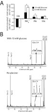
Figure 2. Glucose withdrawal increases mitochondrial glutamine metabolism
A, Glutamine-dependent metabolic rates in the presence and absence of glucose. Averages and SDs of three independent cultures are shown. *: p<0.005. B, 13C NMR spectra of metabolites from cells cultured in medium containing L-[3-13C]-glutamine plus or minus unlabeled glucose. The inset shows an enlargement of the glutamate C4 resonance at 34 ppm. The doublet (d) corresponds to glutamate labeled both at C4 (from acetyl-CoA) and at C3 (from glutamine-derived OAA), and the singlet (s) corresponds to glutamate molecules labeled at C4 but not C3. The experiment was performed twice for each condition. Abbreviation: Ala, alanine.
Figure 3
Figure 3. Glucose withdrawal activates glutamate dehydrogenase
A, Cells were cultured in decreasing glucose concentrations, and the rates of glucose consumption and ammonia production were measured. B, Total ammoniagenesis was measured in cells cultured with or without glucose, and with increasing concentrations of methyl-pyruvate (CH3-Pyr). The average and SD of three independent cultures are shown. C, Glutaminase activity was measured by following transfer of 15N from L-[γ-15N]-glutamine to 15NH4+ in the presence or absence of glucose and CH3-Pyr. The average and SD of three independent cultures are shown. #: p<0.05. D, Cells were cultured in L-[α-15N]-glutamine and the accumulation of 15NH4+ (GDH activity) and of 15N-alanine (ALT activity) was measured in the presence or absence of glucose and CH3-Pyr. The average and SD of three independent cultures are shown. *: p<0.0005
Figure 4
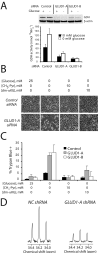
Figure 4. Glioblastoma cells require GDH to survive glucose deprivation
A, Cells were transfected with a control siRNA or siRNAs directed against the GLUD1 transcript. The effect on GDH protein abundance (top) and flux (bottom) was determined in the presence and absence of glucose. The average and SD of three independent cultures are shown. B-C, Cells transfected with the control or GLUD1 siRNAs were subjected to glucose withdrawal, and effects on morphology and viability were determined. The metabolites CH3-Pyr and dm-αKG were tested for their ability to rescue viability in glucose-deprived cells. The average and SD of three independent cultures are shown for each condition. D, Glucose-deprived cells expressing a control (NC) shRNA or an shRNA against the GLUD1 transcript (GLUD1-A) were cultured with L-[3-13C]-Gln. Labeling in carbon 4 of glutamate was analyzed by 13C NMR.
Figure 5
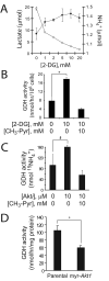
Figure 5. Inhibition of glycolysis or Akt signaling activates GDH
A, Glioblastoma cells were cultured in increasing concentrations of 2-deoxyglucose (2-DG) and the release of lactate (○) and NH4+ (●) was measured. B, Cells were cultured in 4 mM L-[α-15N]-glutamine and the accumulation of 15NH4+ was measured in the presence or absence of 2-DG and CH3-Pyr. C, Cells were cultured with an Akt inhibitor previously shown to suppress glycolysis in these cells (9). The effect on GDH activity in the absence and presence of CH3-Pyr was determined. D, GDH activity was compared between immortalized mouse astrocytes lacking or containing a constitutively-active Akt allele (myr-Akt1). In all experiments, the average and SD of three independent cultures are shown for each condition. #:p<0.05; *: p<0.005.
Figure 6
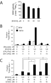
Figure 6. A GDH inhibitor sensitizes glioblastoma cells to glucose withdrawal and to inhibitors of glycolysis and Akt signaling
A, The effect of EGCG on GDH activity was determined in glucose-deprived cells. B, Glioblastoma cells were cultured in the presence or absence of glucose, EGCG, CH3-Pyr and dm-αKG. The effects of these treatments, alone or in combination, were determined on cell viability at 8 and 16 hours. C, Glioblastoma cells were cultured in the presence or absence of EGCG, an Akt inhibitor or 2-DG, alone or in combination. After 24 hours, viability was determined by Trypan blue staining. In all experiments, the average and SD of three independent cultures are shown for each condition. *: p≤0.005.

Similar articles

Cited by

References

    1. Hsu PP, Sabatini DM. Cancer cell metabolism: Warburg and beyond. Cell. 2008;134:703–7. - PubMed
    1. Kim JW, Dang CV. Cancer's molecular sweet tooth and the Warburg effect. Cancer Res. 2006;66:8927–30. - PubMed
    1. Medina MA. Glutamine and cancer. J Nutr. 2001;131:2539S–42S. discussion 50S-1S. - PubMed
    1. DeBerardinis RJ, Sayed N, Ditsworth D, Thompson CB. Brick by brick: metabolism and tumor cell growth. Curr Opin Genet Dev. 2008;18:54–61. - PMC - PubMed
    1. DeBerardinis RJ, Mancuso A, Daikhin E, et al. Beyond aerobic glycolysis: transformed cells can engage in glutamine metabolism that exceeds the requirement for protein and nucleotide synthesis. Proc Natl Acad Sci U S A. 2007;104:19345–50. - PMC - PubMed

Publication types

MeSH terms