. 2010 Feb;58(2):141-56.
doi: 10.1369/jhc.2009.954891.

Tissue distibution of murine Muc19/smgc gene products

Affiliations

Tissue distibution of murine Muc19/smgc gene products

Biswadip Das et al. J Histochem Cytochem. 2010 Feb.

Abstract

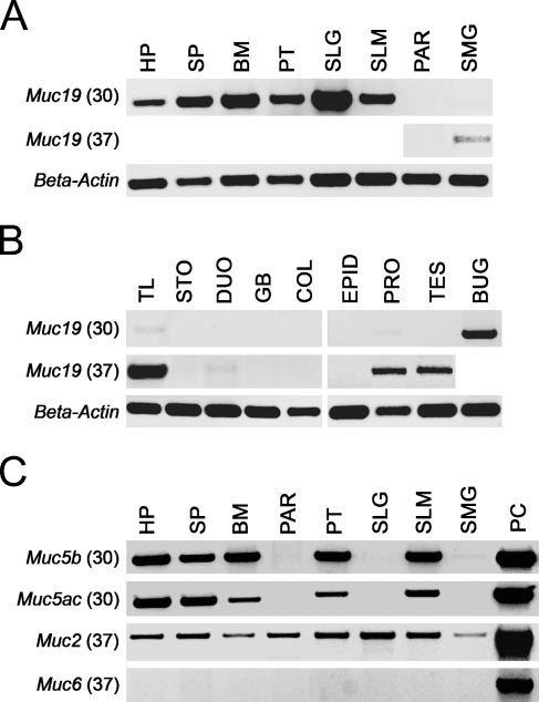
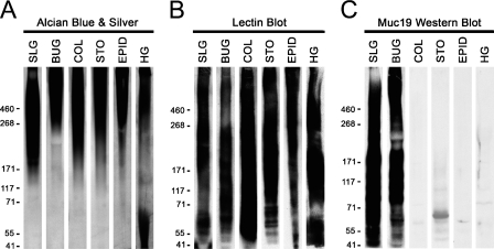
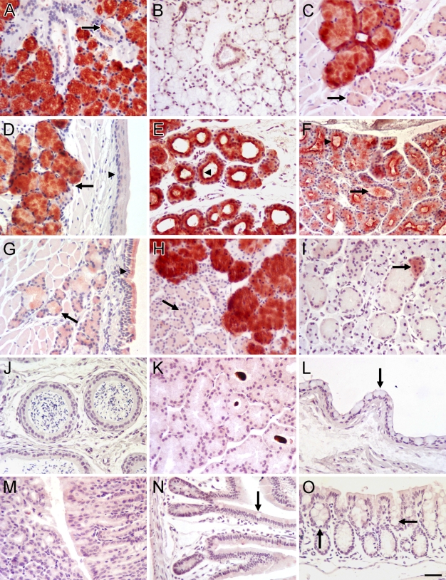
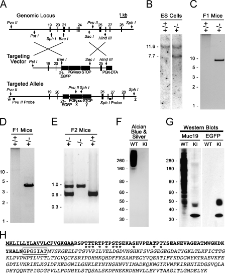
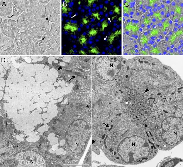
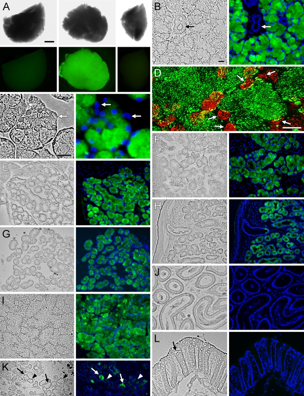
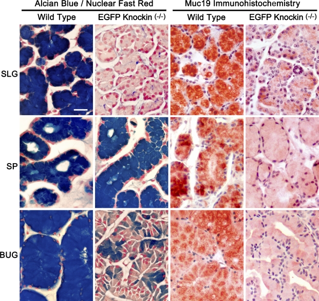
The recently identified gene Muc19/Smgc encodes two diverse splice variants, Smgc (submandibular gland protein C) and Muc19 (mucin 19). Muc19 is a member of the large gel-forming mucin family and is an exocrine product of sublingual mucous salivary glands in mice. SMGC is a transiently expressed secretion product of developing rodent submandibular and sublingual glands. Little is known about the expression of Muc19/Smgc gene products in other murine salivary and non-salivary tissues containing the mucous cell phenotype. Muc19 expression was therefore initially assessed by RT-PCR and immunohistochemistry. As a complementary approach, we developed a knockin mouse model, Muc19-EGFP, in which mice express a fusion protein containing the first 69 residues of Muc19 followed by enhanced green fluorescent protein (EGFP) as a marker of Muc19 expression. Results from both approaches are consistent, with preferential Muc19 expression in salivary major and minor mucous glands as well as submucosal glands of the tracheolarynx and bulbourethral glands. Evidence also indicates that individual mucous cells of minor salivary and bulbourethral glands produce another gel-forming mucin in addition to Muc19. We further find tissue expression of full-length Smgc transcripts, which encode for SMGC, and are restricted to neonatal tracheolarynx and all salivary tissues.


Figures

Figure 1
Figure 1
Expression of Muc19 transcripts in mouse tissues. (A) Upper panel: RT-PCR products using 50 ng of random primed cDNA at 30 cycles from major and minor salivary glands. HP, hard palate; SP, soft palate; BM, buccal mucosa; PT, posterior tongue; SLG, sublingual gland; SLM, sublingual mucosa; PAR, parotid gland; SMG, submandibular gland. Middle panel: tissues with no products at 30 cycles were re-tested at 37 cycles. Lower panel: β-actin–positive controls for each tissue sample (30 cycles, 349-bp product). (B) RT-PCR results for non-salivary tissues (upper and middle panels) as described for A. TL, tracheolarynx; STO, stomach mucosa; DUO, duodenum; GB, gall bladder; COL, colon; EPID, epididymis; PRO, prostate; TES, testes; BUG, bulbourethral gland. (C) Expression of transcripts for the gel-forming mucins Muc5b, Muc5ac, Muc2, and Muc6 in salivary tissues. Shown are RT-PCR products using 50 ng of cDNA at either 30 cycles (Muc5b and Muc5ac) or 37 cycles (Muc2 and Muc6). Abbreviations are as given in A. PC, positive control tissue used for each mucin, tracheolarynx (Muc5b), stomach mucosa (Muc5ac and Muc6), and small intestine (Muc2).
Figure 2
Figure 2
Testing the specificity against gel-forming mucins of antiserum raised against purified rat Muc19. Homogenates from select mouse tissues that express Muc19 and/or other gel-forming mucins were subjected to SDS-PAGE (3–8% gradient gels). One gel (A) was stained directly with Alcian Blue and subsequent silver enhancement to detect highly glycosylated glycoproteins. A second gel (B) was blotted, and lanes were probed with either wheat germ agglutinin (BUC, COL, STO) or peanut agglutinin (SLG, EPID, HG) to verify transfer of high-molecular-mass glycoproteins. A third gel (C) was subjected to Western blotting using rabbit anti-rat sublingual mucin. Lanes were loaded with homogenates from the following original wet weight of tissue: 0.4 mg sublingual gland (SLG), 0.5 mg bulbourethral glands (BUG), 1.4 mg colon mucosa (COL), 2.9 mg stomach mucosa (STO), 0.9 mg epididymis (EPID), and 3.7 mg Harderian glands (HG). In all three cases (A–C), samples were either stained or developed simultaneously for the same period of time.
Figure 3
Figure 3
Immunohistochemical localization of Muc19 in mouse tissues. (A) Sublingual gland; all mucous cells are reactive. Arrow, ductal lumen. (B) Sublingual gland probed with preimmune serum. (C) Posterior tongue; mucous cells are reactive. Arrow, unstained serous acini. (D) Soft palate: oral mucosa; arrow, stained mucous cells; arrowhead, oral epithelium. (E) Hard palate; mucous cells are reactive. Arrowhead, intense staining of apical cytoplasm of mucous cells lining ducts. (F) Minor mucous glands within buccal mucosa; arrow, stained secretions within ductal lumen; arrowhead, apical cytoplasmic staining of mucous tubules. (G) Soft palate: nasal mucosa; arrow, light staining of mucous cell; arrowhead, light staining within ciliated epithelial cells. (H) Bulbourethral gland; mucous cells are reactive. Arrow, unstained serous acini. (I) Submandibular gland; granular convoluted ducts and most seromucous acini are unreactive. Arrow, a few acini are stained. (J) Epididymis. (K) Harderian gland. (L) Conjunctiva epithelium; arrow, surface mucous goblet cells. (M) Stomach mucosa in cross-section (left) and longitudinal-section (right). (N) Ileum; arrow, goblet cell. (O) Colon; arrows, mucous goblet cells within crypts. Paraffin sections (5 μm) were probed with rabbit anti-rat sublingual mucin antiserum at dilutions of 1:1000 (A,C,D,G,H) and 1:300 (B,E,F,I–O). Vectastain ABC-HRP Kit and Nova Red substrate with light hematoxylin counterstain. Bar in O = 20 μm for all panels.
Figure 4
Figure 4
Production and initial characterization of Muc19-EGFP knockin mice. (A) Strategy for targeted disruption of Muc19 transcripts and insertion of enhanced green fluorescent protein (EGFP) in-frame within exon 21 of gene Muc19/Smgc (the fourth exon utilized for Muc19 transcripts). The targeted genomic locus contains gene exons 19–28. The targeting vector contains EGFP coding sequence (in-frame with the Muc19 coding sequence of exon 21), neomycin under control of the phosphoglycerate kinase promoter (PGKneo), and a STOP cassette to further prevent transcriptional read-through. The PGKneo sequence is flanked by loxP sites (not shown). The homology arms are defined by restriction sites PstI/EaeI (5′ arm) and SacI/HindIII (3′ arm). Diphtheria toxin A (DTA) under control of the PGK promoter is for negative selection in embryonic stem (ES) cells. The targeted EGFP knockin recombinant allele is shown with restriction sites (PvuII and SphI) and probes used for Southern blot analyses as well as sites of PCR primers w–z. See Materials and Methods for details. (B) Southern blot analysis of SphI-digested DNA from ES cells indicates non-recombinant cells (+/+) and cells correctly targeted at the 3′ end of the locus (+/−). (C) PCR-based genotyping of F1 agouti mice to distinguish the absence (+/+) or presence (+/−) of germline transmission and correct insertion of the 3′ end of the targeted allele using primers w and x (as shown in A). (D) Same as in C, but using primers y and z to detect germline transmission and correct insertion of the 5′ end of the targeted allele. (E) Genotyping of progeny from intercrossing F1-recombinant mice. The PCR reaction includes one forward primer within intron 20 and two reverse primers, one in intron 21 (595-bp wild-type allele) and the other within EGFP (856-bp targeted allele). (F) Sublingual gland homogenates (150 μg wet weight) from wild-type (WT) and homozygous Muc19-EGFP knockin (KI) mice subjected to SDS-PAGE (4–12% gradient gel) and stained with Alcian Blue and subsequent silver enhancement to detect highly glycosylated glycoproteins. (G) Homogenates as in F were run on a similar gel, blotted, and probed with either anti-rat Muc19 (Muc19) or anti-GFP (EGFP). Loaded homogenates were 150 (Muc19) and 400 μg wet weight (EGFP). (H) Predicted translation of the Muc19-EGFP fusion construct. Residues in bold are from Muc19, boxed residues are encoded by the polylinker, and italic residues are EGFP. Predicted signal peptide is underlined. Asterisks denote predicted potential sites of O-glycosylation.
Figure 5
Figure 5
Muc19-EGFP fluorescence in sublingual mucous cells from homozygous Muc19-EGFP knockin mice and comparison of cell ultrastructure to wild-type acinar cells. (A–C) Cryosection (5 μm) of a sublingual gland from a homozygous Muc19-EGFP knockin mouse shown under brightfield (A) and fluorescent (B) microscopy, with an overlay image shown in C. Nuclei were counterstained with 4′,6-diamidino-2-phenylindole (DAPI) (blue). EGFP fluorescence is predominant within the apical cytoplasm of tubular mucous cells, whereas punctate fluorescence is sparsely scattered within the more-basal regions. The larger granules of serous demilune cells are readily visible under brightfield microscopy, but these cells do not exhibit EGFP fluorescence (arrows, A,B). (D) Transmission electron microscopy (TEM) of a sublingual acinar mucous cell from an 8-week-old male wild-type mouse. Typical mucous cell structure is displayed, including large electron-lucent secretory granules (arrowhead) filling the apical cytoplasm, a basal nucleus (N), and large mitochondria (arrow). (E) TEM of sublingual mucous cells from an age- and sex-matched homozygous Muc19-EGFP knockin mouse. Mucous cells surrounding a small lumen (white arrow) contain small, dense apical secretory granules (arrowhead). Nuclei (N) are basally oriented, but Golgi complexes are more prominent in the absence of large mucous granules. Mitochondria, arrow. Bars: A = 20 μm; D = 5 μm for D,E.
Figure 6
Figure 6
EGFP fluorescence in tissues of heterozygous Muc19-EGFP knockin mice. (A) Glands from a wild-type mouse (left panels) and a heterozygous Muc19-EGFP mouse (center and right panels) shown under brightfield (upper panels) and fluorescent (lower panels) microscopy. Center panels are each of a whole sublingual gland, whereas the right panels are of a quarter portion from an adjoining submandibular gland. (B–L) Cryosections (5-μm) from heterozygous Muc19-EGFP mice. Tissues were initially fixed via vasculature perfusion with 4% paraformaldehyde in PBS. In all cases except panel D, sections were counterstained with DAPI (blue) and both brightfield (left panels) and fluorescent (right panels) images are shown. (B) All mucous cells of a sublingual gland are fluorescent, whereas ductal cells are negative (arrows). (C) Sublingual gland serous demilune cells (arrows) do not display EGFP fluorescence. (D) Confocal z-projection of 40 optical slices (0.2-μm) through a 10-μm frozen section of a gland. Nuclei are stained red with DRAQ5. White arrows indicate basal nuclei of individual mucous cells of a single acinus. EGFP fluorescence is localized to irregularly-shaped granular structures throughout the cytoplasm of mucous cells. (E–H) EGFP fluorescence within mucous cells of minor salivary mucous glands of the sublingual mucosa (E), posterior tongue (F), hard palate (G), and soft palate (H). (I) Bulbourethral gland with EGFP fluorescence within mucous cells. (J) Epididymis. (K) Submucosal glands in tracheolarynx. Some mucous cells display bright EGFP fluorescence (arrows), whereas others exhibit non- or very low–fluorescent mucous cells (arrowheads). (L) Colon. Arrow in left panel indicates a mucous cell. Bars: A = 1 mm; B,C = 15 μm; D = 7 μm; E = 70 μm for E–L.
Figure 7
Figure 7
Comparisons of Alcian Blue staining with Nuclear Fast Red counterstain and anti-Muc19 immunohistochemistry in paraffin sections (7-μm) of mucous glands from wild-type and homozygous Muc19-EGFP knockin mice. SLG, sublingual gland; SP, soft palate salivary glands; BUG, bulbourethral gland. Bar = 20 μm.
Figure 8
Figure 8
Comparative expression of Smgc and Muc19 in murine tissues. (A) Expression in 3-day-old mice of Smgc and Muc19 transcripts in select salivary, digestive, reproductive, and ocular tissues. Upper panel: RT-PCR of 10 ng of random primed cDNA for 35 cycles using primers to exons 1 and 18 of Smgc. The upper band (2637 bp) represents full-length Smgc, whereas the lower band (832 bp) represents the small splice variant, t-Smgc. Middle panel: RT-PCR products from the same tissue cDNA samples, except after 30 cycles and with primers to the 3′ end of Muc19 (349-bp product). Lower panel: β-actin positive controls for each tissue sample (30 cycles, 349-bp product). Products were run on agarose gels of 1.0% (Smgc) and 1.5% (Muc19 and β-actin). SMG, submandibular gland; SLG, sublingual gland; PAR, parotid gland; PT, posterior tongue; SLM, sublingual mucosa; BM, buccal mucosa; HP, hard palate; BUG, bulbourethral gland; TL, tracheolarynx; STO, stomach mucosa; COL, colon; HAR, Harderian gland; CON, conjunctiva; PAN, pancreas; PRO, prostate. Left side of upper panel, mobilities of 1-kb ladder. Results are representative of three separate preparations of each tissue. (B) Tissue expression of Smgc and Muc19 transcripts under the same conditions as in A, but with tissues from 6- to 8-week-old mice. Tissues that displayed no detectable Smgc or Muc19 transcripts in 3-day-old mice, as shown in A, were also negative in adult tissues (not shown). All labeling is the same as in A. Results are representative of three to four separate preparations of each tissue. (C) Western blot of SMGC in whole-tissue homogenates run on a 4%–12% SDS-PAGE gel. Far left lane (asterisk) contains the equivalent of 10 μg wet weight of 3-day-old sublingual glands as a positive control for full-length SMGC (∼105 kDa). Remaining lanes contain 300 μg wet weight of tissues from mice 6 to 8 weeks old. Tissue labels are as in A. Mobilities of molecular mass markers (kDa) are shown on the left. Results are representative of two separate preparations of each tissue.

Similar articles

Cited by

References

    1. Ball WD, Hand AR, Moreira JE, Johnson AO (1988) A secretory protein restricted to type I cells in neonatal rat submandibular glands. Dev Biol 129:464–475 - PubMed
    1. Ball WD, Redman RS (1984) Two independently regulated secretory systems within the acini of the submandibular gland of the perinatal rat. Eur J Cell Biol 33:112–122 - PubMed
    1. Bendtsen JD, Nielsen H, von Heijne G, Brunak S (2004) Improved prediction of signal peptides: SignalP 3.0. J Mol Biol 340:783–795 - PubMed
    1. Chen J, Sun M, Hurst LD, Carmichael GG, Rowley JD (2005) Genome-wide analysis of coordinate expression and evolution of human cis-encoded sense-antisense transcripts. Trends Genet 21:326–329 - PubMed
    1. Culp DJ, Latchney LR, Fallon M, Denny PA, Couwenhoven PC, Sally RI, Chuang S (2004) The gene encoding mouse Muc19: cDNA, genomic organization and relationship to Smgc. Physiol Genomics 19:303–318 - PubMed

Publication types