. 2009 Oct 20;106(42):17626-33.
doi: 10.1073/pnas.0903801106. Epub 2009 Oct 13.

Minimal membrane docking requirements revealed by reconstitution of Rab GTPase-dependent membrane fusion from purified components

Affiliations

Minimal membrane docking requirements revealed by reconstitution of Rab GTPase-dependent membrane fusion from purified components

Christopher Stroupe et al. Proc Natl Acad Sci U S A. .

Abstract

Rab GTPases and their effectors mediate docking, the initial contact of intracellular membranes preceding bilayer fusion. However, it has been unclear whether Rab proteins and effectors are sufficient for intermembrane interactions. We have recently reported reconstituted membrane fusion that requires yeast vacuolar SNAREs, lipids, and the homotypic fusion and vacuole protein sorting (HOPS)/class C Vps complex, an effector and guanine nucleotide exchange factor for the yeast vacuolar Rab GTPase Ypt7p. We now report reconstitution of lysis-free membrane fusion that requires purified GTP-bound Ypt7p, HOPS complex, vacuolar SNAREs, ATP hydrolysis, and the SNARE disassembly catalysts Sec17p and Sec18p. We use this reconstituted system to show that SNAREs and Sec17p/Sec18p, and Ypt7p and the HOPS complex, are required for stable intermembrane interactions and that the three vacuolar Q-SNAREs are sufficient for these interactions.


Conflict of interest statement

The authors declare no conflict of interest.

Figures
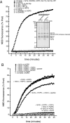
Fig. 1.
Fig. 1.
Ypt7p-dependent lipid mixing. (A) Lipid mixing of direct-method proteoliposomes requires Ypt7p, HOPS, Sec17p/Sec18p, ATP hydrolysis, and SNAREs. Fusion reactions (see Methods) used proteoliposomes of indicated composition and lacked the indicated soluble factors; any omitted components were replaced by their buffers. (Inset) Acceptor and donor proteoliposomes with SNAREs, with or without Ypt7p, were analyzed by SDS/PAGE and Sypro Ruby staining (5 nmol of total lipids per lane). (B) Anti-Ypt7p antibodies block lipid mixing. Anti-Ypt7p (1.2 M final; ▴), a mixture of anti-Ypt7p peptide (12 μM final; ⧫), Ypt7p peptide alone (12 M final; ▾), or RB150 (all others) were preincubated with the indicated proteoliposomes for 10 min at 27 °C. MgCl2, ATP, Sec17p, Sec18p, and HOPS complex or HOPS buffer, as indicated, were then added and reactions were carried out as described in Methods.
Fig. 2.
Fig. 2.
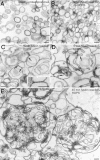
Electron microscopy analysis of proteoliposomes and fusion reactions. Donor proteoliposomes or donor-only fusion reactions were prepared as described. After 5 or 45 min at 27 °C for the reactions, and without incubation for the proteoliposomes alone, glutaraldehyde was added to a final concentration of 0.1% from a 2% stock in 0.1 M sodium phosphate, pH 7.3. Reactions were incubated at room temperature for 30 min, then centrifuged for 15 min at 14,000 rpm in an Eppendorf (Hamburg, Germany) 5415C microcentrifuge at 4 °C. Pellets were covered with 450 μL of 1% low melting point agarose, then processed for transmission electron microscopy as described (79). (A) Starting proteoliposomes not incubated under fusion conditions (mean size = 114 ± 3 nm). (B) Proteoliposomes incubated for 5 min in a fusion reaction without HOPS complex. (C and D) Proteoliposomes incubated for 5 min in a fusion reaction with HOPS. Liposomes are larger and pleomorphic, possibly as a result of incipient fusion. In C, the arrows point to regions where juxtaposed liposomes have established a close contact. The area delimited by two arrows is shown at higher magnification in the Inset. Note that the external leaflets of the proteoliposome membranes in this region have apparently merged into a single osmiophilic line resulting in the formation of a pentalaminar structure suggestive of a fusion event. (D) Large liposomes are frequently endowed with membrane infoldings, resulting in the formation of tubular structures (arrows). (E) Proteoliposomes incubated for 45 min in the fusion reaction with HOPS. Fused liposomes have generated tangled skeins of tubular membranes. (Scale bars, 100 nm.)
Fig. 3.
Fig. 3.
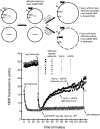
Lipid mixing is not accompanied by lysis. Fusion reactions (see Methods) used proteoliposomes with SNAREs, with Ypt7p (filled symbols) or without Ypt7p (open symbols). Sodium dithionite (40 mM; Sigma) was prepared by addition of solid sodium dithionite to ice-cold RB150+, frozen immediately in aliquots, stored at −80 °C, and thawed just before use. At t = 0, one set of proteoliposomes in RB150+ (13.2 μL; circles and triangles) received freshly thawed sodium dithionite (2 μL) and was incubated at 27 °C. A second set of proteoliposomes (13.2 μL; squares and diamonds) was incubated at 27 °C without sodium dithionite. At t = 30 min, freshly thawed sodium dithionite (2 μL) was added, at room temperature, to this second set of proteoliposomes, and reactions were returned to 27 °C. At t = 37 min, MgCl2, ATP, Sec17p, Sec18p, and HOPS complex (circles and squares) or HOPS buffer (triangles and diamonds) were added, in a total volume of 4.8 μL, at room temperature. Reactions were incubated at 27 °C for 60 min, followed by addition of 2 μL of 1% Thesit and 5-min incubation at 27 °C. Raw fluorescence units are shown.
Fig. 4.
Fig. 4.
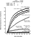
Lipid mixing requires GTP-bound Ypt7p. Fusion reactions (see Methods) used proteolipsomes with SNAREs, with or without Ypt7p as indicated. During the preincubation, reactions received RB150 (squares and circles), GTPγS (100 μM final; triangles), or UTPγS (100 μM final; diamonds), and RB150 (filled symbols) or Gyp1–46 (2 μM final; open symbols). The symbols for reactions without Ypt7p are behind the filled squares and show no detectable increase in NBD fluorescence.
Fig. 5.
Fig. 5.
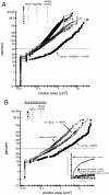
Proteoliposome clustering requires Ypt7p, HOPS complex, Sec17p/Sec18p, and SNAREs. Proteoliposome fusion reactions, including Sec17p, Sec18p, and ATP, were prepared as in Methods, except that only donor proteoliposomes were used. After 20 min at 27 °C, 3 μL of each reaction was mixed on a microscope slide (Gold Seal no. 3051) with 5 μL of a mock reaction without proteoliposomes or HOPS. These mixtures were covered with 22-mm cover slips (Corning no. 2870-22) and randomized, and random fields were imaged with an Olympus BX51 microscope with a 100-W mercury arc lamp (Olympus), 3% U-RSL6 UV/IR filter (Olympus), TRITC/DiI filter set (Chroma Technologies), 1.4 NA Plan Aprochromat ×60 objective (Olympus), Sensicam QE CCD camera (Cooke), and IPLab software (Scanalytics). Measurement of cluster sizes was done with ImageJ using an intensity threshold of 50. At least two images from each reaction were used for measurement. Representative images used for A are depicted in Fig. S4A. (A) Ypt7p, HOPS complex, and SNAREs all are required for proteoliposome clustering. A cumulative distribution plot showing proteoliposome cluster sizes is shown. Proteoliposomes were with Ypt7p (squares and circles) or without Ypt7p (triangles and diamonds) and with SNAREs (filled symbols) or without SNAREs (open symbols). Reactions received HOPS complex (squares and triangles) or HOPS buffer (circles and diamonds) as indicated. The distribution for the reaction with + Ypt7p + SNARE proteoliposomes with added HOPS complex is significantly different (P < 0.0001) from all other distributions by the Wilcoxon-Mann–Whitney test (Kaleidagraph). (B) The three vacuolar Q-SNAREs suffice for Ypt7p- and HOPS complex-dependent proteoliposome clustering. A cumulative distribution plot showing proteoliposome cluster sizes is shown. Reactions contained proteoliposomes with Vti1p, Vam7p, and Vam3p, with Ypt7p (squares) or without Ypt7p (circles), and with added HOPS complex (filled symbols) or HOPS buffer (open symbols). The distribution for the reaction using Ypt7p-bearing proteoliposomes, with HOPS complex, is significantly different (P < 0.0001) from the distribution for the reaction containing Ypt7p-bearing proteoliposomes, but lacking HOPS complex, by the Wilcoxon-Mann–Whitney test (Kaleidagraph). The distributions for the two reactions using proteoliposomes lacking Ypt7p are not significantly different (P = 0.4645) by the same test. The larger clusters in these distributions therefore derive from intrinsic aggregation and are not HOPS complex-dependent. (Inset) Nyv1p is required for proteoliposome fusion. Donor proteoliposomes with Ypt7p and the 3 Q-SNAREs were mixed with acceptor proteoliposomes with Ypt7p and with the three Q-SNAREs (squares) or all four SNAREs (circles), with HOPS (filled symbols) or without HOPS (open symbols), under fusion conditions (see Methods).
Fig. 6.
Fig. 6.
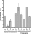
HOPS complex binding to proteoliposomes. Donor-only proteoliposome fusion reactions (5× scale) containing the indicated components were incubated for 20 min at 27 °C then transferred to ice, mixed with 100 μL of 2 M sucrose in RB150+ in 5 × 41-mm ultracentrifuge tubes (Beckman no. 344090), and covered with 200 μL of 0.8 and 0.6 M sucrose in RB150+, then with 10 μL of RB150+. Gradients were centrifuged for 2 h and 30 min at 50,000 rpm at 4 °C in a SW-55 rotor (Beckman, Palo Alto, CA) using the appropriate inserts, and 20 μL of proteoliposomes was harvested from the top interface. Lipid yield was estimated by fluorescence (λexem 540/586 nm) and samples containing 4 nmol of lipids were analyzed by SDS/PAGE and Sypro Ruby staining. Bound HOPS complex was estimated by using a standard curve of purified HOPS.

Comment in

  • Organelle docking: R-SNAREs are late.
    Verhage M. Verhage M. Proc Natl Acad Sci U S A. 2009 Nov 24;106(47):19745-6. doi: 10.1073/pnas.0911425106. Epub 2009 Nov 18. Proc Natl Acad Sci U S A. 2009. PMID: 19923425 Free PMC article. No abstract available.

Similar articles

Cited by

References

    1. Pfeffer SR. Rab GTPases: Specifying and deciphering organelle identity and function. Trends Cell Biol. 2001;11:487–491. - PubMed
    1. Carroll KS, et al. Role of Rab9 GTPase in facilitating receptor recruitment by TIP47. Science. 2001;292:1373–1376. - PubMed
    1. Pagano A, Crottet P, Prescianotto-Baschong C, Spiess M. In vitro formation of recycling vesicles from endosomes requires adaptor protein-1/clathrin and is regulated by rab4 and the connector rabaptin-5. Mol Biol Cell. 2004;15:4990–5000. - PMC - PubMed
    1. Bahadoran P, et al. Rab27a: A key to melanosome transport in human melanocytes. J Cell Biol. 2001;152:843–850. - PMC - PubMed
    1. Mayer A, Wickner W. Docking of yeast vacuoles is catalyzed by the Ras-like GTPase Ypt7p after symmetric priming by Sec18p (NSF) J Cell Biol. 1997;136:307–317. - PMC - PubMed

Publication types

MeSH terms

Substances