Primary dystonia: molecules and mechanisms
- PMID: 19826400
- PMCID: PMC2856083
- DOI: 10.1038/nrneurol.2009.160
Primary dystonia: molecules and mechanisms
Abstract
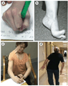
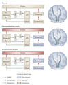
Primary dystonia is characterized by abnormal, involuntary twisting and turning movements that reflect impaired motor system function. The dystonic brain seems normal, in that it contains no overt lesions or evidence of neurodegeneration, but functional brain imaging has uncovered abnormalities involving the cortex, striatum and cerebellum, and diffusion tensor imaging suggests the presence of microstructural defects in white matter tracts of the cerebellothalamocortical circuit. Clinical electrophysiological studies show that the dystonic CNS exhibits aberrant plasticity--perhaps related to deficient inhibitory neurotransmission--in a range of brain structures, as well as the spinal cord. Dystonia is, therefore, best conceptualized as a motor circuit disorder, rather than an abnormality of a particular brain structure. None of the aforementioned abnormalities can be strictly causal, as they are not limited to regions of the CNS subserving clinically affected body parts, and are found in seemingly healthy patients with dystonia-related mutations. The study of dystonia-related genes will, hopefully, help researchers to unravel the chain of events from molecular to cellular to system abnormalities. DYT1 mutations, for example, cause abnormalities within the endoplasmic reticulum-nuclear envelope endomembrane system. Other dystonia-related gene products traffic through the endoplasmic reticulum, suggesting a potential cell biological theme underlying primary dystonia.
Figures


References
-
- Clarimon J, et al. Torsin A haplotype predisposes to idiopathic dystonia. Ann. Neurol. 2005;57:765–767. - PubMed
-
- Clarimon J, et al. Assessing the role of DRD5 and DYT1 in two different case–control series with primary blepharospasm. Mov. Disord. 2007;22:162–166. - PubMed
-
- Fahn S, Bressman SB, Marsden CD. Classification of dystonia. Adv. Neurol. 1998;78:1–10. - PubMed
-
- Mink JW. The basal ganglia and involuntary movements: impaired inhibition of competing motor patterns. Arch. Neurol. 2003;60:1365–1368. - PubMed
Publication types
MeSH terms
Substances
Grants and funding
LinkOut - more resources
Full Text Sources
Other Literature Sources

