. 2009 Dec 10;28(49):4326-43.
doi: 10.1038/onc.2009.299.

Matrix density-induced mechanoregulation of breast cell phenotype, signaling and gene expression through a FAK-ERK linkage

Affiliations

Matrix density-induced mechanoregulation of breast cell phenotype, signaling and gene expression through a FAK-ERK linkage

P P Provenzano et al. Oncogene. .

Abstract

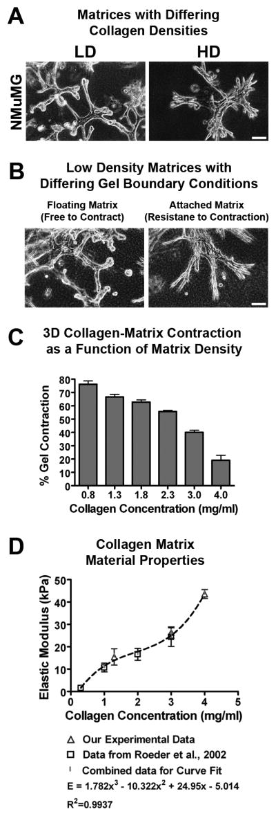
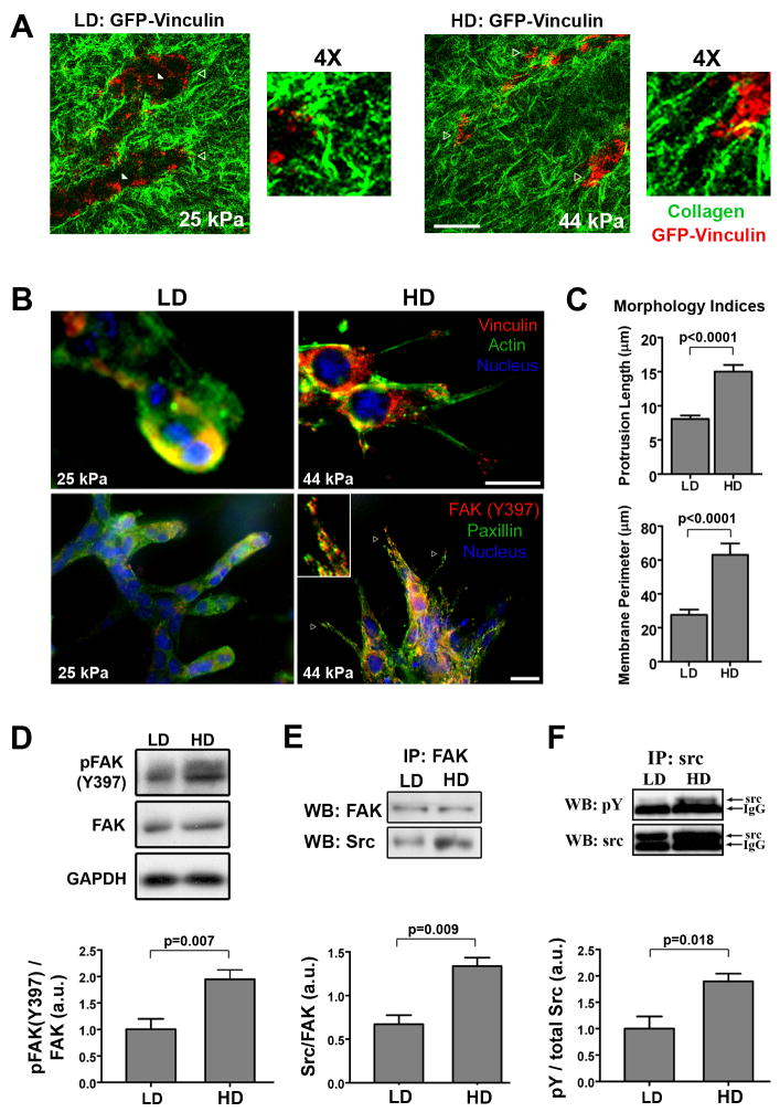
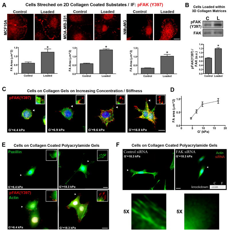
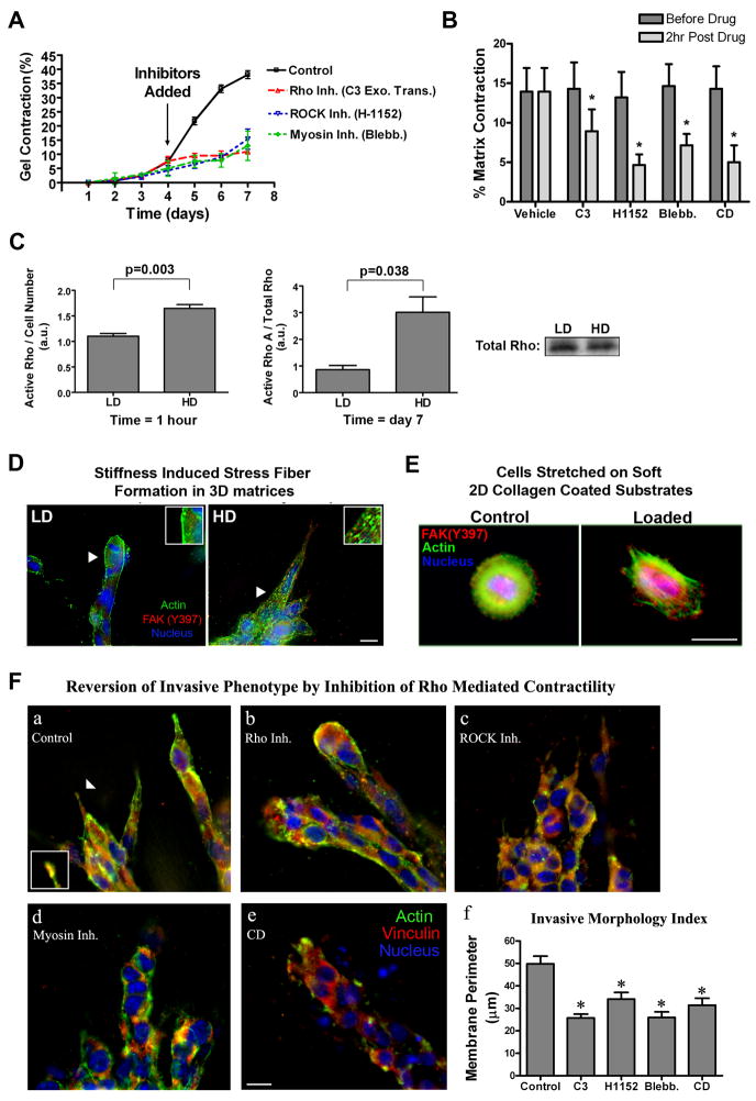
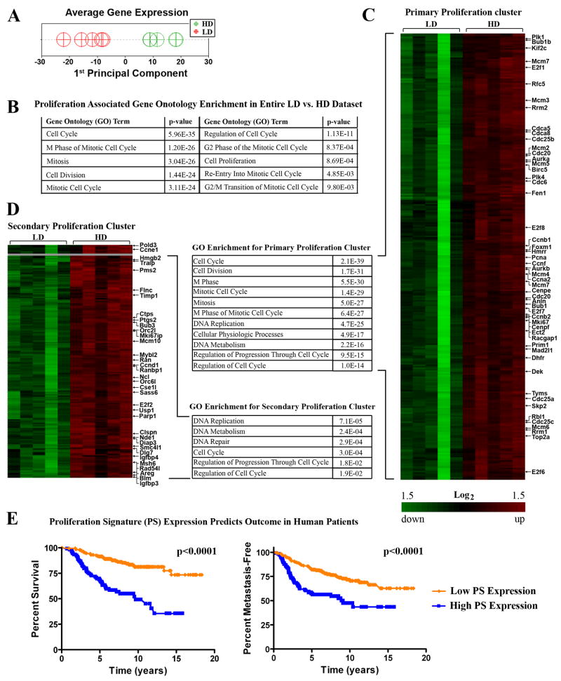
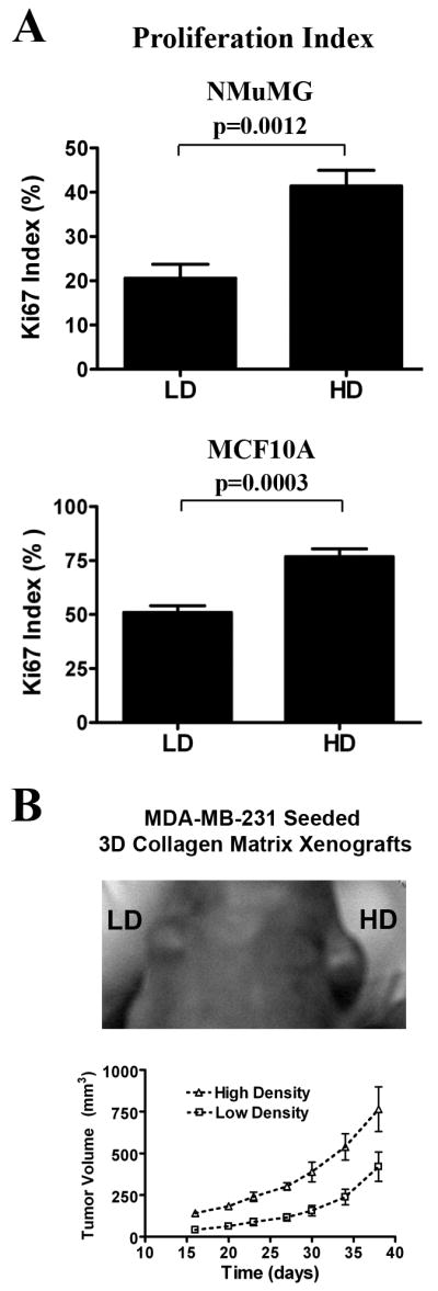
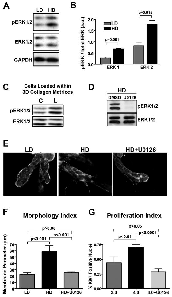
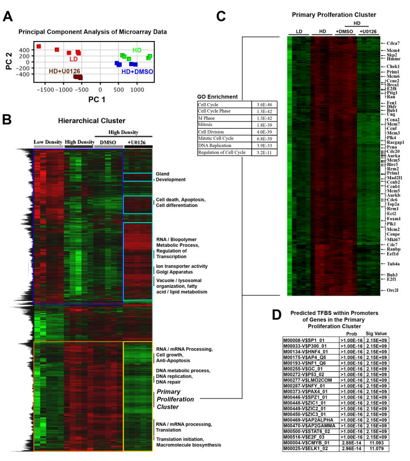
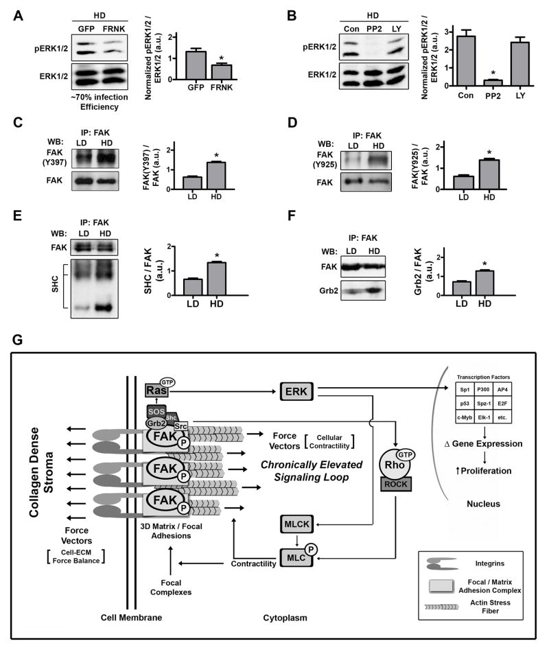
Mammographically dense breast tissue is one of the greatest risk factors for developing breast carcinoma, yet the associated molecular mechanisms remain largely unknown. Importantly, regions of high breast density are associated with increased stromal collagen and epithelial cell content. We set out to determine whether increased collagen-matrix density, in the absence of stromal cells, was sufficient to promote proliferation and invasion characteristic of a malignant phenotype in non-transformed mammary epithelial cells. We demonstrate that increased collagen-matrix density increases matrix stiffness to promote an invasive phenotype. High matrix stiffness resulted in increased formation of activated three-dimensional (3D)-matrix adhesions and a chronically elevated outside-in/inside-out focal adhesion (FA) kinase (FAK)-Rho signaling loop, which was necessary to generate and maintain the invasive phenotype. Moreover, this signaling network resulted in hyperactivation of the Ras-mitogen-activated protein kinase (MAPK) pathway, which promoted growth of mammary epithelial cells in vitro and in vivo and activated a clinically relevant proliferation signature that predicts patient outcome. Hence, the current data provide compelling evidence for the importance of the mechanical features of the microenvironment, and suggest that mechanotransduction in these cells occurs through a FAK-Rho-ERK signaling network with extracellular signal-regulated kinase (ERK) as a bottleneck through which much of the response to mechanical stimuli is regulated. As such, we propose that increased matrix stiffness explains part of the mechanism behind increased epithelial proliferation and cancer risk in human patients with high breast tissue density.


Conflict of interest statement

CONFLICT OF INTEREST DISCLOSURE

The authors declare no conflict of interest.

Figures

Figure 1
Figure 1. Matrix density-induced stiffness regulates epithelial cell phenotype
A. Representative micrographs of NMuMG mammary epithelial cells in low-density (LD) and high-density (HD) collagen matrices (density optimized for each cell type as described in the Materials and Methods section). Bar: = 100 μm B. Representative micrographs of NMuMG cells seeded into LD collagen matrices allowed to freely contract and undergo tubulogenesis (left) or constrained to resist cell-mediated matrix contraction and disrupt differentiation (right). Bar = 100 μm C. NMuMG-mediated matrix contraction as a function of collagen density after 7 days (n = 6; mean ± SEM). D. Elastic modulus of collagen matrices at densities used in this study (n = 4; mean ± s.d.), compared to the data of Roeder et al. (2002) for matched collagen matrix conditions.
Figure 2
Figure 2. Matrix stiffness promotes 3D-matrix adhesions and FAK signaling
A. Representative micrographs of live cell multiphoton excitation (MPE) and second harmonic generation (SHG) imaging of NMuMG cells expressing GFP-Vinculin (MPE = pseudo-colored red) cultured in low density (LD) and high density (HD) collagen matrices (SHG = pseudo-colored green) for 7 days. Note the differential vinculin localization as a function of matrix stiffness, with increased 3D-matrix adhesion clustering at the cell-ECM interface in stiffer HD matrices (open arrowheads). Bottom right (LD) and farthest right (HD) open arrowheads indicate the regions magnified 4X. Bar = 25 μm B. Immunofluorescence analysis of NMuMG cells in LD and HD collagen matrices showing increased localization of two hallmark focal adhesion proteins, vinculin (top) and paxillin (bottom), to 3D-matrix adhesions in HD matrices; with FAK phosphorylated at Y397 (red) co-localized with paxillin in 3D-matrix adhesions under HD conditions (representative of n ≥ 5). Bar = 10 μm C. Morphometric analysis of NMuMG cells after differentiating in LD or HD matrices, showing significantly increased cell protrusion into HD matrices (nlength ≥ 70, nperimeter ≥ 22 from n ≥ 8 matrices/conditions; mean ± SEM). D. Western blot analysis and densitometry of pFAK(Y397) levels in lysates from NMuMG cells cultured in LD or HD matrices for 7 days (n ≥ 6; mean ± SEM). E. Western blot analysis of Src protein that co-precipitated with immunoprecipitated FAK, showing increased FAK-Src association in cells within HD matrices (n = 3; mean ± SEM). F. Western blot analysis and densitometry of phosphorylated Src levels following immunoprecipitation of Src from NMuMG cell lysates after being cultured in LD or HD matrices for 7 days (n = 3; mean ± SEM).
Figure 3
Figure 3. Application of exogenous force and increased substrate stiffness promotes FAK phosphorylation and a FAK-dependent protrusive phenotype
A. Substrate strain-induced cell deformation of mammary epithelial cells adhered to type I collagen-coated (soft) silicone elastomer substrates. Static equibiaxial substrate deformation (10%) was applied for 20 minutes. Subsequently, immunofluorescent analysis was performed to detect and quantify changes in pFAK(Y397) positive focal adhesions (nFA ≥ 440 for NMuMG and MDA-MB-231 cells; nFA ≥ 146 for MCF10A cells; * p=0.006 for MCF10A; p=0.0001 for NMuMG and MDA-MB-231; mean ± SEM). Bar = 10μm B. NMuMG cells cultured within a 3D collagen matrix (3mg/mL) were loaded by deforming (10% axial strain) collagen matrices for 20 minutes. Subsequently, Western blot and densitometry analysis was used to examine significantly (*p=0.0006) increased FAK(Y397) phosphorylation levels following matrix deformation (n ≥ 4). C. Immunofluorescent analysis for FAK(Y397) phosphorylation (red), actin cytoskeleton architecture (green), and the nucleus (blue) in NMuMG cells cultured on collagen matrices of increasing substrate stiffness (shear modulus was increased by increasing collagen matrix density, range = 1 to 4 mg/mL). Bar = 10 μm D. pFAK(Y397)-positive focal adhesion area increasing as a function of increasing substrate stiffness (G′; nFA ≥ 125; mean ± SEM) E. Immunofluorescent analysis of paxillin (top) or FAK(Y397) phosphorylation and actin cytoskeleton architecture (bottom) in NMuMG cells cultured on type I collagen-coated (30 μg/mL) polyacrylamide gels of increasing stiffness (polyacrylamide gels were crosslinked with varying concentrations of bisacrylamide to control stiffness). The nucleus is shown in blue. Bar = 10 μm F. Fluorescent localization of the actin stress fibers (green) in NMuMG cells cultured on type I collagen-coated (30 μg/mL) polyacrylamide gels of equal stiffness transfected with either non-silencing control siRNA (left) or siRNA targeting FAK (right). siRNA was labeled with Cy3 (red) to ensure examination of siRNA transfected cells. The nucleus is shown in blue. A representative Western blot demonstrating FAK siRNA knockdown (consistently greater than 85%) is shown in the right panel. Arrowheads indicate the regions magnified in panels C, E, and F. Bar = 10 μm.
Figure 4
Figure 4. Increased matrix stiffness promotes Rho-mediated contractility
A. Inhibition of Rho with cell-permeable C3 exoenzyme transferase (10 μg/mL), ROCK (which promotes cellular contractility by directly phosphorylating MLC and/or inhibiting MLC phosphatase) with H1152 (2.5 μM), or myosin-based contractility with blebbistatin (10 μM) significantly suppressed NMuMG-mediated matrix contraction (n = 3; mean ± SEM). B. Inhibition of Rho, ROCK, myosin-based contractility, or the actin cytoskeleton as described in A for 2 hours in contracted matrices resulted in a significant relaxation of the collagen matrix (n = 4; mean ± SEM; *p<0.01). C. Quantitative analysis of RhoA activation in NMuMG cells in low density (LD) and high density (HD) collagen matrices after 1 hour (normalized by cell number: left) or 7 days (normalized by total Rho protein: right; n ≥ 3, mean ± SEM).. A representative Western blot for total Rho after 7 days is shown (bottom right) D. Fluorescence staining analysis of NMuMG cells in LD and HD collagen matrices showing increased actin stress fiber formation (green) under HD conditions. Bar = 10 μm E. Fluorescence staining analysis of substrate strain-induced cell deformation of MCF10A cells (as described in Figure 3) was performed to detect changes in the actin cytoskeleton (green). Immunofluorescent analysis of FAK(Y397) phosphorylation is shown in red and the nucleus in blue. Arrows indicate the regions magnified in the subpanels. Bar = 10 μm F. Inhibition of Rho with C3 (a, 10 μg/mL), ROCK with H1152 (b, 2.5 μM), myosin-based contractility with blebbistatin (c, 10 μM) or disruption of the actin cytoskeleton with cytochalasin D (d, 1 μM) for 2 hours in cells that has already developed the HD-induced invasive phenotype caused the invasive phenotype to be significantly reverted (f) when compared to controls (a; *p<0.01; mean ± SEM). Bar = 10 μm
Figure 5
Figure 5. Matrix density-induced stiffness promotes expression of clinically relevant proliferation-signature genes
A. Principal Component Analysis (PCA) of density regulated genes to reduce the dimensionality of the data sets and examine the extent to which loss matrix density influences the pattern of expression. Data were mean centered then PCA performed from all experimental samples (LD, n=5; HD, n=5), with the 1st Principal Component a measure of average expression. B. Gene Ontology analysis of the entire LD vs. HD data set showing enriched transcripts associated with proliferation. C and D. Proliferation node gene clusters generated by hierarchically clustering (significantly) differentially expressed genes from NMuMG cells within LD and HD matrices showing increased expression of genes associated with proliferation. Also shown is Gene Ontology analysis of the primary (C) and secondary (D) clusters demonstrating enrichment for proliferation associated transcripts. E. Prognostic value of the Proliferation Signature (PS) transcripts shown in Table 1. Using publicly available data from van de Vijver et al., (van de Vijver et al., 2002) we found that the 41 genes divide human breast cancer patients into two main clusters (cluster analysis provided in Supplementary Figure S5,). Kaplan-Meier survival analysis of the two groups shows that the patients differ significantly in both survival and metastasis-free outcome. Average expression in patients with poorer outcome (blue line) was higher for the PS genes while patients with better outcome (orange line) presented lower expression of these genes.
Figure 6
Figure 6. Matrix density-induced stiffness promotes proliferation of mammary epithelial cells
A. Ki-67 index of proliferation potential in NMuMG (top) and MCF10A (supplemented with 50ng/mL HGF to induce tubulogenesis; bottom) cells cultured in LD and HD matrices. Data are mean ± SEM obtained from >300 cells per condition in n ≥ 5 samples per group. B. Tumor growth in a xenograft model is increased by high matrix density. Human MDA-MB-231 cells were seeded into LD and HD matrices prior to inoculation into nude mice. Each mouse received a LD and HD cell-seeded matrix transplanted into contralateral sides above the 4th inguinal mammary glands. Increased tumor growth resulted from HD xenografts when compared to the LD control (n = 6; mean ± SEM, p<0.01 for each time point after day 15).
Figure 7
Figure 7. Matrix stiffness induces ERK phosphorylation and results in ERK-dependent membrane protrusion and cell proliferation
A and B. Western blot analysis (A) and densitometry (B) of phosphorylated ERK1/2 levels (using a phospho-specific antibody that recognizes phosphorylation of the threonine (Thr183) and tyrosine (Tyr185) residues in the activation loops of ERK1 and ERK2) in lysates from NMuMG cells cultured in LD or HD matrices for 7 days (n ≥ 3; mean ± SEM). C. NMuMG (and MCF10A = data not shown) cells loaded by deforming the collagen matrices as described in Figure 3 show increased ERK1/2 phosphorylation following application of matrix strain (representative of n ≥ 4 samples per condition). D. Representative Western blot analysis of phosphorylated ERK levels in lysates from NMuMG cells cultured in HD matrices following treatment with 10 μm U0126 or DMSO (control). E and F. Inhibition of ERK phosphorylation with the MEK inhibitor U0126 (10 μM) for 24 hours in cells that developed the HD-induced invasive phenotype (middle) results in cells that were significantly (F) reverted (right) to the LD phenotype (left). A color version of panel E is available online as Supp. Fig. S9. G. Ki-67 index of proliferation potential in NMuMG cells cultured in LD and HD matrices. Cells in HD matrices were either treated with DMSO (control) or 10 μm U0126 for 24 hours in cells that had developed the HD-induced invasive phenotype. Data are mean ± SEM obtained from >250 cells per condition in n ≥ 4 samples per group.
Figure 8
Figure 8. ERK mediated regulation of the HD-induced transcriptome shift
A. Principal Component Analysis (PCA) of ERK1/2 regulated genes in order to reduce the dimensionality of the data sets as well as determine the extent to which loss of ERK1/2 activity influences the pattern of expression. Data were mean centered then PCA performed from all experimental samples: LD, n=5; HD, n=5; HD+DMSO, n=5; HD+U0126 (10 μM for 24 hours, as described in Figure 7), n=5 arrays. The 1st Principal Component, which may be a measure of average expression, indicates that inhibition of ERK phosphorylation shifts the transcriptome back to near LD levels. B. Hierarchical cluster of transcripts differentially expressed due to HD matrix conditions over the entire data set (described in panel A) showing that ~70% of the transcripts that are regulated by increased matrix density/stiffness are reverted toward LD levels following ERK inhibition. The majority of the transcripts that were repressed (blue box) due to high matrix density were reverted by inhibition of ERK phosphorylation (turquoise boxes), while the majority of the transcripts that were induced (orange box) due to high matrix density were also reverted by inhibition of ERK phosphorylation (yellow box). C. Proliferation node gene cluster generated by hierarchically clustering (B) showing reversion of proliferation-associated transcripts following ERK inhibition. The accompanying Gene Ontology analysis of the cluster demonstrates enrichment for proliferation associated transcripts. D. Computationally predicted transcription factor binding sites (TFBS) that are enriched in the proliferation cluster (C).
Figure 9
Figure 9. Ras pathway activation of ERK is dependent on FAK phosphorylation
A and B. Western blot analysis and densitometry of ERK phosphorylation levels in lysates from NMuMG cells cultured in HD matrices for 7 days. A: 24 hours before lysis, cells were infected with either Adeno-GFP or Adeno-FRNK-GFP (n ≥ 4; *p<0.02; mean ± SEM). B: Two hours before lysis, Src or PI3K were inhibited with PP2 (10 μM) or LY294002 (25 μM; n = 3; *p<0.005; mean ± SEM). C and D. Western blot analysis and densitometry of (C) pFAK(Y397) and (D) pFAK(Y925) phosphorylation levels following immunoprecipitation of FAK from NMuMG cell lysates after being cultured in LD or HD matrices for 7 days (n = 3; *p<0.001; mean ± SEM). E and F. Western blot analysis of (E) SHC and (F) Grb2 proteins that co-precipitated with immunoprecipitated FAK, showing increased FAK-SHC and FAK-Grb2 association in cells within HD matrices (n = 3; *p<0.004; mean ± SEM). G. Model for the regulation of epithelial growth by matrix density-induced increases in ECM stiffness. As mammary epithelial cells encounter exogenous mechanical force or increased resistance to cellular contractility from stiff high density matrices they respond in a FAK-dependent manner by developing mature focal or 3D-matrix adhesions. Sustained mechanical signaling results in chronic upregulation of a FAK-Rho signaling loop that produces hyperactivation of related pathways, such as the Ras-MAPK pathway. Elevated ERK1/2 phosphorylation feeds back to regulate epithelial phenotype, controls the majority of the mechanically-induced transcriptome shift, and induces transcription of clinically-relevant proliferation associated genes, which results in increased epithelial proliferation.

References

    1. Alowami S, Troup S, Al-Haddad S, Kirkpatrick I, Watson PH. Breast Cancer Res. 2003;5:R129–35. - PMC - PubMed
    1. Boyd NF, Lockwood GA, Byng JW, Tritchler DL, Yaffe MJ. Cancer Epidemiol Biomarkers Prev. 1998;7:1133–44. - PubMed
    1. Boyd NF, Martin LJ, Stone J, Greenberg C, Minkin S, Yaffe MJ. Curr Oncol Rep. 2001;3:314–21. - PubMed
    1. Burridge K, Chrzanowska-Wodnicka M. Annu Rev Cell Dev Biol. 1996;12:463–519. - PubMed
    1. Burridge K, Fath K, Kelly T, Nuckolls G, Turner C. Annu Rev Cell Biol. 1988;4:487–525. - PubMed

Publication types

MeSH terms

Substances