

Side population cells have the characteristics of cancer stem-like cells/cancer-initiating cells in bone sarcomas
- PMID: 19826427
- PMCID: PMC2768447
- DOI: 10.1038/sj.bjc.6605330
Side population cells have the characteristics of cancer stem-like cells/cancer-initiating cells in bone sarcomas
Abstract
Background: Several human cancers have been found to contain cancer stem-like cells (CSCs) having cancer-initiating ability. However, only a few reports have shown the existence of CSCs in bone and soft tissue sarcomas. In this study, we identified and characterised side population (SP) cells that showed drug-resistant features in human bone sarcoma cell lines.
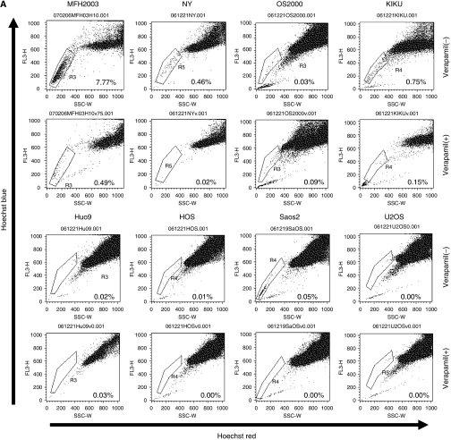
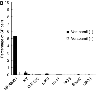
Methods: In seven osteosarcoma cell lines (OS2000, KIKU, NY, Huo9, HOS, U2OS and Saos2) and in one bone malignant fibrous histiocytoma (MFH) cell line (MFH2003), the frequency of SP cells was analysed. Tumourigenicity of SP cells was assessed in vitro and in vivo. Gene profiles of SP cells and other populations (main population; MP) of cells were characterised using cDNA microarrays.
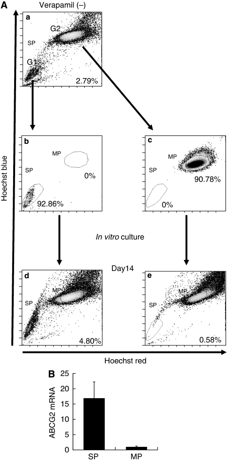
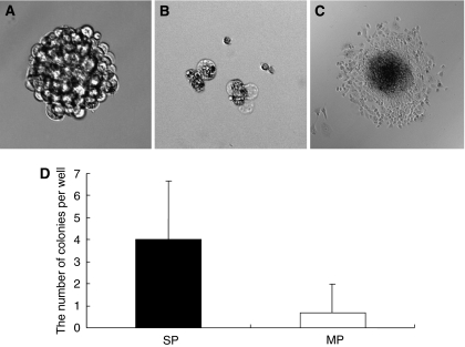
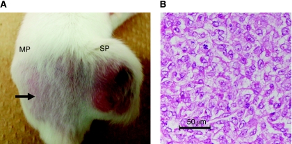
Results: SP cells were found in NY (0.31%) and MFH2003 (5.28%). SP cells of MFH2003 formed spherical colonies and re-populated into SP and MP cells. In an NOD/SCID mice xenograft model, 1 x 10(3) sorted SP cell-induced tumourigenesis. cDNA microarray analysis showed that 23 genes were upregulated in SP cells.
Conclusions: We showed that SP cells existed in bone sarcoma cell lines. SP cells of MFH2003 had cancer-initiating ability in vitro and in vivo. The gene profiles of SP cells could serve as candidate markers for CSCs in bone sarcomas.
Figures
Similar articles
-
Autologous CTL response against cancer stem-like cells/cancer-initiating cells of bone malignant fibrous histiocytoma.Cancer Sci. 2011 Aug;102(8):1443-7. doi: 10.1111/j.1349-7006.2011.01962.x. Epub 2011 May 31. Cancer Sci. 2011. PMID: 21518139
-
HDAC2 depletion promotes osteosarcoma's stemness both in vitro and in vivo: a study on a putative new target for CSCs directed therapy.J Exp Clin Cancer Res. 2018 Dec 3;37(1):296. doi: 10.1186/s13046-018-0978-x. J Exp Clin Cancer Res. 2018. PMID: 30509303 Free PMC article.
-
Aberrant Wnt/β-catenin signaling and elevated expression of stem cell proteins are associated with osteosarcoma side population cells of high tumorigenicity.Mol Med Rep. 2015 Oct;12(4):5042-8. doi: 10.3892/mmr.2015.4025. Epub 2015 Jul 2. Mol Med Rep. 2015. PMID: 26134785 Free PMC article.
-
Perspectives on cancer stem cells in osteosarcoma.Cancer Lett. 2013 Sep 10;338(1):158-67. doi: 10.1016/j.canlet.2012.05.028. Epub 2012 May 29. Cancer Lett. 2013. PMID: 22659734 Free PMC article. Review.
-
Cancer stem cells in osteosarcoma: recent progress and perspective.Acta Oncol. 2011 Nov;50(8):1142-50. doi: 10.3109/0284186X.2011.584553. Epub 2011 Jul 1. Acta Oncol. 2011. PMID: 21718210 Review.
Cited by
-
Isolation and identification of cancer stem-like cells from side population of human prostate cancer cells.J Huazhong Univ Sci Technolog Med Sci. 2012 Oct;32(5):697-703. doi: 10.1007/s11596-012-1020-8. Epub 2012 Oct 18. J Huazhong Univ Sci Technolog Med Sci. 2012. PMID: 23073799
-
Osteosarcoma: Cells-of-Origin, Cancer Stem Cells, and Targeted Therapies.Stem Cells Int. 2016;2016:3631764. doi: 10.1155/2016/3631764. Epub 2016 Jun 5. Stem Cells Int. 2016. PMID: 27366153 Free PMC article. Review.
-
Osteosarcoma: mouse models, cell of origin and cancer stem cell.Postdoc J. 2014 Feb;2(2):19-30. doi: 10.14304/surya.jpr.v2n2.3. Postdoc J. 2014. PMID: 27617267 Free PMC article.
-
SDCBP Modulates Stemness and Chemoresistance in Head and Neck Squamous Cell Carcinoma through Src Activation.Cancers (Basel). 2021 Oct 1;13(19):4952. doi: 10.3390/cancers13194952. Cancers (Basel). 2021. PMID: 34638436 Free PMC article.
-
Cancer Stem Cells and Pediatric Solid Tumors.Cancers (Basel). 2011 Jan 14;3(1):298-318. doi: 10.3390/cancers3010298. Cancers (Basel). 2011. PMID: 21394230 Free PMC article.
References
-
- Chen S, Dai Y, Harada H, Dent P, Grant S (2007) Mcl-1 down-regulation potentiates ABT-737 lethality by cooperatively inducing Bak activation and Bax translocation. Cancer Res 67: 782–791 - PubMed
-
- Collins AT, Berry PA, Hyde C, Stower MJ, Maitland NJ (2005) Prospective identification of tumorigenic prostate cancer stem cells. Cancer Res 65: 10946–10951 - PubMed
-
- Dean M, Fojo T, Bates S (2005) Tumour stem cells and drug resistance. Nat Rev Cancer 5: 275–284 - PubMed
Publication types
MeSH terms
LinkOut - more resources
Full Text Sources
Other Literature Sources
Medical
