. 2010 Feb;30(2):299-310.
doi: 10.1038/jcbfm.2009.210. Epub 2009 Oct 14.

Phosphodiesterase III inhibition promotes differentiation and survival of oligodendrocyte progenitors and enhances regeneration of ischemic white matter lesions in the adult mammalian brain

Affiliations

Phosphodiesterase III inhibition promotes differentiation and survival of oligodendrocyte progenitors and enhances regeneration of ischemic white matter lesions in the adult mammalian brain

Nobukazu Miyamoto et al. J Cereb Blood Flow Metab. 2010 Feb.

Abstract

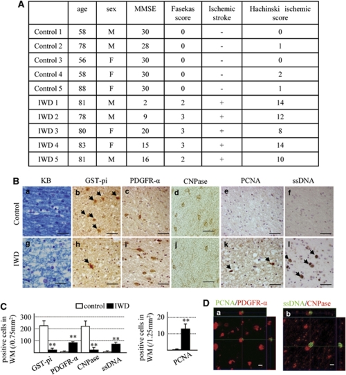
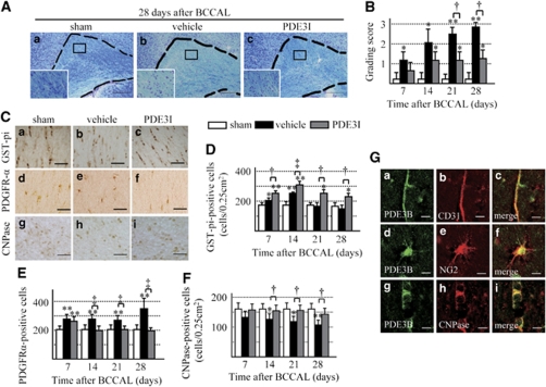
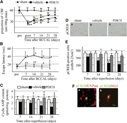
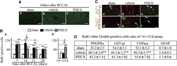
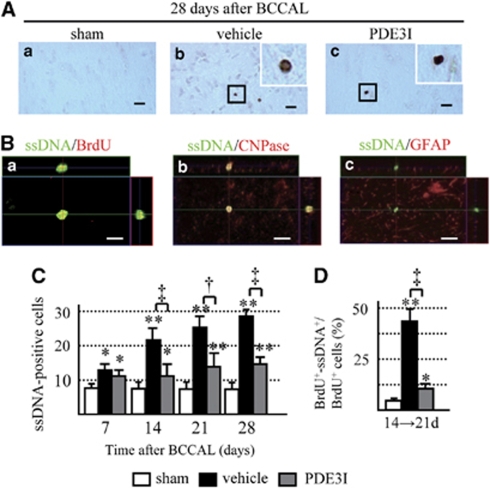
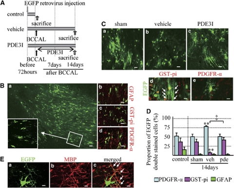
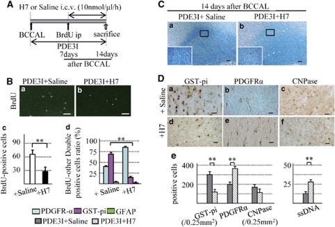
Vascular dementia is caused by blockage of blood supply to the brain, which causes ischemia and subsequent lesions primarily in the white matter, a key characteristic of the disease. In this study, we used a chronic cerebral hypoperfusion rat model to show that the regeneration of white matter damaged by hypoperfusion is enhanced by inhibiting phosphodiesterase III. A rat model of chronic cerebral hypoperfusion was prepared by bilateral common carotid artery ligation. Performance at the Morris water-maze task, immunohistochemistry for bromodeoxyuridine, as well as serial neuronal and glial markers were analyzed until 28 days after hypoperfusion. There was a significant increase in the number of oligodendrocyte progenitor cells in the brains of patients with vascular dementia as well as in rats with cerebral hypoperfusion. The oligodendrocyte progenitor cells subsequently underwent cell death and the number of oligodendrocytes decreased. In the rat model, treatment with a phosphodiesterase III inhibitor prevented cell death, markedly increased the mature oligodendrocytes, and promoted restoration of white matter and recovery of cognitive decline. These effects were cancelled by using protein kinase A/C inhibitor in the phosphodiesterase III inhibitor group. The results of our study indicate that the mammalian brain white matter tissue has the capacity to regenerate after ischemic injury.


Figures

Figure 1
Figure 1
Human studies. (A) Clinical characteristics of the patients and normal control and white matter lesions. MMSE: mini-mental scale (cutoff 24/30). Fasekas score and Hachinski ischemic score were determined on the basis of the methods described by Fazekas et al (1988) and Hachinski et al (1975). (B) Photomicrographs of Klüver–Barrera (a, g)-, GST-pi (b, h; arrowheads, positive cells)-, PDGFR-α (c, i)-, CNPase (d, j)-, PCNA (e, k; arrowheads, positive cells)-, and ssDNA (f, l; arrowheads, positive cells)-stained white matter sections. Control group (a–f) and ischemic white-matter diseases (IWD) group (g–l). Bars=50 μm. (C) Number of GST-pi-, PDGFR-α-, CNPase-, and PCNA-positive cells in white matter (WM). Data are mean±s.e.m. of control group (n=6) and ischemic white-matter diseases group (n=5). (D) Immunofluorescence staining of PDGFR-α (red)/PCNA (green) (a) and CNPase (red)/ssDNA (green) (b) in the white matter of the ischemic white-matter diseases group of Z-stack images. Bars=10 mm. **P<0.001, compared with the control group.
Figure 2
Figure 2
(A) Photomicrographs of Klüver–Barrera-stained sections of the experimental rat white matter (marked by dotted line). Sham (a), vehicle (b), and PDE3I (c) at 28 days after BCCAL. Insets: magnified views. Magnification, × 100. Bar=150 μm. (B) Grading-score histograms of the sham, vehicle, and PDE3I white-matter lesion groups. (C) Photomicrographs of GST-pi (a–c)-, PDGFR-α (d–f)-, and CNPase (g–i)-stained cells in the white matter of sham (a, d, g), vehicle (b, e, h), and PDE3I (c, f, i) groups at 28 days after BCCAL. Bars=40 μm. (DF) Proportion of GST-pi (panel D)-, PDGFR-α (panel E)-, and CNPase (panel F)-positive cells in the white matter. (G) Double immunofluorescent staining of PDE3B(green,(a, d, g)), CD31 (red, b), CNPase (red, e), NG2 (red, h), and merged images (c, f, i) in the white matter of sham group. Bars=10 μm. Values are mean±s.e.m. of 5 rats in each group. *P<0.05, **P<0.001, compared with the sham-operated group; P<0.05, P<0.001, compared with the vehicle group.
Figure 3
Figure 3
(A) Temporal changes in cerebral blood flow (CBF). Pre: before BCCAL; post: immediately after BCCAL, and at 7, 14, 21, and 28 days after BCCAL. (B) Effect of ischemic insult on learning deficit as measured by the Morris water-maze task. (C) cAMP content of white matter. (D) Photomicrographs of pCREB-stained cells in the white matter of sham (a), vehicle (b), and PDE3I (c) groups at 28 days after BCCAL. Bars=40 μm. (E) Proportion of pCREB-positive cells in the white matter. (F) Double immunofluorescent staining of pCREB (green), CNPase (red, a) and NG2 (red, b). Bars=10 μm. Values are mean±s.e.m. of 5 rats in each group. *P<0.05, **P<0.001 compared with the sham-operated group; P<0.05, P<0.001 compared with the vehicle group.
Figure 4
Figure 4
(A) 5-Bromodeoxyuridine (BrdU) immunofluorescence staining in the white matter of vehicle- and PDE3I-treated rats. Sham (a), vehicle (b), and PDE3I (c) rats at day 14 after BCCAL. Bar=50 μm. (B) Number of BrdU-positive cells in the white matter. (C) Double immunofluorescent staining of BrdU, GST-pi, and PDGFR-α. Representative white matter of the sham, vehicle-, and PDE3I-treated groups at 14 days after BCCAL (green, BrdU; red, GST-pi (a–c); PDGFR-α (d–f); a, d; sham; b, e; vehicle; and c, f; PDE3I group, arrowheads; merged cells). Scale bar=20 μm. (D) Proportion of BrdU-positive cells double-labeled with glial cell markers (PDFGR-α, GST-pi, CNPase, and GFAP) in white matter. Data in panel B and D are mean±s.e.m. of 5 rats in each group. *P<0.05, **P<0.001, compared with the sham-operated group; P<0.05, P<0.001, compared with the vehicle group.
Figure 5
Figure 5
(A) Photomicrographs showing ssDNA staining in the white matter of sham (a), vehicle (b)-, and PDE3I (c)-treated rats 28 days after BCCAL. Bar=20 μm. Insets in b and c: magnified views. Magnification, × 400. (B) Colocalization of ssDNA-positive cells in the vehicle group at days 14 → 21 after BCCAL. (C) Number of ssDNA-positive cells. Double immunofluorescence BrdU staining (red (a)), CNPase (red (b)), GFAP (red (c)), and ssDNA (green (a–c)). Bar=10 μm. (D) Percentage of BrdU/ssDNA-double-positive cells relative to BrdU-positive cells. BrdU+: BrdU-positive cells, ssDNA+: ssDNA-positive cells. Data in panels B and D are mean±s.e.m. of 5 rats in each group. *P<0.05, **P<0.001, compared with sham-operated group; P<0.05, P<0.001, compared with vehicle group.
Figure 6
Figure 6
(A) EGFP-retrovirus injection protocol (sham, n=4; vehicle, n=6; cilostazol, n=4). (B) (a) Photomicrographs showing GFP staining in the white matter 72 h after injection. Bar=40 μm. (b–d) Colocalization of GFP-positive cells in the white matter 72 h after injection (red, b; GFAP, c; GST-pi, d; PDGFR-α). Bars=20 μm. (C) GFP immunofluorescence staining in the white matter of sham (a), vehicle (b)-, and PDE3I (c)-treated rats at day 14 after BCCAL. Bars=20 μm. Double immunofluorescence staining for GST-pi (red (d)), PDGFR-α (red (e)), GFP (green (d, e)). Bars=10 μm. (D) Proportion of GFP-positive cells double labeled with glial cell markers (PDFGR-α, GST-pi, and GFAP) in the white matter of sham, sham-operated group; veh, vehicle group; pde, PDE3I-treated group. Data are mean±s.e.m. **P<0.001, compared with the sham-operated group; P<0.001, compared with the vehicle group. (E) Colocalization of GFP-positive cells in the white matter of PDE3I group at day 14 after BCCAL. Double immunofluorescence staining for GFP (green (a)), MBP (red (b)), and merged (yellow (c), arrowheads; merged dendrites). Bar=10 μm.
Figure 7
Figure 7
(A) Osmotic mini-pump protocol. (B) a, b; 5-bromodeoxyuridine (BrdU) immunofluorescence staining in the white matter of PDE3I+Saline group (a) and +H7 group (b) 14 days after BCCAL. Bars=50 μm. (c) Number of BrdU-positive cells in the white matter. (d) Proportion of BrdU-positive cells double labeled with glial cell markers (PDFGR-α, GST-pi, and GFAP). (C) Photomicrographs of Klüver–Barrera staining of the white matter of PDE3I+Saline (a) and PDE3I+H7 group (b) at 14 days after BCCAL. (D) Photomicrographs of GST-pi (a, d) and PDGFR-α (b, e), CNPase (c, f) in white matter of the PDE3I+Saline (a–c) and PDE3I+H7 (d–f) groups 14 days after BCCAL. Bars=20 μm. (e) Proportion of GST-pi-, PDGFR-α-, CNPase- and ssDNA-positive cells in the white matter. Data in Bc and De are mean±s.e.m. of 5 rats in each group, *P<0.05; **P<0.001 compared with the other groups.

References

    1. Arvidsson A, Collin T, Kirik D, Kokaia Z, Lindvall O. Neuronal replacement from endogenous precursors in the adult brain after stroke. Nat Med. 2002;8:963–970. - PubMed
    1. Bansal R, Pfeiffer SE. FGF-2 converts mature oligodendrocytes to a novel phenotype. J Neurosci Res. 1997;50:215–228. - PubMed
    1. Barnett AH, Bradbury AW, Brittenden J, Crichton B, Donnelly R, Homer-Vanniasinkam S, Mikhailidis DP, Stansby G. The role of cilostazol in the treatment of intermittent claudication. Curr Med Res Opin. 2004;20:1661–1670. - PubMed
    1. Brown WR, Moody DM, Thore CR, Challa VR. Cerebrovascular pathology in Alzheimer's disease and leukoaraiosis. Ann N Y Acad Sci. 2000;903:39–45. - PubMed
    1. Butt AM. Structure and Function of Oligodendrocytes. New York, USA: Oxford University Press; 2004.

Publication types

MeSH terms