Review
. 2010:72:95-121.
doi: 10.1146/annurev-physiol-021909-135811.

Chloride channels: often enigmatic, rarely predictable

Affiliations
Review

Chloride channels: often enigmatic, rarely predictable

Charity Duran et al. Annu Rev Physiol. 2010.

Abstract

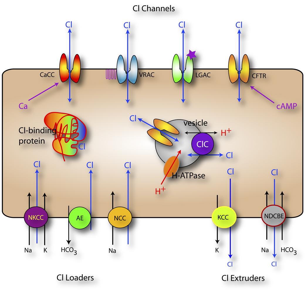
Until recently, anion (Cl(-)) channels have received considerably less attention than cation channels. One reason for this may be that many Cl(-) channels perform functions that might be considered cell-biological, like fluid secretion and cell volume regulation, whereas cation channels have historically been associated with cellular excitability, which typically happens more rapidly. In this review, we discuss the recent explosion of interest in Cl(-) channels, with special emphasis on new and often surprising developments over the past five years. This is exemplified by the findings that more than half of the ClC family members are antiporters, and not channels, as was previously thought, and that bestrophins, previously prime candidates for Ca(2+)-activated Cl(-) channels, have been supplanted by the newly discovered anoctamins and now hold a tenuous position in the Cl(-) channel world.


Figures

Figure 1
Figure 1. Cellular Chloride Signaling
Cells actively transport Cl across the plasma membrane by transporters that accumulate Cl intracellularly (Cl loaders including the Na+-K+-Cl cotransporters NKCC, Cl-HCO3 exchangers AE, and Na+-Cl cotransporters NCC) or pump Cl out of the cell (K+-Cl cotransporters KCC and Na+-dependent Cl - HCO3 exchanger NDCBE). Cl flows passively across a variety of Cl channels in the plasma membrane including Ca-activated Cl channels (CaCC), cAMP-activated Cl channels (CFTR), cell-volume regulated anion channels (VRAC), and ligand-gated anion channels (GABAA and glycine receptors). In addition, Cl channels and transporters are found in intracellular membranes, such as the endosomal-lysosomal pathway, and play a role in regulating intra-vesicular pH and Cl concentration. Intravesicular pH and [Cl] are important in vesicular trafficking (183). Finally, many proteins are regulated by Cl as depicted by the Cl binding protein.
Figure 2
Figure 2. Relationship of Biophysically Identified Cl channels to genes
Various kinds of Cl channels that have been described in native cells by electrophysiological analysis are shown at the left. Candidate gene families are shown on the right. Lines show proposed relationships between the native channels and candidate genes. In many cases, a biophysically identified channel has been linked to multiple genes.
Figure 3
Figure 3. Ion flux through a single subunit of a ClC protein
A common architecture (far left) is able to accommodate ion channel activity (upper panels), or Cl:H+ exchange (lower panels). For ClC channels, proton entry into the permeation pathway is driven by a change in membrane potential, causing protonation of the conserved fast gate glutamate, leading to channel opening and passive Cl diffusion. For ClC transporters, proton entry is driven by the pH gradient. Protons are transported via an internal proton transfer site and the conserved gating glutamate, leading to the exchange of 2 Cl ions per proton.
Figure 4
Figure 4. ClC protein structure
(A). Membrane topology of a ClC protein. Each ClC protein is composed of two identical subunits containing 18 helical domains per subunit. Blue: CBS domains. Green: membrane segments. Re-drawn from (75). (B) Crystal structure of the S. Typhimurium ClC protein shown from the side. One subunit is shown in blue, while the other is shown in green. Bound chloride ions are shown as yellow spheres. Residues comprising the selectivity filter (S106, E1148, and Y445, and the intracellular proton binding site E203 are shown as red spheres (C). Crystal structure of the C-terminal domain of ClC-0. (D) The proposed Cl and H+ conductance pathways of a single subunit are illustrated with the selectivity filter and intracellular proton sites shown in stick representation. Some helices have been removed for clarity. Images in B–D were generated using Pymol based in the PDB entries 1KPL (B,D) and 2D4Z (C)
Figure 5
Figure 5. Cartoon of Ano1 transmembrane topology
(A) Anoctamins are thought to have 8 transmembrane domains and a re-entrant loop between transmembrane domain 5 and 6 with cytoplasmic C- and N- termini. The structure is based on experimental studies on Ano7 from (103). (B) Alignment of the putative pore domain of Ano1 and Ano2 showing conserved basic amino acids at positions 621, 645, and 668 (numbering relative to Ano1 sequence used by Yang et al. (93).
Figure 6
Figure 6. Regulation of hBest1
hBest1 is regulated directly by Ca2+ binding to a region immediately after the last transmembrane and by PKC-dependent phosphorylation at S358 in the C-terminus. In addition, hBest1 can regulate voltage-gated Ca2+ channels via an SH3-binding domain in the C-terminus. Effects of cell volume on hBest1 are thought to be regulated by phosphorylation - dephosphorylation of S358 via a protein phosphatase that is likely to be protein phosphatase 2A via a pathway that may involve ceramide.

References

Literature Cited

    1. Hille B. Ionic Channels of Excitable Membranes. Sunderland, MA: Sinaur Associates Inc.; 1984.
    1. Keynes RD. Chloride in the squid giant axon. The Journal of Physiology Online. 1963;169:690–705. - PMC - PubMed
    1. Hogben CA. Physiochemical aspects of hydrochloric acid formation. Am J Dig.Dis. 1959;4:184–193. - PubMed
    1. Coombs JS, Eccles JC, Fatt P. The specific ionic conductances and the ionic movements across the motoneuronal membrane that produce the inhibitory post-synaptic potential. The Journal of Physiology Online. 1955;130:326–373. - PMC - PubMed
    1. Hille B. Ionic Channels of Excitable Membranes. Sunderland, MA: Sinaur Associates Inc.; 1992.

Related Resources

    1. Alvarez-Leefmans FJ, Delpire E, editors. Physiology and Pathology of Chloride Transporters and Channels in the Nervous System: From Molecules to Diseases. Elsevier; 2009. (in press, expected August, 2009)
    1. Bracamontes JR, Steinbach JH. Steroid interaction with a single potentiating site is sufficient to modulate GABA-A receptor function. Mol Pharmacol. 2009;75:973–981. - PMC - PubMed
    1. Fuller CM, editor. Calcium-activated Chloride Channels. Current Topics in Biomembranes. Volume 53. Academic Press; 2002. 441 pages ISBN-13: 978-0-12-153353-3.
    1. Lynch JW. Native glycine receptor subtypes and their physiological roles. Neuropharmacology. 2009;56:303–309. - PubMed
    1. Olsen RW, Sieghart W International Union of Pharmacology. LXX. Subtypes of γ-Aminobutyric AcidA Receptors: Classification on the Basis of Subunit Composition, Pharmacology, and Function. Update. Pharmacol Rev. 2008;60:243–260. - PMC - PubMed

LinkOut - more resources