

Cloning of Bovine herpesvirus type 1 and type 5 as infectious bacterial artifical chromosomes
- PMID: 19828032
- PMCID: PMC2770474
- DOI: 10.1186/1756-0500-2-209
Cloning of Bovine herpesvirus type 1 and type 5 as infectious bacterial artifical chromosomes
Abstract
Background: Bovine herpesviruses type 1 (BoHV1) and type 5 (BoHV5) are two closely related pathogens of cattle. The identity of the two viruses on the amino acid level averages 82%. Despite their high antigenetic similarities the two pathogens induce distinctive clinical signs. BoHV1 causes respiratory and genital tract infections while BoHV5 leads to severe encephalitis in calves.
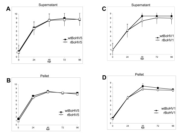
Findings: The viral genomes of BoHV1 and BoHV5 were cloned as infectious bacterial artificial chromosomes (BACs). First, recombinant viruses carrying the genetic elements for propagation in bacteria were generated. Second, DNA from these recombinant viruses were transferred into prokaryotic cells. Third, DNA from these bacteria were transferred into eukaryotic cells. Progeny viruses from BAC transfections showed similar kinetics as their corresponding wild types.
Conclusion: The two viral genomes of BoHV1 and BoHV5 cloned as BACs are accessible to the tools of bacterial genetics. The ability to easily manipulate the viral genomes on a molecular level in future experiments will lead to a better understanding of the difference in pathogenesis induced by these two closely related bovine herpesviruses.
Figures


Similar articles
-
Isolation and characterization of bovine herpes virus 5 (BoHV5) from cattle in India.PLoS One. 2020 Apr 24;15(4):e0232093. doi: 10.1371/journal.pone.0232093. eCollection 2020. PLoS One. 2020. PMID: 32330151 Free PMC article.
-
Comparative analysis of the replication of bovine herpesvirus 1 (BHV1) and BHV5 in bovine-derived neuron-like cells.Arch Virol. 2015 Nov;160(11):2683-91. doi: 10.1007/s00705-015-2537-5. Epub 2015 Aug 5. Arch Virol. 2015. Retraction in: Arch Virol. 2020 May;165(5):1261. doi: 10.1007/s00705-020-04571-0. PMID: 26239341 Retracted.
-
First report of isolation and molecular characterization of bubaline herpesvirus 1 (BuHV1) from Argentinean water buffaloes.Arch Virol. 2014 Nov;159(11):2917-23. doi: 10.1007/s00705-014-2146-8. Epub 2014 Jun 18. Arch Virol. 2014. PMID: 24938487
-
Cloning of herpesviral genomes as bacterial artificial chromosomes.Rev Med Virol. 2003 Mar-Apr;13(2):111-21. doi: 10.1002/rmv.380. Rev Med Virol. 2003. PMID: 12627394 Review.
-
Recent advances in cloning herpesviral genomes as infectious bacterial artificial chromosomes.Cell Cycle. 2011 Feb 1;10(3):434-40. doi: 10.4161/cc.10.3.14708. Epub 2011 Feb 1. Cell Cycle. 2011. PMID: 21245660 Free PMC article. Review.
Cited by
-
Efficient strategy for constructing duck enteritis virus-based live attenuated vaccine against homologous and heterologous H5N1 avian influenza virus and duck enteritis virus infection.Vet Res. 2015 Apr 16;46(1):42. doi: 10.1186/s13567-015-0174-3. Vet Res. 2015. PMID: 25889564 Free PMC article.
-
Glycoprotein D of bovine herpesvirus 5 (BoHV-5) confers an extended host range to BoHV-1 but does not contribute to invasion of the brain.J Virol. 2010 Jun;84(11):5583-93. doi: 10.1128/JVI.00228-10. Epub 2010 Mar 10. J Virol. 2010. PMID: 20219909 Free PMC article.
-
The genome of Chelonid herpesvirus 5 harbors atypical genes.PLoS One. 2012;7(10):e46623. doi: 10.1371/journal.pone.0046623. Epub 2012 Oct 2. PLoS One. 2012. PMID: 23056373 Free PMC article.
-
Herpesvirus BACs: past, present, and future.J Biomed Biotechnol. 2011;2011:124595. doi: 10.1155/2011/124595. Epub 2010 Oct 27. J Biomed Biotechnol. 2011. PMID: 21048927 Free PMC article. Review.
References
-
- Wyler R, Engels M, Schwyzer M. Infectious bovine rhinotracheitis/vulvovaginitis (BHV-1) Hingham, Mass.: Kluwer Academic Publishers; 1989.
LinkOut - more resources
Full Text Sources
