. 2009 Oct 14:9:218.
doi: 10.1186/1471-2180-9-218.

A novel inhibitor of Chlamydophila pneumoniae protein kinase D (PknD) inhibits phosphorylation of CdsD and suppresses bacterial replication

Affiliations

A novel inhibitor of Chlamydophila pneumoniae protein kinase D (PknD) inhibits phosphorylation of CdsD and suppresses bacterial replication

Dustin L Johnson et al. BMC Microbiol. .

Abstract

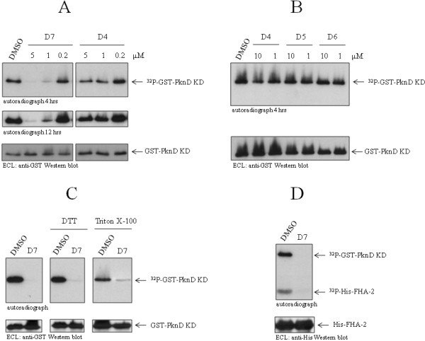
Background: We have shown previously that Chlamydophila pneumoniae contains a dual-specific Ser/Thr protein kinase that phosphorylates CdsD, a structural component of the type III secretion apparatus. To further study the role of PknD in growth and development we sought to identify a PknD inhibitor to determine whether PknD activity is required for replication.

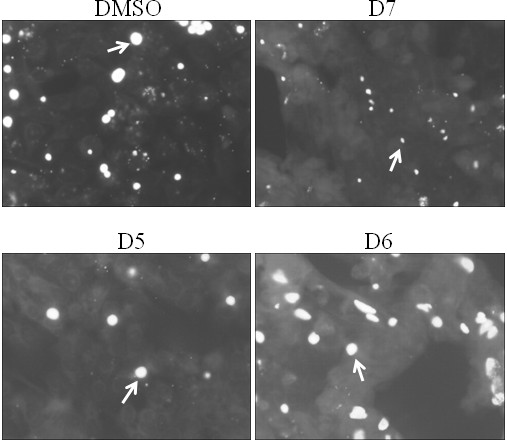
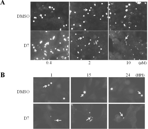
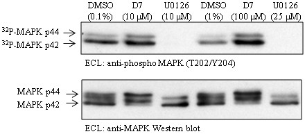
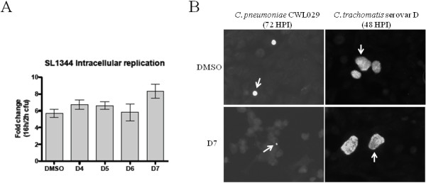
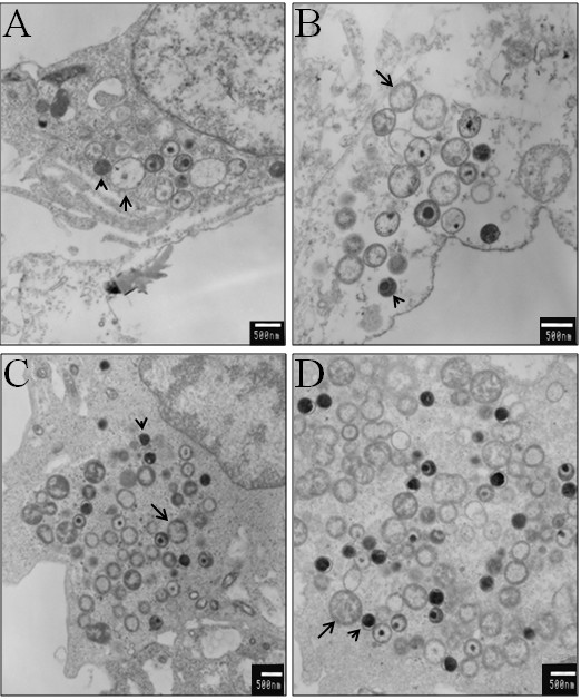
Results: Using an in vitro kinase assay we screened 80 known eukaryotic protein kinase inhibitors for activity against PknD and identified a 3'-pyridyl oxindole compound that inhibited PknD autophosphorylation and phosphorylation of CdsD. The PknD inhibitor significantly retarded the growth rate of C. pneumoniae as evidenced by the presence of very small inclusions with a reduced number of bacteria as seen by electron microscopy. These inclusions contained the normal replicative forms including elementary bodies (EB), intermediate bodies (IB) and reticulate bodies (RB), but lacked persistent bodies (PB), indicating that induction of persistence was not the cause of reduced chlamydial growth. Blind passage of C. pneumoniae grown in the presence of this PknD inhibitor for 72 or 84 hr failed to produce inclusions, suggesting this compound blocks an essential step in the production of infectious chlamydial EB. The compound was not toxic to HeLa cells, did not block activation of the MEK/ERK pathway required for chlamydial invasion and did not block intracellular replication of either Chlamydia trachomatis serovar D or Salmonella enterica sv. Typhimurium suggesting that the inhibitory effect of the compound is specific for C. pneumoniae.

Conclusion: We have identified a 3'-pyridyl oxindole compound that inhibits the in vitro kinase activity of C. pneumoniae PknD and inhibits the growth and production of infectious C. pneumoniae progeny in HeLa cells. Together, these results suggest that PknD may play a key role in the developmental cycle of C. pneumoniae.


Figures

Figure 1
Figure 1
Inhibition of PknD by compound D7. A: compound D7, but not compound D4 or DMSO, substantially inhibited PknD autophosphorylation (32P-GST-PknD KD) at 5 and 1 μM, as seen by autoradiography. B: related compounds D5 and D6 did not inhibit PknD at 1 or 10 μM. C: 1 mM DTT and 1% Triton X-100 did not decrease inhibition of PknD by compound D7 (used at 10 μM in all panels). DMSO (0.1%) is shown as control. D: compound D7 inhibited phosphorylation of the FHA-2 domain (32P-His-FHA-2) of CdsD by PknD. Western blotting showed equivalent amounts of protein in each autoradiograph (lower panels).
Figure 2
Figure 2
Compound D7 inhibits the growth of C. pneumoniae in HeLa cells. Detection of inclusions at 72 hpi by IF microscopy revealed very small inclusions when C. pneumoniae-infected HeLa cells were exposed to 10 μM of compound D7, but not when exposed to DMSO (0.1%) or 10 μM of compounds D5 or D6. Arrows indicate representative inclusions. Inclusions were stained with FITC-conjugated anti-LPS monoclonal antibody containing Evan's Blue counterstain.
Figure 3
Figure 3
C. pneumoniae growth inhibition by compound D7 is dose-dependent. A: compound D7 at 0.4 μM exhibited no inhibition of chlamydial growth (normal size inclusions), 2 μM exhibited partial inhibition (smaller inclusions), and 10 μM had a significant inhibitory effect (significantly reduced inclusion size) (bottom panels, left to right, respectively). DMSO controls at 0.004, 0.02, and 0.1% (top panels, left to right, respectively) did not restrict growth as indicated by inclusion size. Arrows indicate representative inclusions. B: Addition of 10 μM compound D7 to C. pneumoniae-infected HeLa cells at 1, 15 or 24 hpi resulted in small inclusions at 72 hpi. Inclusions were stained with FITC-conjugated anti-LPS monoclonal antibody containing Evan's Blue counterstain.
Figure 4
Figure 4
Compound D7 does not reduce HeLa cell viability. A: subconfluent HeLa cell monolayers incubated in MEM containing either DMSO (0.1%) or compound D7 (10 μM) with 2 μg/mL cycloheximide (+), were collected by trypsinization and the cell density was measured by absorbance at 800 nM at the times indicated. Compound D7 did not significantly alter HeLa cell number compared to DMSO alone. B: cell culture supernatant adenylate kinase activity from the samples in (A). Exposure of HeLa cells to 10 μM compound D7 for 44 hours was not more cytotoxic than cells exposed to DMSO. At 66 hours there was a small increase in HeLa cell release of adenylate kinase in the D7-exposed group. Error bars represent means plus 2 standard deviations.
Figure 5
Figure 5
Compound D7 does not block activation of the MEK/ERK pathway in EGF-stimulated HeLa cells. HeLa cells incubated with DMSO, compound D7 or U0126 were activated with EGF and the levels of MAP kinase phosphorylation were determined by Western blot using anti-phospho ERK1/2 antibody. Compound D7 at 10 and 100 μM, and DMSO at 0.1 and 1%, did not prevent phosphorylation of MAP kinase following EGF stimulation of HeLa cells. U0126 at 10 and 25 μM completely prevented phosphorylation of MAP kinase. Blots were probed with antibody to phosphorylated MAPK (upper panel), and with antibody to total MAPK (lower panel).
Figure 6
Figure 6
Compound D7 does not inhibit the growth of Salmonella enterica sv. Typhimurium or C. trachomatis serovar D in HeLa cells. A: compounds D4, D5, D6 and D7 (10 μM) or DMSO (0.1%), did not prevent replication of Salmonella enterica sv. Typhimurium SL1344 in HeLa cells. Compounds were added to the media 2 hours after host cell infection, and bacteria harvested at both 2 and 16 hpi in order to plot the fold change in colony forming units. B: compound D7 did not inhibit the growth of Chlamydia trachomatis serovar D. Compound D7 (10 μM) was added to cell monolayers 1 hpi and inclusions were stained at 48 hpi. Large inclusions were seen in both D7- (bottom right panel) and DMSO-exposed (0.1%; top right panel) cells while small inclusions were seen for C. pneumoniae in D7-exposed cells. Arrows indicate representative inclusions. The monoclonal antibody contained Evan's Blue counterstain for detection of host cells.
Figure 7
Figure 7
Normal developmental forms of C. pneumoniae are found within compound D7-exposed inclusions. At 48 hpi, infected HeLa cells incubated in MEM containing 10 μM of either compound D6 or D7 were observed by TEM. A, B: inclusions in D7-exposed cells are smaller and contain fewer bacteria, but all three developmental forms (EB, IB and RB) of C. pneumoniae are present. C, D: C. pneumoniae inclusions exposed to compound D6 are normal in size and contain the same normal developmental forms. Size bars are indicated in white (500 nm). Representative micrographs indicating RB (arrows) and EB (arrow heads) are shown.
Figure 8
Figure 8
Compound D7 reduces the number and infectivity of C. pneumoniae progeny. HeLa cells were infected with C. pneumoniae (MOI of 5) and MEM containing either DMSO (0.1%) or D7 (10 μM) was added at 1 hpi. Cells were lysed at 72 hpi and chlamydial lysates diluted 10-1 and 10-2 and used to infect fresh HeLa cell monolayers. Infected cells were then incubated for 72 hours in MEM (without D7 or DMSO) and inclusions were stained with FITC-conjugated anti-LPS monoclonal antibody. C. pneumoniae harvested from DMSO-exposed HeLa cells produced many inclusions of normal size upon subsequent passage (top panels; dilution of passage indicated). A substantial reduction in both the number and size of inclusions was seen with chlamydiae harvested from HeLa cells exposed to compound D7 (bottom panels). Similar results were obtained with undiluted chlamydial lysates and with lysates harvested at 84 hpi (data not shown).

Similar articles

Cited by

References

    1. Hahn DL, Azenabor AA, Beatty WL, Byrne GI. Chlamydia pneumoniae as a respiratory pathogen. Front Biosci. 2002;7:e66–e76. doi: 10.2741/hahn. - DOI - PubMed
    1. Paldanius M, Juvonen R, Leinonen M, Bloigu A, Silvennoinen-Kassinen S, Saikku P. Asthmatic persons are prone to the persistence of Chlamydia pneumoniae antibodies. Diagn Microbiol Infect Dis. 2007;59:117–122. doi: 10.1016/j.diagmicrobio.2007.04.004. - DOI - PubMed
    1. Sutherland ER, Martin RJ. Asthma and atypical bacterial infection. Chest. 2007;132:1962–1966. doi: 10.1378/chest.06-2415. - DOI - PubMed
    1. Campbell LA, Kuo CC, Grayston JT. Chlamydia pneumoniae and cardiovascular disease. Emerg Infect Dis. 1998;4:571–579. doi: 10.3201/eid0404.980407. - DOI - PMC - PubMed
    1. Grayston JT. Background and current knowledge of Chlamydia pneumoniae and atherosclerosis. J Infect Dis. 2000;181(Suppl 3):S402–S410. doi: 10.1086/315596. - DOI - PubMed

Publication types

MeSH terms

LinkOut - more resources