Review
. 2010 Feb 5;1217(6):902-24.
doi: 10.1016/j.chroma.2009.09.073. Epub 2009 Oct 2.

Porous polymer monoliths: amazingly wide variety of techniques enabling their preparation

Affiliations
Review

Porous polymer monoliths: amazingly wide variety of techniques enabling their preparation

Frantisek Svec. J Chromatogr A. .

Abstract

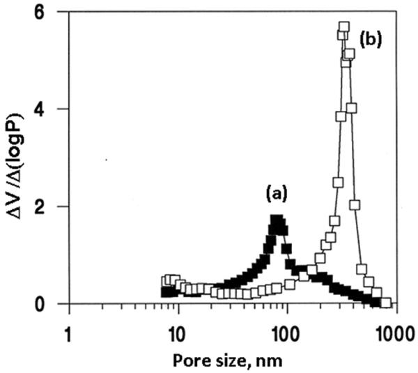
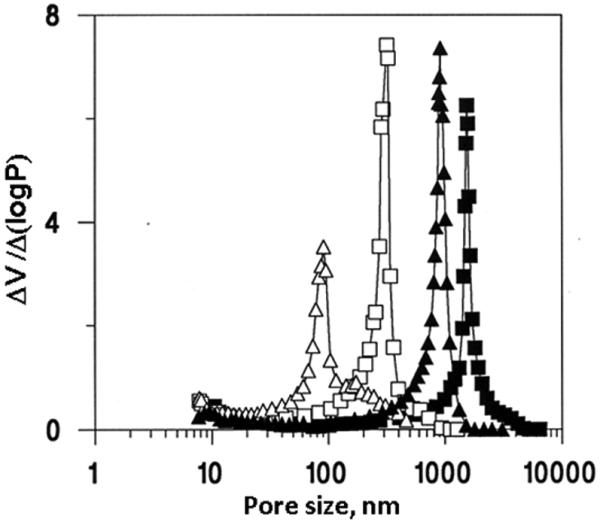
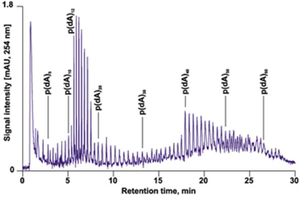
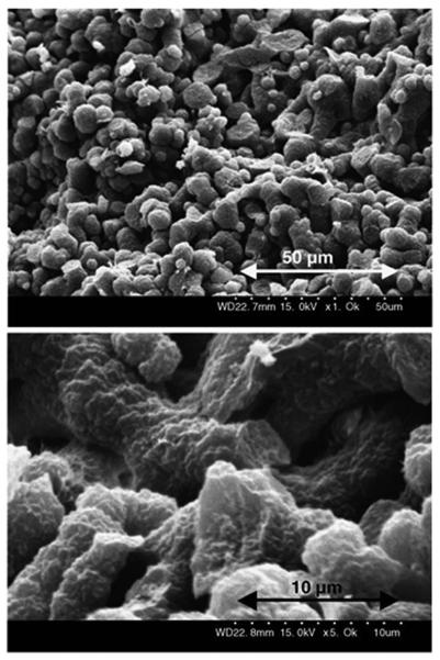
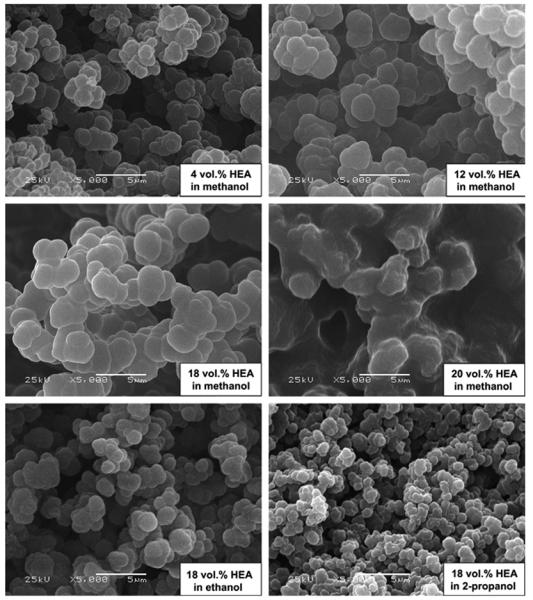
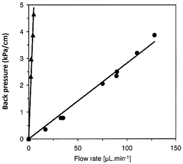
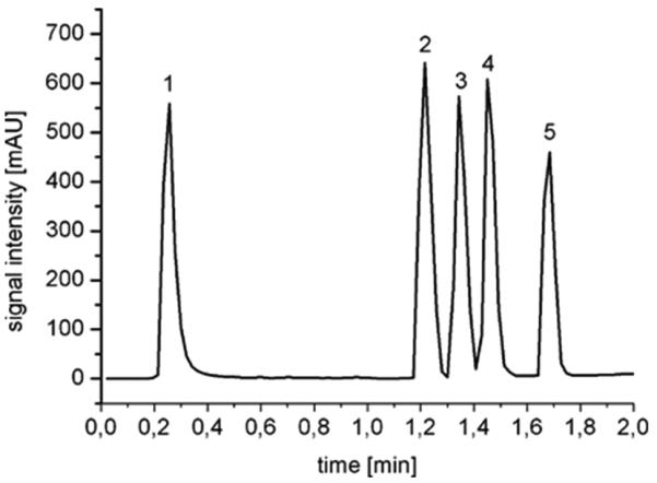
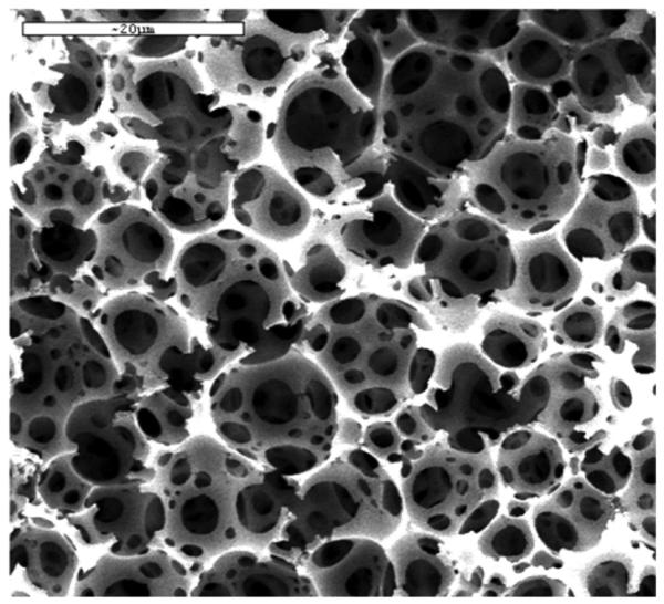
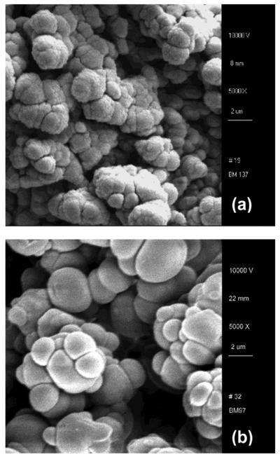
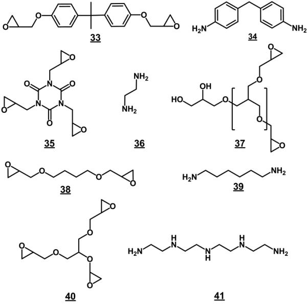
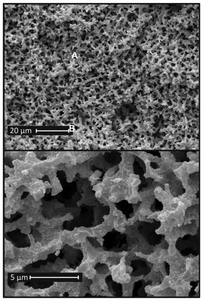
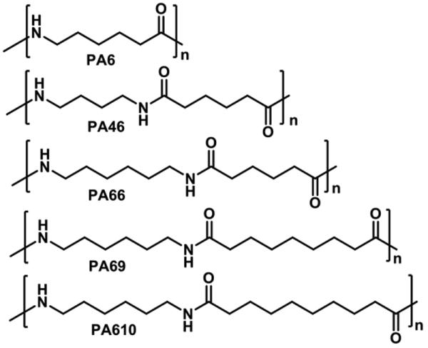
The porous polymer monoliths went a long way since their invention two decades ago. While the first studies applied the traditional polymerization processes at that time well established for the preparation of polymer particles, creativity of scientists interested in the monolithic structures has later led to the use of numerous less common techniques. This review article presents vast variety of methods that have meanwhile emerged. The text first briefly describes the early approaches used for the preparation of monoliths comprising standard free radical polymerizations and includes their development up to present days. Specific attention is paid to the effects of process variables on the formation of both porous structure and pore surface chemistry. Specific attention is also devoted to the use of photopolymerization. Then, several less common free radical polymerization techniques are presented in more detail such as those initiated by gamma-rays and electron beam, the preparation of monoliths from high internal phase emulsions, and cryogels. Living processes including stable free radicals, atom transfer radical polymerization, and ring-opening metathesis polymerization are also discussed. The review ends with description of preparation methods based on polycondensation and polyaddition reactions as well as on precipitation of preformed polymers affording the monolithic materials.


Figures

Fig. 1
Fig. 1
Mercury porosimetry differential pore size distribution profiles of the poly(glycidyl methacrylate-co-ethylene dimethacrylate) beads and monolith prepared via suspension (a) and bulk (b) polymerization at a temperature of 70 °C from a polymerization mixture comprising azobisisobutyronitrile(1% with respect to monomers), 24% glycidyl methacrylate, 16% ethylene dimethacrylate, cyclohexanol 48%, and dodecanol 12%. Reproduced from ref. [43] with permission.
Fig. 2
Fig. 2
Mercury porosimetry differential pore size distribution curves of the poly(glycidyl methacrylate-co-ethylene dimethacrylate) monoliths prepared from mixtures containing 6 (triangles) and 12% dodecanol (squares) using polymerization at a temperature of 55 (closed points) and 70 °C (open points). Reproduced from ref. [43] with permission.
Fig 3
Fig 3
High-resolution of phosphorylated and non-phosphorylated oligonucleotides in a monolithic capillary column. Conditions: Poly(styrene-co-divinylbenzene) monolith, 60 × 0.2-mm i.d, gradient, 1-7 % acetonitrile in water in 5 min, 7-8% in 5 min, 8-9% in 6 min, 9-10.4% in 14 min in 100 mmol/L triethylammonium acetate, flow rate 2.1 μL/min, 50 °C. Reproduced from ref. [67] with permission
Fig. 4
Fig. 4
Morphology of poly(glycerol dimethacrylate) monolith prepared using solution of polystyrene with a molecular mass of 3 840 000 in chlorobenzene. Reproduced from ref. [80] with permission
Fig. 5
Fig. 5
Examples of crosslinking monomers used for the preparation of porous polymer monoliths
Fig. 6
Fig. 6
Separation of phenol derivatives using 80 × 0.2 mm I.D. monolithic poly(4-methylstyrene-co-1,2-(4-vinylphenyl)ethane) capillary column polymerized for 45 min. Conditions: column 80 × 0.2 mm I.D., mobile phase 25% acetonitrile in 0.1% aqueous trifluoroacetic acid, flow rate 4 μL/min, UV detection at 210 nm. Reproduced from ref. [100] with permission.
Fig.7
Fig.7
Examples of monomers used for the preparation of porous polymer monoliths.
Fig. 8
Fig. 8
Rapid reversed-phase separation of proteins at a flow-rate of 10 ml/min using 50×4.6 mm I.D. poly(styrene-divinylbenzene) monolithic column. Conditions: Mobile phase gradient: 42% to 90% acetonitrile in water with 0.15% trifluoroacetic acid in 0.35 min. Detection: UV 280 nm. Peaks: ribonuclease (1), cytochrome c (2), bovine serum albumin (3), carbonic anhydrase (4), chicken egg albumin (5).
Fig. 9
Fig. 9
Examples of modification of typical porous polymer monoliths containing glycidyl methacrylate (A) and chloromethylstyrene (B) units.
Fig. 10
Fig. 10
Scheme of the two-step sequential photografting procedure. Reproduced from ref. [122] with permission.
Fig. 11
Fig. 11
Separation of a mixture of carbohydrates by anion-exchange chromatography using an optimized latex-coated polymeric monolithic capillary column. Conditions: Column size 10 cm ×250 μm i.d, pore size 0.97 μm. Flow rate 13 μL/min Mobile phase aqueous ammonium hydroxide 64 mmol/L (pH 12.8). Peaks: D(+)galactose (1), D(+)glucose (2), D(+)xylose (3), D(+)mannose (4), maltose (5), D(−)fructose (6), sucrose (7). Reproduced from ref. [127] with permission.
Fig. 12
Fig. 12
Chromatographic separation of three model proteins using monolithic poly(butyl methacrylate-co-ethylene dimethacrylate) capillary column prepared by thermally (A) and photochemically (B) initiated polymerization. Conditions: column 20 cm × 100 μm I.D.; mobile phase A 2% formic acid in 98:2 water:acetonitrile mixture, mobile phase B 2% formic acid in acetonitrile; gradient from 100% A to 50% B in A in 4 min; flow rate 4 μL/min. Peaks: (1) ribonuclease A (2 pmol), (2) cytochrome c (1 pmol), and (3) myoglobin (0.3 pmol). Dashed line represents the overall back pressure in the system. Reproduced from ref. [143] with permission.
Fig. 13
Fig. 13
SEM micrographs of poly(2-hydroxyethyl acrylate-co-diethylene glycol dimethacrylate) monoliths prepared in presence of different porogenic solvents. Irradiation temperature 25 °C, dose rate of 16 kGy/h, total dose of 30 kGy. Reproduced from ref. [155] with permission.
Fig. 14
Fig. 14
Back pressure per unit length of a 1mm I.D. monolithic capillary as a function of methanol flow rate for poly(2-hydroxyethyl methacrylate-co-ethylene dimethacrylate) monoliths polymerized using electron beam at a dose of 50 (●) and 100 kGy (▲) Reproduced from ref. [158] with permission.
Fig. 15
Fig. 15
Separation of lysozyme (1), ribonuclease A (2), insulin (3), cytochrome c (4), and albumin (5) using poly(ethyl methacrylate-co-trimethylolpropane trimethacrylate) monolith prepared using electron beam initiated polymerization. Glass column 100×3 mm i.d.; mobile phase A: 95% water + 5% acetonitrile + 0.1% TFA; mobile phase B: 20% water + 80% acetonitrile + 0.1% TFA; linear gradient, 10-90% B in 2 min; flow rate, 3 mL/min; UV detection at 200 nm. Reproduced from ref. [162] with permission.
Fig. 16
Fig. 16
Scanning electron micrograph of a typical polyHIPE polymer. Reproduced from ref. [168] with permission.
Fig. 17
Fig. 17
Fast anion exchange separation of lysozyme l (1), bovine serum albumin (2), ovalbumin (3), and pepsin (4) using a monolithic polyHIPE column with 2-hydroxy-3-(diethylamino)-propyl functionalities. Conditions: mobile phase: buffer A 10 mmol/L TrisHCl buffer, pH 7.6; buffer B 1 mol/L NaCl in A; gradient time 45 s; flow rate 6 mL/min; UV detection at 280 nm. Reproduced from ref. [172] with permission.
Fig. 18
Fig. 18
Scheme of formation of cryogels. Reproduced from ref.[185] with permission.
Fig. 19
Fig. 19
SEM of the dextran-based cryogel prepared at −20 °C and conventional dextran gel prepared at room temperature. Reproduced from ref. [177] with permission.
Fig. 20
Fig. 20
Structures of stable free radicals. 21 2,2,6,6-tetramethyl-1-piperidyloxy (TEMPO); 22 2,2,5-trimethyl-3-(1-phenylethoxy)-4-phenyl-3-azahexane (TPPA); 23 3-carboxy-2,2,5,5-tetramethylpyrrolidinyl-1-oxy (3-carboxy-PROXYL); 24 4-carboxy-2,2,6,6-etramethylpiperidinyl-1-oxy (4-carboxy-TEMPO).
Fig. 21
Fig. 21
Integral pore size distribution profiles of porous polymer monoliths prepared by a typical polymerization at 70 °C (1) and in the presence (2) and the absence (3) of TEMPO at 130 °C. Conditions: polymerization mixture–styrene 20 wt%; divinylbenzene 20wt %; 1-dodecanol 60 wt%; benzoyl peroxide 0.5 wt % (with respect to monomers); TEMPO 1.2 molar excess with respect to benzoyl peroxide. Reproduced from ref. [47] with permission.
Fig. 22
Fig. 22
SEC calibration curves of 50 × 8 mm I.D. poly(styrene-co-divinylbenzene) monoliths prepared at a temperature of 130 °C using 85:15 PEG-decanol porogen and different stable free radicals at a molar ratio of 1.3 to 1 with respect to benzoyl peroxide. Reproduced from ref. [205] with permission.
Fig. 23
Fig. 23
Scanning electron microscopy images of poly(divinylbenzene) monoliths prepared with increasing percentage of polymer porogen in the polymerization mixture composed of divinylbenzene, 1,3,5-trimethylbenzene, dimethylsiloxane, TEMPO, acetic anhydride, and benzoyl peroxide. Reproduced from ref. [206] with permission.
Fig. 24
Fig. 24
Examples of tellurium containing initiators.
Fig 25
Fig 25
Examples of monomers used in the ring opening metathesis polymerization affording porous monoliths.
Fig. 26
Fig. 26
SEM micrographs of monolith prepared using ROMP process in a 3 mm I.D. column and 200 μm I.D. capillary. Reproduced from ref. [220] with permission.
Fig. 27
Fig. 27
Examples of reaction partners used for the preparation of monoliths using polycondensation reaction.
Fig. 28
Fig. 28
SEM micrographs of epoxy monoliths prepared from 45% organic phase consisting of epoxide monomer mixture of 1,4-butanediol diglycidyl ether, bisphenol A diglycidyl ether glyceryl triglycidyl ether (4.5:4.5:1) and diethylene glycol dibutyl ether, dispersed in the aqueous phase containing 20% diethylene glycol diethyl ether as the cosurfactant and 80% of the stoichiometric amount of diaminohexane and tetraethylenepentamine (9.7:1) dissolved in 0.1 calcium chloride solution. Reproduced from ref. [233] with permission.
Fig. 29
Fig. 29
Structures of soluble polyamides used for the preparation of monoliths via phase separation.
Fig. 30
Fig. 30
SEM micrographs of monolith prepared from polyamides. Reproduced from ref. [235] with permission.

Comment in

References

    1. Tsvett MS. Ber. Deut. Botan. Gessel. 1906;24:322.
    1. Hjertén S. Ind. Eng. Chem. Res. 1999;38:1205.
    1. Regnier FE. HRC-J. 2000;23:19.
    1. Svec F, Peters EC, Sýkora D, Fréchet JMJ. J. Chromatogr. 2000;887:3. - PubMed
    1. Josic D, Buchacher A, Jungbauer A. J. Chromatogr. B. 2001;752:191. - PubMed

Publication types

LinkOut - more resources