. 2009 Oct 27;106(43):18225-30.
doi: 10.1073/pnas.0907635106. Epub 2009 Oct 14.

Mechanism for the allosteric regulation of phosphodiesterase 2A deduced from the X-ray structure of a near full-length construct

Affiliations

Mechanism for the allosteric regulation of phosphodiesterase 2A deduced from the X-ray structure of a near full-length construct

Jayvardhan Pandit et al. Proc Natl Acad Sci U S A. .

Abstract

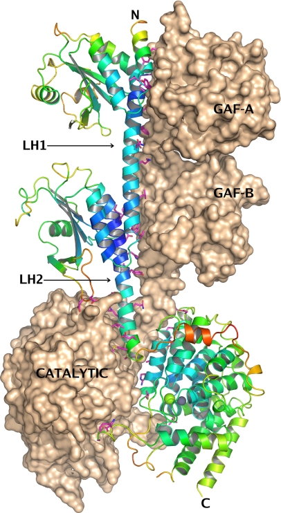
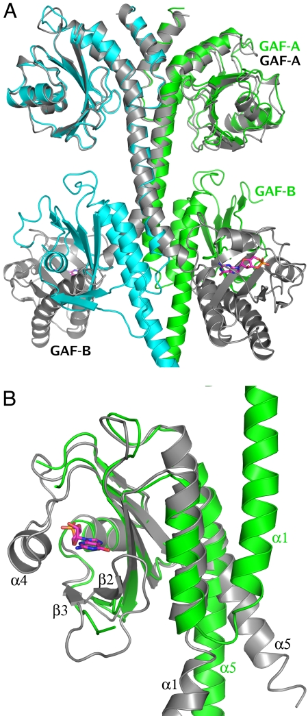
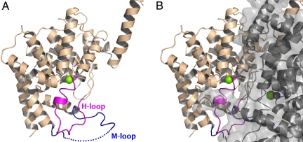
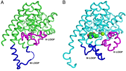
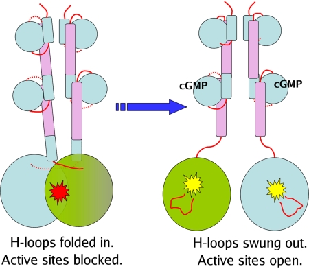
We report the X-ray crystal structure of a phosphodiesterase (PDE) that includes both catalytic and regulatory domains. PDE2A (215-900) crystallized as a dimer in which each subunit had an extended organization of regulatory GAF-A and GAF-B and catalytic domains connected by long alpha-helices. The subunits cross at the GAF-B/catalytic domain linker, and each side of the dimer contains in series the GAF-A and GAF-B of one subunit and the catalytic domain of the other subunit. A dimer interface extends over the entire length of the molecule. The substrate binding pocket of each catalytic domain is occluded by the H-loop. We deduced from comparisons with structures of isolated, ligand-bound catalytic subunits that the H-loop swings out to allow substrate access. However, in dimeric PDE2A (215-900), the H-loops of the two catalytic subunits pack against each other at the dimer interface, necessitating movement of the catalytic subunits to allow for H-loop movement. Comparison of the unliganded GAF-B of PDE2A (215-900) with previous structures of isolated, cGMP-bound GAF domains indicates that cGMP binding induces a significant shift in the GAF-B/catalytic domain linker. We propose that cGMP binding to GAF-B causes movement, through this linker region, of the catalytic domains, such that the H-loops no longer pack at the dimer interface and are, instead, free to swing out to allow substrate access. This increase in substrate access is proposed as the basis for PDE2A activation by cGMP and may be a general mechanism for regulation of all PDEs.


Conflict of interest statement

The authors declare no conflict of interest.

Figures

Fig. 1.
Fig. 1.
Structure of hPDE2A (215–900). The asymmetric unit contains two molecules, A and B, related by a noncrystallographic 2-fold axis of symmetry, which, in this view, is roughly parallel to the plane of the paper in the vertical direction. The three domains are labeled in the figure, as well as the linker helices LH1 and LH2 that connect them. Molecule B is shown in surface representation, and molecule A is shown in a ribbons representation, with the ribbons colored by crystallographic B-factor, blue being low and red being high. Regions of the structure with higher B-factors, such as the linker between the GAF-B domain and the catalytic domain, are expected to be more flexible. All side chains from molecule A that are within 3.5 Å of molecule B are shown as magenta sticks, The dimer interface extends over the surface of the entire molecule. The two catalytic sites in the vicinity of the Zn2+ and Mg2+ ions (shown as gray and green spheres) mutually occlude each other at the dimer interface. All figures showing the structure were generated with PyMOL (www.pymol.org).
Fig. 2.
Fig. 2.
Comparison of PDE2A (215–900) with the structure of the cGMP-bound GAF domains from mouse PDE2A (PDB ID code 1MC0) (26). (A) Superposition of the two dimers [1mc0, gray; PDE2A (215–900), green and cyan] using just the Cα atoms from the GAF-A domain of one subunit shows the GAF-A dimer interface to be nearly identical in the two structures, while the GAF-B domains do not overlap at all. GAF-A and GAF-B domains from one subunit of each structure have been labeled. (B) Superposition of just the GAF-B domains from the two structures [1mc0, gray; PDE2A (215–900), green] shows the conformational changes between the cGMP-bound and apo GAF-B domain. There are significant differences (A) in the immediate vicinity of the cGMP binding site—α4 helix and β2-β3 loop, which interact with the bound cyclic nucleotide (shown in magenta sticks), are completely disordered in the PDE2A (215–900) structure, and (B) in the orientation of α1 and α5, the two helices that define the N and C terminii of this domain are rotated by nearly 180° and 30°, respectively.
Fig. 3.
Fig. 3.
The two catalytic sites mutually occlude each other at the dimer interface. (A) The catalytic domain of a single subunit of the PDE2A (215–900) dimer is shown in ribbons representation, with the H-loop colored in magenta and the M-loop colored in blue. The active site, whose location can be inferred from the position of the Zn2+ and Mg2+ ions (shown as gray and green spheres) is partially occupied by residues from the H-loop. Residues 840–850 of the M-loop have not been modeled due to disorder and are indicated by a dotted line. (B) The second subunit is shown as a semitransparent gray surface, as well as in ribbons representation, keeping exactly the same orientation as in panel A. The H-loop is blocked from swinging out of the active site by the dimer interface.
Fig. 4.
Fig. 4.
Crystal structures of the isolated catalytic domains also show “open” and “closed” conformations of the H-loop. The Cα trace of the isolated catalytic domain (579–919) is shown as green tubes. The H-loop (700–723) and M-loop (830–856) are colored magenta and blue, respectively. (A) Structure of the unliganded catalytic domain that shows the H-loop folded into the catalytic site and displacing the Mg2+ ion. The Zn2+ ion is shown as a gray sphere. (B) Structure of the catalytic domain co-crystallized with IBMX, showing the H-loop swung out. Zn2+ and Mg2+ ions, shown as gray and green spheres, are both visible in this structure.
Fig. 5.
Fig. 5.
Proposed mechanism of activation. Cyclic nucleotide binding to the GAF-B domain is accompanied by an ordering of the α4 helix, which “closes down” on the nucleotide, and by a relative rotation of the two linker helices, which causes the two catalytic domains to swing out relative to each other. The H-loop, which was held in a position to occlude the substrate binding site in the catalytic domain, now swings out, making the substrate binding site accessible.

References

    1. Conti M, Beavo J. Biochemistry and physiology of cyclic nucleotide phosphodiesterases: Essential components in cyclic nucleotide signaling. Annu Rev Biochem. 2007;76:481–511. - PubMed
    1. Huai Q. Three-dimensional structures of PDE4D in complex with roliprams and implication on inhibitor selectivity. Structure. 2003;11:865–873. - PubMed
    1. Wang H, et al. Kinetic and structural studies of phosphodiesterase-8A and implication on the inhibitor selectivity. Biochemistry. 2008;47:12760–12768. - PMC - PubMed
    1. Huai Q, Colicelli J, Ke H. The crystal structure of AMP-bound PDE4 suggests a mechanism for phosphodiesterase catalysis. Biochemistry. 2003;42:13220–13226. - PubMed
    1. Huai Q, Liu Y, Francis SH, Corbin JD, Ke H. Crystal structures of phosphodiesterases 4 and 5 in complex with inhibitor 3-isobutyl-1-methylxanthine suggest a conformation determinant of inhibitor selectivity. J Biol Chem. 2004;279:13095–13101. - PubMed

Substances