. 2009 Dec 11;284(50):34793-808.
doi: 10.1074/jbc.M109.065979. Epub 2009 Oct 14.

Melanoma antigen gene protein-A11 (MAGE-11) F-box links the androgen receptor NH2-terminal transactivation domain to p160 coactivators

Affiliations

Melanoma antigen gene protein-A11 (MAGE-11) F-box links the androgen receptor NH2-terminal transactivation domain to p160 coactivators

Emily B Askew et al. J Biol Chem. .

Abstract

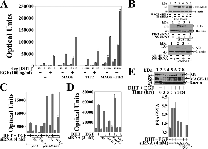
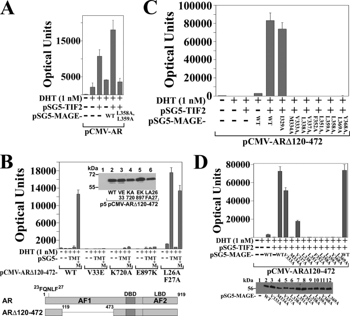
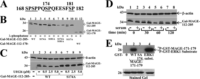
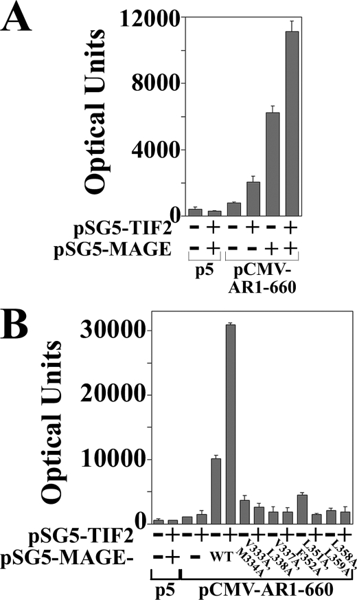
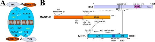
Androgen-dependent transcriptional activity by the androgen receptor (AR) and its coregulators is required for male reproductive development and function. In humans and other primates, melanoma antigen gene protein-A11 (MAGE-11) is an AR selective coregulator that increases AR transcriptional activity. Here we show that the interaction between AR and MAGE-11 is mediated by AR NH(2)-terminal FXXLF motif binding to a highly conserved MAGE-11 F-box in the MAGE homology domain, and is modulated by serum stimulation of mitogen-activated protein kinase phosphorylation of MAGE-11 Ser-174. The MAGE-11-dependent increase in AR transcriptional activity is mediated by a direct interaction between MAGE-11 and transcriptional intermediary factor 2 (TIF2) through the NH(2)-terminal region of TIF2, and by a MAGE-11 FXXIF motif interaction with an F-box-like region in activation domain 1 of TIF2. The results suggest that MAGE-11 functions as a bridging factor to recruit AR coactivators through a novel FXX(L/I)F motif-F-box interaction paradigm.


Figures

FIGURE 1.
FIGURE 1.
Dependence of AR transactivation on MAGE-11 and TIF2. A, CWR-R1 cells were transfected with 0.1 μg of MMTV-Luc with or without 0.1 μg of pSG5-MAGE-11 or pSG5-TIF2. Cells were incubated in the absence and presence of DHT (shown as -log DHT concentration) and 0.1 μg/ml of EGF. B, COS cells were transfected using Lipofectamine 2000 with 1 μg of pSG5 or pSG5-MAGE (top panel), 1 μg of pSG5 or pSG5-TIF2 (middle panel), and 0.5 μg of pCMV5 or pCMV-AR (bottom panel) in the absence and presence of 1 nm nonspecific (NS) siRNA, MAGE-11 siRNA-2 and -3, TIF2 siRNA-3, and AR siRNA-3. Cell extracts were analyzed by immunoblot for MAGE-11 (10 μg of protein/lane), TIF2 (40 μg/lane), AR (30 μg/lane), and β-actin. C, CWR-R1 cells were transfected with 0.1 μg of MMTVΔ-(−421–−364)-Luc, 0.2 μg of pSG5 or pSG5-MAGE in the absence and presence of 4 nm nonspecific or TIF2 siRNA-3, and incubated in the absence and presence of 0.1 nm DHT and 0.01 μg/ml of EGF. D, LAPC-4 cells were transfected with 0.1 μg of MMTVΔ-(−421–−364)-Luc and 3 nm siRNA and incubated in the absence and presence of 0.1 nm DHT and 0.1 μg/ml EGF. E, top panel, CWR-R1 cells incubated for 48 h in serum-free medium were treated for the indicated times with 0.1 nm DHT and 10 ng/ml of EGF. Cell extracts in IB lysis buffer (200 μg of protein/lane) (lanes 2–8) were analyzed by immunoblot using 10 μg/ml of MAGE-Ab-(13–26), -(59–79), and -(94–108) antibodies overnight at 4 °C, and AR32 and β-actin antibodies (33). Extracts of COS cells expressing pCMV-AR (30 μg of protein) and pSG5-MAGE (0.25 μg of protein) served as controls (lane 1). Bottom panel, CWR-R1 cells were transfected with 4 nm siRNA and incubated for 6 h with or without 10 nm DHT and 10 ng/ml of EGF. RNA was extracted and analyzed by quantitative reverse transcription-PCR as described under “Experimental Procedures” for endogenous PSA mRNA relative to peptidylprolyl isomerase A.
FIGURE 2.
FIGURE 2.
Requirement for a MAGE-11 F-box in AR transactivation. A, schematic diagram of full-length MAGE-11 showing the MAGE homology domain (MHD) with F-box residues 329–369, phosphorylation site Thr-360 within the F-box, Lys-240 and Lys-245 monoubiquitinylation (Ub) sites outside the F-box, and the NH2-terminal nuclear localization signal (NLS). B, sequence homology between MAGE-11 F-box residues 329–369, cyclin F F-box residues 35–73, and MAGE family members, with conserved acidic residues in blue and hydrophobic residues in red. C, alanine mutagenesis of the MAGE F-box at conserved hydrophobic residues. D, Ishikawa cells were transfected with pCMV-AR (10 ng/well), 0.1 μg/well of PSA-Enh-Luc, and 0.1 μg of pSG5 empty vector (—), pSG5-MAGE wild-type (WT), or the indicated F-box mutant. Cells were incubated in the absence and presence of 1 nm DHT with or without 0.1 μg/ml of EGF. Bottom panel, Ishikawa cells were transfected with 2 μg of pSG5 (—), pSG5-MAGE WT, and the indicated F-box mutant. Cells were extracted in IB lysis buffer and 30 μg of protein/lane analyzed on immunoblots probed with antibody raised against FLAG-tagged human MAGE-11 (2 μg/ml). E, FLAG-b empty vector (—) or FLAG-AR (4 μg) were expressed in COS cells with pSG5-HA-MAGE-(112–429) WT or Δ329–369 F-box deletion (0.5 μg of DNA/10-cm dish). Cells were treated in the absence and presence of 10 nm DHT and 0.1 μg/ml EGF, lysed in IP lysis buffer, and incubated with FLAG resin overnight at 4 °C. Immunoprecipitates and cell extracts (25 μg of protein/lane) were analyzed by immunoblot using FLAG and HA antibodies.
FIGURE 3.
FIGURE 3.
Requirement for MAGE-11 F-box in AR transactivation by TIF2. A, CV1 cells were transfected with 0.1 μg of pCMV-AR and 5 μg of MMTV-Luc, 2 μg of pSG5-TIF2, 2 μg of pSG5-MAGE wild-type (WT) or L358A/L359A F-box mutant, and incubated in the absence and presence of 1 nm DHT. B, CV1 cells were transfected with 5 μg of MMTV-Luc and 0.1 μg of pCMV-ARΔ120–472 WT or the indicated mutant, 2 μg of pSG5-TIF2 (pSG5-T) and/or 2 μg of pSG5-MAGE (pSG5-M). Cells were incubated in the absence and presence of 1 nm DHT. Inset, pCMV-ARΔ120–472 WT and mutants (5 μg) were expressed in COS cells because expression was too low in CV1 cells. Cell extracts (30 μg of protein/lane) were analyzed on immunoblots probed with AR52 antibody. Bottom panel, schematic diagram of full-length human AR amino acid residues 1–919 with the NH2-terminal FXXLF motif 23FQNLF27, activation function 1 (AF1), DNA binding domain (DBD), activation function 2 (AF2) in the ligand binding domain (LBD), and the ARΔ120–472 deletion mutant that lacks AF1. C, pCMV-ARΔ120–472 (0.1 μg) was expressed in CV1 cells with 5 μg of MMTV-Luc, 2 μg of pSG5-TIF2 and/or 2 μg of pSG5-MAGE WT or the indicated F-box mutant. Cells were incubated in the absence and presence of 1 nm DHT. D, pCMV-ARΔ120–472 (0.1 μg) was expressed in CV1 cells with 5 μg of MMTV-Luc in the absence and presence of 2 μg of pSG5-TIF2 and/or 2 μg of pSG5-MAGE WT or single residue F-box mutant. Cells were incubated in the absence and presence of 1 nm DHT. Bottom panel, CV1 cells were transfected with pSG5 (8 μg/10-cm dish) (—), pSG5-MAGE WT or the indicated single residue F-box mutant. Cell extracts in IB lysis buffer (95 μg of protein/lane) were analyzed on immunoblots probed with antibody raised against FLAG-tagged MAGE-11.
FIGURE 4.
FIGURE 4.
Interaction between MAGE-11 F-box and AR FXXLF motif. A, GAL-MAGE wild-type (WT), F-box mutant, or GAL-MAGE-K236A/K240A/K245A (KA) (0.05 μg) were transfected in HeLa cells with 0.1 μg of 5×GAL4Luc3 and 0.1 μg of VP16 empty vector (—), VP-AR-(1–660) or VP-AR-(1–660)-L26A,F27A (LFAA) with a mutation in the AR FXXLF motif. Bottom panel, GAL-MAGE F-box or KA mutants, WT and GAL0 (—) (2 μg/10-cm dish) were expressed in HeLa cells. Cell extracts prepared in IB lysis buffer (150 μg of protein/lane) were analyzed on immunoblots probed using GAL antibody (1:100 dilution). B, HeLa cells were transfected with 0.1 μg of 5×GAL4Luc3, 0.05 μg of GAL-AR-(4–52), and 0.1 μg of VP16 (—), VP-MAGE WT, or the indicated mutant. Bottom panel, HeLa cells were transfected with VP-MAGE mutants (lanes 1–8), VP-MAGE WT (lane 9), and VP16 empty vector (—) (lane 10) (2 μg/10-cm dish). Cell extracts in IB lysis buffer (100 μg of protein/lane) were analyzed by immunoblot using VP16 antibody. C, HeLa cells were transfected with 0.05 μg of GAL-AR-(4–52) or GAL-AR-(16–36), 0.1 μg of 5×GAL4Luc3 and 0.1 μg of VP16 empty vector (—) or VP-MAGE WT or the indicated mutant. D, coimmunoprecipitation of endogenous Skp1 with FLAG-MAGE. FLAG empty vector (—) (3 μg), FLAG-MAGE (3 μg), and FLAG-MAGE-(112–429) (6 μg) were expressed in HEK293 cells. The next day cells were treated for 24 h with 100 ng/ml of EGF and 1 μm MG132. IP of an overnight incubation and cell extracts (35 μg of protein/lane) were analyzed using FLAG and Skp1 antibodies.
FIGURE 5.
FIGURE 5.
Serum stimulation of MAP kinase phosphorylation of MAGE-11 Ser-174. A, MAGE-11 Ser-Pro sites Ser-168, Ser-170, Ser-174, and Ser-181 (underlined) include consensus MAP kinase site 172PQSP175. B, GAL-MAGE-(112–205) wild-type (WT) and mutants and GAL-MAGE-(112–170) (5 μg) were expressed in COS cells. Cell extracts in IP lysis buffer without sodium fluoride (35 μg) were incubated in the absence and presence of 800 IU of λ-phosphatase for 1 h at 4 °C, and the immunoblot probed with GAL antibody. C, GAL0 (—), GAL-MAGE-(112–205) WT, or S174A mutant (5 μg) were expressed in COS cells. Immediately after transfection and the next day, serum-free medium containing 1 μm MG132 and U0126 was added. Cells were extracted in IB lysis buffer containing phosphatase mixture inhibitors 1/2. Protein extracts (35 μg of protein/lane) were analyzed on immunoblots probed using GAL and β-actin antibodies. D, GAL-MAGE-(112–205) (5 μg) was expressed in COS cells incubated in the absence (lanes 1, 3, 5, and 7) or presence of 10% fetal bovine serum (lanes 2, 4, 6, and 8). Cells were harvested at 0, 30, 60, and 120 min in IB lysis buffer. Cell extracts (35 μg of protein/lane) were analyzed by immunoblots probed with GAL and β-actin antibodies. E, ERK1 in vitro phosphorylation of MAGE-11 Ser-174. In vitro kinase assay (top panel) was performed as described (19) at 30 °C for 30 min with 0.16 μCi/μl of [γ-32P]ATP, 0.1 μg of active ERK1 kinase and purified GST0 (lane 1), GST-MAGE-(171–179) WT (lane 2), and S174A mutant (lane 3), and ERK1 substrate modeled after myelin basic protein (37) (lane 4). The dried gel was exposed to x-ray film for 16 h, and rehydrated for Coomassie Blue staining to demonstrate equivalent protein loading (bottom panel).
FIGURE 6.
FIGURE 6.
MAGE-11 and TIF2-dependent increase in AR AF1 activity. A, CV1 cells were transfected with 5 μg of PSA-Enh-Luc and 0.05 μg of pCMV5 empty vector (p5) or pCMV-AR-(1–660) in the absence and presence of 0.1 μg of pSG5-MAGE and/or 2 μg of pSG5-TIF2. B, CV1 cells were transfected with 5 μg of PSA-Enh-Luc and 0.05 μg of p5 or 0.05 μg of pCMV-AR-(1–660) in the absence and presence of 2 μg of pSG5-TIF2 and/or 1 μg of pSG5-MAGE WT or the indicated F-box mutant.
FIGURE 7.
FIGURE 7.
Interaction between MAGE-11 and TIF2. A, FLAG-b empty vector (—) (8 μg) and 5 μg of pSG5-HA-MAGE (lane 1), or 8 μg of FLAG-TIF2 and 5 μg of pSG5-HA-MAGE (lane 2) were expressed in COS cells. Cells were incubated overnight in the presence of 0.1 μg/ml EGF and 1 μm MG132, harvested in IP lysis buffer, and incubated with FLAG resin overnight at 4 °C. Immunoprecipitates and cell extracts (40 μg of protein/lane) were analyzed on immunoblots probed with HA and TIF2 antibodies. B, pSG5-TIF2 (8 μg) was expressed with 4 μg of FLAG-b empty vector (—) or 4 μg of FLAG-MAGE in COS cells. Cells were incubated overnight in the presence of 0.1 μg/ml EGF and 1 μm MG132, harvested in IP lysis buffer, and incubated with FLAG resin overnight at 4 °C. Immunoprecipitates and cell extracts (50 μg of protein/lane) were analyzed on immunoblots probed using FLAG and TIF2 antibodies. C, in vitro GST affinity matrix assays using the indicated GST-TIF2 fusion fragments expressed in E. coli and 35S-labeled MAGE-11 expressed from pSG5-MAGE-11 using the TnT T7 Quick Coupled transcription-translation system. Input lane contains 2% of the total 35S-labeled MAGE-11 used in the reactions.
FIGURE 8.
FIGURE 8.
MAGE-11 and TIF2 interaction domains. A, schematic diagram of full-length MAGE-11 and deletion fragments, with nuclear localization signal (NLS, residues 18–22), monoubiquitinylation sites Lys-240 and -245, Ser-174 and Thr-360 phosphorylation sites (19), MAGE homology domain (MHD), and the F-box region. B, FLAG-b empty vector (—) (8 μg) (lanes 1–5) or 8 μg of FLAG-TIF2 (lanes 6–10) were expressed in COS cells with 0.5 μg of pSG5-HA-MAGE-(112–429), 1 μg of pSG5-HA-MAGE-(112–307), or 2 μg of pSG5-HA-MAGE-(165–307), -(112–298), and –(112–276). Cells were incubated with 0.1 μg/ml of EGF and 1 μm MG132, solubilized in IP lysis buffer and immunoprecipitated using FLAG affinity resin overnight at 4 °C. IP (upper panel) and cell extracts (lower panels) (15 μg of protein/lane for HA-MAGE, 55 μg of protein/lane for FLAG-TIF2) were analyzed on immunoblots probed using HA and FLAG antibodies. C, FLAG-b empty vector (lanes 1 and 2) (—), FLAG-TIF2 (lanes 3 and 4), FLAG-TIF2.0-(1–627), or FLAG-TIF2.8-(1011–1179) (8 μg) were expressed in COS cells with 5 μg of pSG5-HA-MAGE or 0.5 μg of pSG5-HA-MAGE-(112–429). Cells were incubated with 0.1 μg/ml of EGF and 1 μm MG132, extracted in IP lysis buffer, and incubated with FLAG affinity resin overnight at 4 °C. Immunoprecipitates (upper panel) and cell extracts (lower panels) (25 μg of protein/lane) were analyzed on immunoblots probed using FLAG and HA antibodies. D, FLAG-b empty vector (—) (8 μg) (lanes 1–3) and FLAG-TIF2.0-(1–627) (lanes 4–6) were expressed in COS cells with 1 μg of pSG5-HA-MAGE-(112–429), 2 μg of pSG5-HA-MAGE-(165–429), or 2 μg of pSG5-HA-MAGE-(112–307). Cells were incubated with 0.1 μg/ml of EGF and 1 μm MG132, extracted in IP lysis buffer, and incubated with FLAG resin for 1 h at 4 °C. Immunoprecipitated proteins (upper panel) and cell extracts (lower panels) (25 μg of protein/lane) were analyzed on immunoblots probed using HA and FLAG antibodies. E, HeLa cells were transfected with 0.05 μg of GAL-MAGE-(85–205) wild-type (WT), S174A or S174D mutant, 0.1 μg of 5×GAL4Luc3, and 0.05 μg of VP16 empty vector (—) or VP-TIF2.0-(1–627). Bottom panel, GAL0 empty vector (—) (2 μg) and 2 μg of GAL-MAGE-(85–205) WT, S174A, or S174D mutant were expressed in HeLa cells. Cell extracts (200 μg of protein/lane) were analyzed on immunoblots probed using a GAL antibody.
FIGURE 9.
FIGURE 9.
MAGE-11 increases TIF2 AD1 activity. A, schematic diagram of GAL-TIF2 fusion proteins showing three LXXLL motifs (LX) beginning at residues 641, 690, and 745, activation domain 1 (AD1) residues 1011–1131, glutamine (Q)-rich region 1131–1179, and activation domain 2 (AD2) residues 1288–1464. B, HeLa cells were transfected with 0.1 μg of 5×GAL4Luc3 and 0.05 μg of GAL-TIF2.0-(1–627), GAL-TIF2.1-(624–1287), and GAL-TIF2.3-(624–1179)-m123, which contains L644E,L645A,L693A,L694A,L748A,L749A mutations in the LXXLL motifs, or GAL-TIF2.2-(1288–1464), and 0.1 μg of pSG5 empty vector (—), pSG5-HA-MAGE or pSG5-HA-MAGE-(112–429). C, HeLa cells were transfected with 0.1 μg/well of 5×GAL4Luc3 and 2 ng of GAL-TIF2.8-(1011–1179) or GAL-TIF2.12-(940–1131), which contain AD1 and 0.1 μg of pSG5 (—), pSG5-HA-MAGE, or pSG5-HA-MAGE-(112–429). D, GAL-TIF2.8-(1011–1179) (2 ng), which contains AD1 was expressed in HeLa cells with 0.1 μg/well of 5×GAL4Luc3, 0.1 μg of pSG5 (—), or 0.1 μg of pSG5-HA-MAGE-(112–429) wild-type (WT) or the indicated F-box mutant. Bottom panel, HeLa cells were transfected with 2 μg of pSG5, pSG5-HA-MAGE-(112–429) WT, and the indicated F-box mutant. Cell extracts (150 μg of protein/lane) were analyzed on immunoblots probed with HA antibody. E, HeLa cells were transfected with 0.1 μg/well of 5×GAL4Luc3, 2 ng of GAL-TIF2.8-(1011–1179), and 0.05 μg of pSG5 (—), pSG5-HA-p300, or pSG5-HA-MAGE-(112–429) WT or V337A/L338A F-box mutant.
FIGURE 10.
FIGURE 10.
FXXLF and F-box related sequences in AR, MAGE-11, TIF2, Skp1, and Skp2. A, FXXLF-like motifs include AR-(15–36) FXXLF motif 23FQNLF27 that mediates the androgen-dependent AR N/C interaction with AF2 (6) and interaction with MAGE-11 (7). MAGE-11-(252–273) FXXIF motif 260FPEIF264 interacts with the AD1 region of TIF2. TIF2-(325–346) FXXIY motif 333FSQIY337 function is unknown. Skp1-(93–114) FXXIL motif 101FELIL105 is in a region that interacts with the Skp2 F-box (43). Acidic and basic residues (blue) flank the FXXLF-like motifs (red). B, F-box related sequences have predicted α-helical structure and relatively conserved spacing of two acidic residues (blue) and multiple hydrophobic residues (red). F-box-like sequences similar to the cyclin F F-box-(35–73) include MAGE-11 F-box-(329–369) that interacts with the AR FXXLF motif, Skp2 F-box-(99–139) that interacts with a region that contains the Skp1 FXXIL motif (43), and TIF2 AD1 F-box-(1073–1112) that interacts with the MAGE-11 FXXIF motif.
FIGURE 11.
FIGURE 11.
MAGE-11 FXXIF motif-dependent interaction with TIF2. A, top panels, FLAG-b empty vector (lane 1) (—) or FLAG-TIF2.8-(1011–1179) (lanes 2–4) (8 μg) were expressed in COS cells with 0.5 μg of pSG5-HA-MAGE-(112–429) wild-type (WT) (lanes 1 and 2) or the indicated mutant (lanes 3 and 4). Cells were incubated with 0.1 μg/ml of EGF and 1 μm MG132 and immunoprecipitated overnight at 4 °C using FLAG resin. IP (upper panel) and cell extracts (25 μg of protein for HA-MAGE, 50 μg of protein for FLAG-TIF2.8, lower panels) were analyzed on immunoblots using HA and FLAG antibodies. Bottom panels, HeLa cells were transfected with 2 μg of pSG5 (—), pSG5-HA-MAGE-(2–429) WT, F260A, or F264A mutant (left panel), and pSG5-HA-MAGE-(112–429) WT, F260A, or F264A mutant, and pSG5 (—) (right panel). Cells extracts in IB lysis buffer (150 μg of protein/lane) were analyzed on immunoblots using HA antibody. B, GAL-TIF2.1-(624–1287) or GAL-TIF2.3m123 (TIF2-(624–1179)-L644E,L645A,L693A,L694A,L748A,L749A) (0.05 μg) were transfected in HeLa cells with 0.1 μg of pSG5 (—), pSG5-HA-MAGE-(112–429) WT or mutant, and 0.1 μg of 5×GAL4Luc3. C, CV1 cells were transfected with 0.1 μg of pCMV-AR and 5 μg of PSA-Enh-Luc with and without 2 μg of pSG5-TIF2 and 1 μg of WT or mutant pSG5-HA-MAGE. Cells were incubated in the absence and presence of 1 nm DHT. Bottom panel, CV1 cells were transfected with 8 μg of pSG5 (–), pSG5-HA-MAGE-(112–429) WT, F260A, or F264A mutant. Cells extracts in IB lysis buffer (200 μg of protein/lane) were analyzed on immunoblots probed with HA antibody. D, CV1 cells were transfected with 0.1 μg of pCMV-ARΔ120–472 with and without 2 μg of pSG5-TIF2, 2 μg of WT or mutant pSG5-HA-MAGE, and 5 μg of MMTV-Luc. Cells were incubated in the absence and presence of 1 nm DHT. Bottom panel, CV1 cells were transfected with 8 μg of pSG5 (—), pSG5-HA-MAGE WT, F151A, F260A, or F264A mutant. Cell extracts in IB lysis buffer (150 μg/lane) were analyzed on immunoblots using HA antibody. E, HeLa cells were transfected with 0.1 μg of 5×GAL4Luc3, 50 ng of GAL0 empty vector (—), WT or mutant GAL-MAGE-(251–272), and 0.1 μg of VP16 empty vector (—), VP-TIF2.0-(1–627), VP-TIF2.1-(624–1287), or VP-TIF2.2-(1288–1464). Bottom panel, HeLa cells were transfected with 2 μg of GAL0 (lane 1), GAL-MAGE-(251–272) WT (lane 2), or F260A,F264A mutant (lane 3). Cell extracts (100 μg of protein/lane) were analyzed on immunoblots using GAL antibody.
FIGURE 12.
FIGURE 12.
Interactions between AR, MAGE-11, and TIF2. A, schematic diagram of dynamic interactions between AR (blue), MAGE-11 (orange), and TIF2 (light blue) in the context of the AR antiparallel dimer bound to DNA. Indicated is the AR NH2-terminal (N) activation function 1 (AF1), AR carboxyl-terminal (C) activation function 2 (AF2), and the AR FXXLF motif interaction site for MAGE-11 and AF2 in the N/C interaction. B, detailed schematic diagram of interactions between MAGE-11 (orange) F-box residues 329–369 in the carboxyl-terminal MAGE homology domain (MHD) with the AR (blue) NH2-terminal FXXLF motif 23FQNLF27, which is modulated by phosphorylation in the MAGE-11 F-box at Thr-360, monoubiquitinylation (Ub) at Lys-240 and -245 (19), and serum stimulation of MAP kinase phosphorylation of MAGE-11 Ser-174 outside the F-box. AR transcriptional activity is increased by MAGE-11 F-box binding of the AR FXXLF motif, which competitively inhibits AR FXXLF motif binding to AF2 in the AR N/C interaction. This exposes AF2 in the AR ligand binding domain (LBD) for TIF2 (light blue) LXXLL (LX) motif binding. MAGE-11 also increases AR transcriptional activity through direct interactions with the TIF2 NH2-terminal region and with AD1 mediated in part by MAGE-11 FXXIF motif 260FPEIF264.

References

    1. Quigley C. A., De Bellis A., Marschke K. B., el-Awady M. K., Wilson E. M., French F. S. (1995) Endocr. Rev. 16, 271–321 - PubMed
    1. Askew E. B., Gampe R. T., Jr., Stanley T. B., Faggart J. L., Wilson E. M. (2007) J. Biol. Chem. 282, 25801–25816 - PMC - PubMed
    1. He B., Gampe R. T., Jr., Hnat A. T., Faggart J. L., Minges J. T., French F. S., Wilson E. M. (2006) J. Biol. Chem. 281, 6648–6663 - PubMed
    1. Newmark J. R., Hardy D. O., Tonb D. C., Carter B. S., Epstein J. I., Isaacs W. B., Brown T. R., Barrack E. R. (1992) Proc. Natl. Acad. Sci. U.S.A. 89, 6319–6323 - PMC - PubMed
    1. He B., Gampe R. T., Jr., Kole A. J., Hnat A. T., Stanley T. B., An G., Stewart E. L., Kalman R. I., Minges J. T., Wilson E. M. (2004) Mol. Cell 16, 425–438 - PubMed

Publication types

MeSH terms