

Review
doi: 10.1056/NEJMra0901217.
Cell death
Affiliations
- PMID: 19828534
- PMCID: PMC3760419
- DOI: 10.1056/NEJMra0901217
Item in Clipboard
Review
Cell death
N Engl J Med.
.
No abstract available
Figures
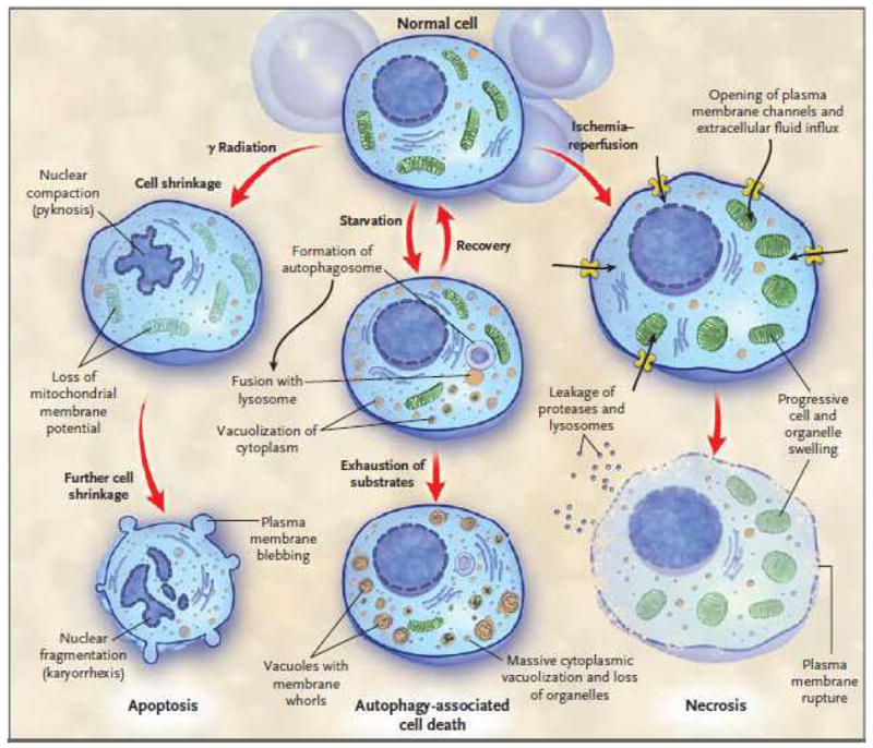
Note the characteristic differences in the 3 types of death. Depending upon the injury and the type of cell, a particular mode of cell death may predominate. Crosstalk between the different types of cell death pathways exists at multiple levels and is not shown.
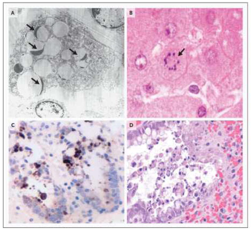
2A. Electron microscopic image of a phagocytic cell that has engulfed multiple apoptotic thymocytes. The compacted thymocyte nuclei have a classic ‘crescent shaped’ appearance, due to ‘layering’ of chromatin along the nuclear membrane. Normal appearing nuclei are present at six and one o’clock in the field of view (uranyl acetate/lead citrate; x2500). Thymic tissue section obtained from a 26 y.o. female who died following a motor vehicle accident complicated by ARDS and sepsis. 2B. A single apoptotic hepatocyte (identified by arrow) contains multiple compacted nuclear fragments indicative of apoptosis (hematoxylin and eosin [H&E]; x1000). Sample from 81 y.o. male in motor vehicle accident complicated by ventilator associated pneumonia. 2C Two adjacent crypts in colon mucosa immunohistochemically stained for cytokeratin 18 cleavage fragments (a positive reaction is brown in this color image). Cytokeratin 18 is cleaved by active caspases in both intrinsic and extrinsic apoptotic pathways. Detached cells in crypt lumens and epithelial cells still integrated into the crypt lining are positive; these cells also have classic apoptotic nuclear morphology. (see also 2D) (cytokeratin 18 immunostain [clone M30]/DAB with hematoxylin counterstain; x600). Sample from a 24 y.o. male who had aortic dissection and bowel ischemia following motor vehicle accident. 2D. A H&E stain of a colon section from a 23 y.o. patient with ischemic injury to bowel following intestinal surgery. The colonic intestinal epithelial cells show characteristic apoptotic features of nuclear compaction and fragmentation and have been sloughed into the bowel lumen (x400).
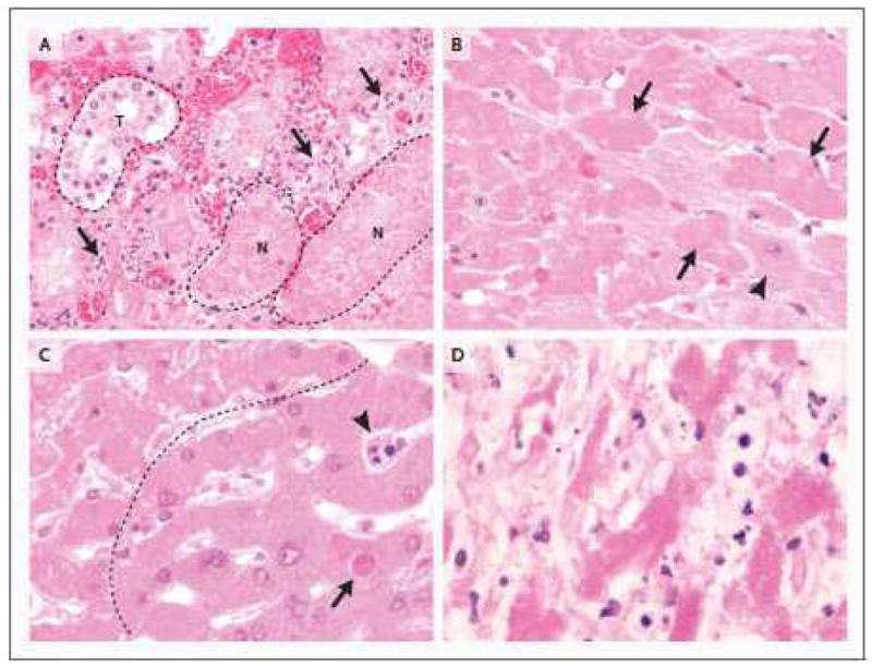
3A. The transition from viable cells to ischemic parenchyma is recapitulated in renal cortical necrosis, in this case from a 42 y.o. male who had undergone renal arterial embolization for renal cell carcinoma (not shown). Hypereosinophilia, loss of distinct nuclear detail and cytoplasmic vacuolization again characterize necrotic cells. Note the relatively intact tubule (t, dashed outline), compared with representative necrotic tubules (n; dashed outline). Note also that in the interface between viable and necrotic tubules (arrows), apoptotic cells (presumably neutrophils and mononuclear inflammatory cells) are abundant (H&E; x600). 3B. At higher magnification, the characteristic features of necrosis are apparent. Compared to viable cardiomyocytes with pale cytoplasm and distinct nuclear features (lower right of image with representative normal cell identified by arrowhead), necrotic cells are hypereosinophilic with uneven cytoplasmic vacuolization and either loss of nuclear detail or nuclear absence (identified by arrows). This morphology typifies an early manifestation of so-called ‘coagulative’ necrosis (H&E; x600). 3C. In this image, hepatocytes above and to the left of the dashed line exhibit changes of early necrosis, including vacuolization of hypereosinophilic cytoplasm and loss of nuclear detail. Heavy and narrow black arrows identify a sinusoidal inflammatory cell and a hepatocyte, respectively, with compacted and fragmented nuclei indicative of apoptosis. (H&E; x600). 53 y.o. male with pneumonia and bacteremia due to Streptococcus pneumoniae. (600X).
3D. Similar to 3C, but from an 81 y.o. male with ventilator associated pneumonia; this liver shows features typical of more advanced necrosis, with remnants of hepatocytes (eosinophilic cords) that lack obvious cell borders or recognizable nuclei. Cells and cell fragments admixed with hepatocytes are products of apoptosis; they most likely represent apoptotic lymphocytes or neutrophils in sinusoids (H&E; x600).
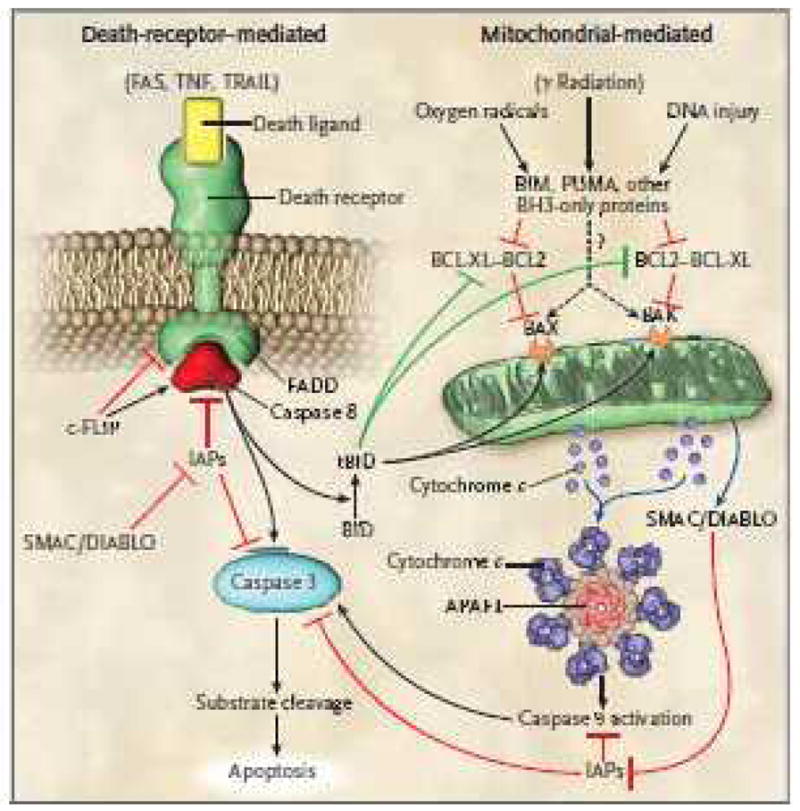
There are two major pathways of apoptosis, i.e, the death receptor pathway which is mediated by activation of death receptors and the Bcl-2 regulated mitochondrial pathway which is mediated by noxious stimuli that ultimately lead to mitochondrial injury. Ligation of death receptors recruits the adaptor protein FADD (Fas associated death domain) via its death domain. FADD in turn recruits caspase-8 which ultimately activates caspase-3, the key ‘executioner’ caspase. c-FLIP can either inhibit or potentiate binding of FADD and caspase-8 depending upon its concentration. In the intrinsic pathway, pro-apoptotic BH3 proteins are activated by noxious stimuli which interact with and inhibit anti-apoptotic Bcl-2 or Bcl-xL. Thus, Bax and Bax are free to induce mitochondrial permeabilization with release of cytochrome-C which ultimately results in activation of caspase-9 (via the apoptosome). Caspase-9 then activates caspase-3. SMAC/DIABLO is also released following mitochondrial permeabilization and acts to block the action of IAPs (inhibitors of apoptosis protein) which inhibit caspase activation. Note that there is potential cross-talk between the 2 pathways which is mediated by tBid that is produced by caspase-8 mediated Bid cleavage. tBid acts to inhibit Bcl-2/Bcl-xL and to activate Bax and Bak. There is debate about whether pro-apoptotic BH3 molecules Bim, PUMA, etc. can act directly on Bax and Bak (indicated by the question mark) to induce mitochondrial permeability or whether they only act on Bcl-2/Bcl-xL.
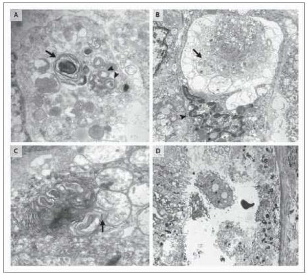
5A. Two large autophagosomes are present and identified by arrows. The autophagosome on the lower right (double arrow) contains mitochondria and other organelles in varying stages of degradation. The autophagosome in the left of the image encompasses organelle fragments with more extensive degradation (uranyl acetate/lead citrate; x40,000). Sample obtained from an 85 y.o. female with peritonitis. 5B. This cell also demonstrates extensive autophagic vacuolization with few remaining intact organelles. The arrow identifies an autophagosome containing mitochondrial fragments. Note that the cell nucleus (identified by lower arrow) has features of nuclear condensation and that it is unlikely that this cell would have remained viable - an example of ‘autophagy-associated cell death’ (uranyl acetate/lead citrate; x10,000). Sample obtained from a 73 y.o. female with urosepsis. 5C. A cell in which the autophagosomes have assumed a more complex appearance of with redundant whorls of membrane derived material. This complex lysosomal structure is juxtaposed to and focally invaginates into an adjacent mitochondrion (uranyl acetate/lead citrate;x30,000). Specimen obtained from a 73 y.o. female with urosepsis. 5D. Image of a necrotic proximal renal tubular from a 44 y.o. male who presented with acute renal failure. The patient had a history of vancomycin toxicity and cirrhosis. The cells show marked organellar and cytoplasmic swelling, loss of brush border and loss of cytoplasmic detail. (uranyl acetate/lead citrate; x5000). Figures 5A, 5B, and 5C are reproduced with permission from Laboratory Investigation (Feb 2. 2009 Epub ahead of print – Watanabe E, et al. Sepsis induces extensive vacuolization in hepatocytes: a clinical and laboratory based study.)
References
Publication types
MeSH terms
Grants and funding
LinkOut - more resources
Full Text Sources
Other Literature Sources