. 2010 Jan;84(1):188-200.
doi: 10.1128/JVI.01377-09.

Role of nuclear factor Y in stress-induced activation of the herpes simplex virus type 1 ICP0 promoter

Affiliations

Role of nuclear factor Y in stress-induced activation of the herpes simplex virus type 1 ICP0 promoter

Anna S Kushnir et al. J Virol. 2010 Jan.

Abstract

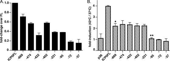
Herpesviruses are characterized by the ability to establish lifelong latent infections and to reactivate periodically, leading to recurrent disease. The herpes simplex virus type 1 (HSV-1) genome is maintained in a quiescent state in sensory neurons during latency, which is characterized by the absence of detectable viral protein synthesis. Cellular factors induced by stress may act directly on promoters within the latent viral genome to induce the transcription of viral genes and trigger reactivation. In order to identify which viral promoters are induced by stress and elucidate the cellular mechanism responsible for the induction, we generated a panel of HSV-1 promoter-luciferase constructs and measured their response to heat shock. Of the promoters tested, those of ICP0 and ICP22 were the most strongly upregulated after heat shock. Microarray analysis of lytically infected cells supported the upregulation of ICP0 and ICP22 promoters by heat shock. Mutagenic analysis of the ICP0 promoter identified two regions necessary for efficient heat-induced promoter activity, both containing predicted nuclear factor Y (NF-Y) sites, at bases -708 and -75 upstream of the transcriptional start site. While gel shift analysis confirmed NF-Y binding to both sites, only the site at -708 was important for efficient heat-induced activity. Reverse transcription-PCR analysis of selected viral transcripts in the presence of dominant-negative NF-Y confirmed the requirement for NF-Y in the induction of the ICP0 but not the ICP22 promoter by heat shock in lytically infected cells. These findings suggest that the immediate-early ICP0 gene may be among the first genes to be induced during the early events in HSV-1 reactivation, that NF-Y is important for this induction, and that other factors induce the ICP22 promoter.


Figures

FIG. 1.
FIG. 1.
Response of selected HSV-1 promoters to heat shock. (A) Viral promoter- and control-luciferase constructs used in these studies. Vero cells were transfected with the indicated HSV-1 promoter-firefly luciferase constructs in duplicate. At 24 h posttransfection, cells were heat shocked for 3 h at 43°C or maintained at 37°C (mock heat shocked). After a 4-h recovery period at 37°C, cells were harvested and subjected to the luciferase reporter assay (Promega). (B) Basal promoter activity (mock heat shocked) presented as relative light units (RLU). (C) Fold induction following heat shock. The data are presented as the fold change versus non-heat-shocked control (43°C/37°C) ± the standard error of the mean (SEM). IE genes are represented by hatched bars, E genes are represented by cross-hatched bars, DE genes are represented by small cross-hatched bars, and L genes are represented by a striped bar (n ≥ 3). The luciferase activity of all samples was at least threefold higher than the background level (mock-transfected cells).
FIG. 2.
FIG. 2.
Effect of heat shock on global HSV-1 transcription during lytic infection. Vero cells were infected with HSV-1 KOS at an MOI of 10 PFU/cell for 30 min, heat shocked for 1 h at 43°C or maintained at 37°C (mock heat shocked), and allowed to recover at 37°C for 1 h. Total RNA was then harvested, labeled, and hybridized to HSV-1 microarrays. Arrays were visualized by chemiluminescence, and images were captured with a charge-coupled device camera and processed with GEArray Expression Analysis Suite 2.0 (SABiosciences). The microarrays were standardized by using the interquartile method. The upper line represents a 1.5-fold increase, the middle line represents no change in expression, and the lower line represents a 1.5-fold decrease relative to the mock-treated control. Red stars indicate data points upregulated by >1.5-fold with treatment, black stars indicate data points not significantly affected by treatment, and green stars indicate data points downregulated by >1.5-fold compared to control. Upregulated genes of interest are indicated by circles and arrows. The figure represents three independent experiments.
FIG. 3.
FIG. 3.
Diagram of the wild-type ICP0 promoter and truncations used in the present study. Truncations of the ICP0 promoter were generated by PCR and named for the location of the truncation relative to the TSS. The full-length promoter construct is shown as ICP0FL. Predicted transcription factor binding sites are shown and the TSS is indicated by an arrow. ICP0FL represents the full-length ICP0 promoter, spanning from −800 to +150 bp relative to the TSS.
FIG. 4.
FIG. 4.
Two regions within the ICP0 promoter are important for efficient upregulation after heat shock. Truncations of the ICP0 promoter were generated by PCR. Vero cells were transiently transfected with the full-length ICP0 promoter (ICP0FL) or truncated constructs in duplicate, heat shocked or mock heat shocked, and allowed to recover. Lysates were prepared, and the luciferase activity was quantitated. (A) Basal activity of the truncated promoters relative to the full-length construct. (B) Heat-induced activity in the fold change versus the mock heat-shocked control (43°C/37°C) ± the SEM (n = 3; one-way ANOVA [P < 0.0001]; Bonferroni's multiple comparison post-test [*, P < 0.001; **, P < 0.01]; HSE induction, 242.8 ± 46.45-fold).
FIG. 5.
FIG. 5.
Fine mapping of the ICP0 promoter to identify elements important for upregulation after heat shock. Truncations in the 5′- and TSS-proximal regions of the ICP0 promoter were generated by PCR. Vero cells were transiently transfected with ICP0FL or truncated constructs in duplicate, heat shocked or mock heat shocked, and allowed to recover. Lysates were prepared, and luciferase activity was quantitated. 5′ truncations are indicated as follows: basal activity of the promoters relative to the full-length construct (A) and heat-induced activity in the fold change versus the mock heat-shocked control (43 or 37°C) ± the SEM (B) (n = 3; one-way ANOVA [P = 0.0002]; Bonferroni's multiple comparison post-test [*, P = 0.05]; HSE induction, 257.5 ± 19.27-fold). TSS-proximal truncations are indicated as follows: basal activity of the promoters relative to the full-length construct (C) and Heat-induced activity in the fold change versus the mock heat-shocked control (43 and 37°C) ± the SEM (D) (n = 3; one-way ANOVA [P = 0.0001]; Bonferroni's multiple comparison post-test [*, P < 0.01]; HSE induction, 328.1 ± 32.17-fold).
FIG. 6.
FIG. 6.
NF-Y associates with the ICP0 promoter at −708 and is important for efficient promoter upregulation after heat shock. (A) Gel shift analysis of NF-Y association with the ICP0 promoter. Vero cell nuclear extracts were incubated with αNF-YA antibody (Santa Cruz) for 1 h on ice. Either wild-type (WT) or mutant 32P-labeled probe was then added, and the binding reaction was allowed to proceed for 30 min on ice. Protein-DNA complexes were resolved on a 4% polyacrylamide gel in 0.5× TBE. The locations of the NF-Y and supershifted complexes and of the unbound probe are indicated by arrows. Lanes: 1, consensus NF-Y probe (Con); 2, consensus probe with NF-YA antibody; 3, wild-type −708 ICP0 probe; 4, wild-type −708 ICP0 probe with NF-YA antibody; 5, −708NFYmut1 probe; 6, −708NFYmut2 probe; 7, −708mut1 probe with NF-YA antibody; 8, specific (S) cold competitor; 9, nonspecific (NS) cold competitor; 10, no nuclear extract; NS, nonspecific band. (B and C) Luciferase assays for ICP0 promoter activity. Vero cells were transiently transfected with the wild-type (ICP0FL and ICP0pAlter) or mutated constructs in duplicate, heat shocked or mock heat-shocked, and allowed to recover. Lysates were prepared, and the luciferase activity was quantitated. (B) Basal activity of the promoters relative to the wild-type construct. (C) Heat-induced activity in the fold change versus the mock heat-shocked control (43 and 37°C) ± the SEM (n = 3; *, one-way ANOVA [P = 0.0005]; HSE induction, 543.7 ± 59.77-fold).
FIG. 7.
FIG. 7.
NF-Y associates with the ICP0 promoter at −75 but is not required for efficient promoter upregulation after heat shock. (A) Gel shift analysis of NF-Y association with the ICP0 promoter. Vero cell nuclear extracts were incubated with αNF-YA antibody (Santa Cruz) for 1 h on ice. Either wild-type (WT) or mutant ICP0 32P-labeled probe was then added to the binding reaction. Binding was allowed to proceed for 30 min on ice. Protein-DNA complexes were resolved on a 4% polyacrylamide gel in 0.5× TBE. The locations of the NF-Y and supershifted complexes are indicated by arrows. Lanes: 1, consensus NF-Y probe (Con); 2, consensus probe with NF-YA antibody; 3, wild-type −75 ICP0 probe; 4, wild-type −75 ICP0 probe with NF-YA antibody; 5, −75NFYmut1 probe; 6, −75NFYmut2 probe; 7, specific (S) cold competitor; 8, nonspecific (NS) cold competitor; 9, no nuclear extract; NS, nonspecific band. (B and C) Luciferase assays for ICP0 promoter activity. Vero cells were transiently transfected with the wild-type (ICP0FL and ICP0pAlter) or mutated constructs in duplicate, heat shocked or mock heat shocked, and allowed to recover. Lysates were prepared, and the luciferase activity was quantitated. (B) Basal activity of the promoters relative to the wild-type construct. (C) Heat-induced activity in the fold change over mock heat-shocked control (43 and 37°C) ± the SEM (HSE induction, 514 ± 51.66-fold; n = 4).
FIG. 8.
FIG. 8.
NF-Y activity is necessary for induction of the ICP0 promoter after heat shock during lytic infection. Vero cells were infected with wild-type, DN NF-YA, or GFP adenoviruses at an MOI of 25 PFU/cell and superinfected with HSV-1 KOS at an MOI of 1 PFU/cell for 30 min, heat shocked for 1 h at 43°C, and allowed to recover for 1 h at 37°C, and the RNA was harvested for RT-PCR. PCRs were performed in triplicate with primers specific for ICP0, ICP22, and β-actin. Reactions were standardized to β-actin. ICP0 (A) and ICP22 (B) transcript levels. The results are presented as the fold induction of transcription in response to heat shock (43 or 37°C) ± the SEM (n = 3).

References

    1. Ackland-Berglund, C. E., D. J. Davido, and D. A. Leib. 1995. The roles of the cAMP-response element and TATA box in expression of the herpes simplex virus type 1 latency-associated transcripts. Virology 210:141-151. - PubMed
    1. Andrews, N. C., and D. V. Faller. 1991. A rapid micropreparation technique for extraction of DNA-binding proteins from limiting numbers of mammalian cells. Nucleic Acids Res. 19:2499. - PMC - PubMed
    1. Ashburner, M., and J. J. Bonner. 1979. The induction of gene activity in Drosophila by heat shock. Cell 17:241-254. - PubMed
    1. Bi, W., L. Wu, F. Coustry, B. de Crombrugghe, and S. N. Maity. 1997. DNA binding specificity of the CCAAT-binding factor CBF/NF-Y. J. Biol. Chem. 272:26562-26572. - PubMed
    1. Cai, W., T. L. Astor, L. M. Liptak, C. Cho, D. M. Coen, and P. A. Schaffer. 1993. The herpes simplex virus type 1 regulatory protein ICP0 enhances virus replication during acute infection and reactivation from latency. J. Virol. 67:7501-7512. - PMC - PubMed

Publication types

Substances

LinkOut - more resources