

Chronic wasting disease (CWD) susceptibility of several North American rodents that are sympatric with cervid CWD epidemics
- PMID: 19828611
- PMCID: PMC2798418
- DOI: 10.1128/JVI.00560-09
Chronic wasting disease (CWD) susceptibility of several North American rodents that are sympatric with cervid CWD epidemics
Abstract
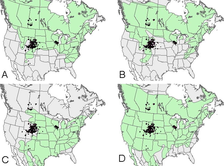
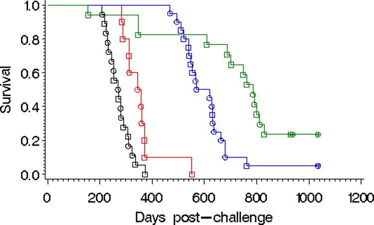
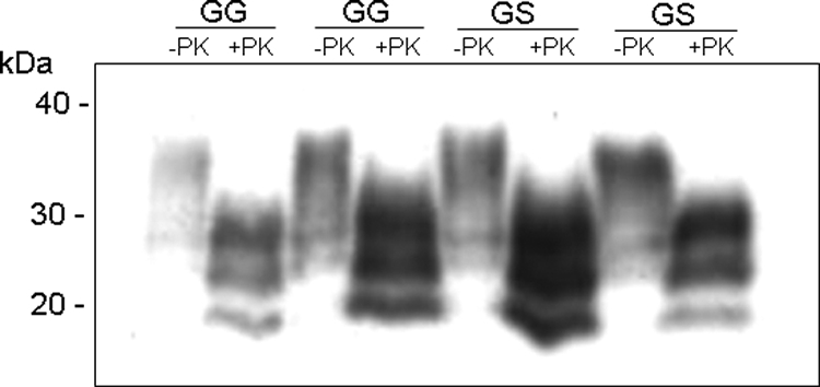
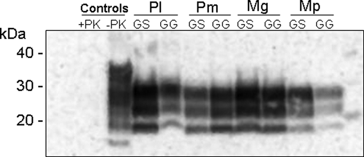
Chronic wasting disease (CWD) is a highly contagious always fatal neurodegenerative disease that is currently known to naturally infect only species of the deer family, Cervidae. CWD epidemics are occurring in free-ranging cervids at several locations in North America, and other wildlife species are certainly being exposed to infectious material. To assess the potential for transmission, we intracerebrally inoculated four species of epidemic-sympatric rodents with CWD. Transmission was efficient in all species; the onset of disease was faster in the two vole species than the two Peromyscus spp. The results for inocula prepared from CWD-positive deer with or without CWD-resistant genotypes were similar. Survival times were substantially shortened upon second passage, demonstrating adaptation. Unlike all other known prion protein sequences for cricetid rodents that possess asparagine at position 170, our red-backed voles expressed serine and refute previous suggestions that a serine in this position substantially reduces susceptibility to CWD. Given the scavenging habits of these rodent species, the apparent persistence of CWD prions in the environment, and the inevitable exposure of these rodents to CWD prions, our intracerebral challenge results indicate that further investigation of the possibility of natural transmission is warranted.
Figures
Similar articles
-
Chronic wasting disease of cervids.Curr Top Microbiol Immunol. 2004;284:193-214. doi: 10.1007/978-3-662-08441-0_8. Curr Top Microbiol Immunol. 2004. PMID: 15148993 Review.
-
Studies in bank voles reveal strain differences between chronic wasting disease prions from Norway and North America.Proc Natl Acad Sci U S A. 2020 Dec 8;117(49):31417-31426. doi: 10.1073/pnas.2013237117. Epub 2020 Nov 23. Proc Natl Acad Sci U S A. 2020. PMID: 33229531 Free PMC article.
-
Deer Prion Proteins Modulate the Emergence and Adaptation of Chronic Wasting Disease Strains.J Virol. 2015 Dec;89(24):12362-73. doi: 10.1128/JVI.02010-15. Epub 2015 Sep 30. J Virol. 2015. PMID: 26423950 Free PMC article.
-
Alteration of the chronic wasting disease species barrier by in vitro prion amplification.J Virol. 2011 Sep;85(17):8528-37. doi: 10.1128/JVI.00809-11. Epub 2011 Jun 22. J Virol. 2011. PMID: 21697475 Free PMC article.
-
The role of genetics in chronic wasting disease of North American cervids.Prion. 2012 Apr-Jun;6(2):153-62. doi: 10.4161/pri.19640. Epub 2012 Apr 1. Prion. 2012. PMID: 22460693 Free PMC article. Review.
Cited by
-
Occurrence, transmission, and zoonotic potential of chronic wasting disease.Emerg Infect Dis. 2012 Mar;18(3):369-76. doi: 10.3201/eid1803.110685. Emerg Infect Dis. 2012. PMID: 22377159 Free PMC article. Review.
-
Spontaneous generation of rapidly transmissible prions in transgenic mice expressing wild-type bank vole prion protein.Proc Natl Acad Sci U S A. 2012 Feb 28;109(9):3498-503. doi: 10.1073/pnas.1121556109. Epub 2012 Feb 13. Proc Natl Acad Sci U S A. 2012. PMID: 22331873 Free PMC article.
-
In vitro prion protein conversion suggests risk of bighorn sheep (Ovis canadensis) to transmissible spongiform encephalopathies.BMC Vet Res. 2013 Aug 9;9:157. doi: 10.1186/1746-6148-9-157. BMC Vet Res. 2013. PMID: 23938169 Free PMC article.
-
Is the prevalent human prion protein 129M/V mutation a living fossil from a Paleolithic panzootic superprion pandemic?Prion. 2014 Jan-Feb;8(1):2-10. doi: 10.4161/pri.27601. Prion. 2014. PMID: 24398570 Free PMC article.
-
Review on PRNP genetics and susceptibility to chronic wasting disease of Cervidae.Vet Res. 2021 Oct 7;52(1):128. doi: 10.1186/s13567-021-00993-z. Vet Res. 2021. PMID: 34620247 Free PMC article. Review.
References
-
- Agrimi, U., R. Nonno, G. Dell'Omo, M. A. Di Bari, M. Conte, B. Chiappini, E. Esposito, G. Di Guardo, O. Windl, G. Vaccari, and H. P. Lipp. 2008. Prion protein amino acid determinants of differential susceptibility and molecular feature of prion strains in mice and voles. PLoS Pathog. 4:e1000113. - PMC - PubMed
-
- Agrimi, U., R. Nonno, M. A. Di Bari, P. Fazzi, M. Conte, P. Frassanito, S. Simson, C. Parisi, and G. Vaccari. 2006. Efficient transmission and characterization of chronic wasting disease in bank voles. Prion2006, Turin, Italy. http://www.neuroprion.org/resources/pdf_docs/conferences/prion2006/abstr..., p.246.
-
- Baeten, L. A., B. E. Powers, J. E. Jewell, T. R. Spraker, and M. W. Miller. 2007. A natural case of chronic wasting disease in a free-ranging moose (Alces alces shirasi). J. Wildl. Dis. 43:309-314. - PubMed
-
- Bartz, J. C., R. F. Marsh, D. I. McKenzie, and J. M. Aiken. 1998. The host range of chronic wasting disease is altered upon passage in ferrets. Virology 251:297-301. - PubMed
-
- Bruce, M., A. Chree, E. S. Williams, and H. Fraser. 2000. Perivascular PrP amyloid in the brains of mice infected with chronic wasting disease. Brain Pathol. 10:662-663 (Abstr. C32-08.)
MeSH terms
Substances
LinkOut - more resources
Full Text Sources
Other Literature Sources
Miscellaneous
