. 2010 Jan;84(1):227-42.
doi: 10.1128/JVI.00481-09.

Interactions and oligomerization of hantavirus glycoproteins

Affiliations

Interactions and oligomerization of hantavirus glycoproteins

Jussi Hepojoki et al. J Virol. 2010 Jan.

Abstract

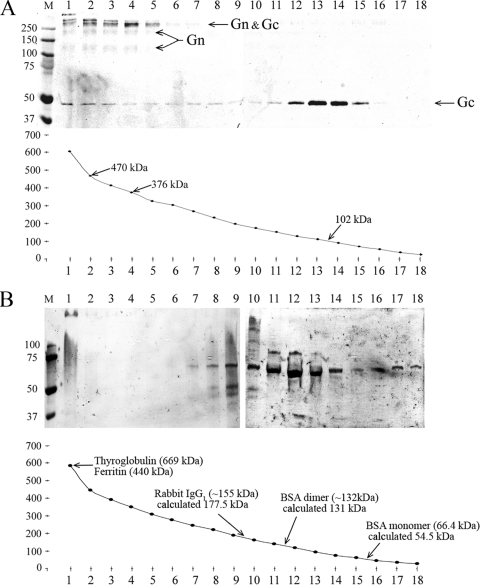
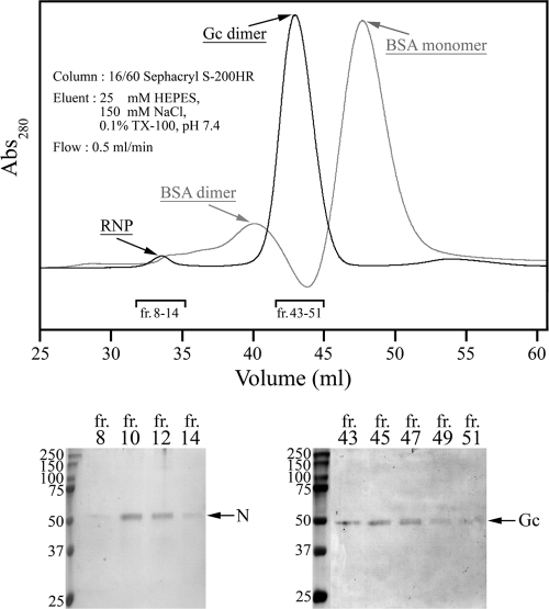
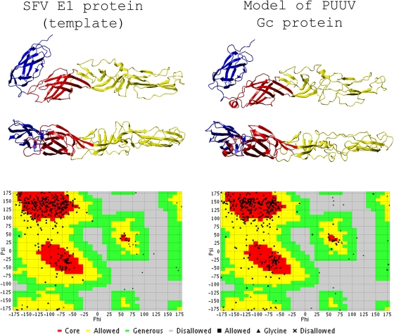
In this report the basis for the structural architecture of the envelope of hantaviruses, family Bunyaviridae, is systematically studied by the interactions of two glycoproteins N and C (Gn and Gc, respectively) and their respective disulfide bridge-mediated homo- and heteromeric oligomerizations. In virion extracts Gn and Gc associated in both homo- and hetero-oligomers which were, at least partially, thiol bridge mediated. Due to strong homo-oligomerization, the hetero-oligomers of Gn and Gc are likely to be mediated by homo-oligomeric subunits. A reversible pH-induced disappearance of a neutralizing epitope in Gc and dissociation of the Gn-Gc complex at pH values below 6.2 provide proteochemical evidence for the fusogenicity of Gc. Incomplete inactivation of virions at acidic pH indicates that additional factors are required for hantavirus fusion, as in the case of pestiviruses of the Flaviviridae. Based on similarities to class II fusion proteins, a structure model was created of hantavirus Gc using the Semliki Forest virus E1 protein as a template. In total, 10 binding regions for Gn were found by peptide scanning, of which five represent homotypic (Gn(I) to Gn(V)) and five represent heterotypic (Gc(I) to Gc(V)) interaction sites that we assign as intra- and interspike connections, respectively. In conclusion, the glycoprotein associations were compiled to a model wherein the surface of hantaviruses is formed of homotetrameric Gn complexes interconnected with Gc homodimers. This organization would create the grid-like surface pattern described earlier for hantaviruses in negatively stained electron microscopy specimens.


Figures

FIG. 1.
FIG. 1.
Effect of low-pH treatment on PUUV Gn-Gc complex in immunoprecipitation of virion lysates. (A) Gn-specific immunoprecipitation with MAb 5A2. Autoradiography of MAb 5A2-Sepharose-bound, [35S]cysteine-labeled Gn and Gc after washing at indicated pH values. The control lane C represents background binding to protein G Sepharose at pH 8.0 without MAb. SDS-PAGE was run under reducing conditions with 2-mercaptoethanol as the reducing agent. The band labeled (Gn)n may also contain Gc. (B) Loss of PUUV Gc recognition by Gc-specific MAb 4G2. Gc in nonlabeled PUUV lysate was bound to MAb 4G2-Sepharose in buffers at the indicated pH. Proteins were separated by reducing SDS-PAGE and immunoblotted with anti-Gc serum. (C) MAb 4G2 binding to Gc prevents dissociation at low pH. Nonlabeled PUUV lysate was bound to MAb 4G2-Sepharose at pH 8.0 and washed in buffers at the indicated pH. The material remaining bound was separated by reducing SDS-PAGE and immunoblotted with anti-Gc serum. C1 and C2 represent controls; C1 is a virus preparation used in immunoprecipitation, and C2 is MAb 4G2-Sepharose without addition of virus. IgG(γ) (∼50 kDa) and IgG(κ/λ) (25 kDa; migrating at front) chains are indicated. (D) The low-pH-induced dissociation of Gn-Gc complex is reversible. Lysates of metabolically ([35S]cysteine) labeled PUUV treated at pH 5.8 or kept at pH 8.0, as indicated, were immunoprecipitated with Gc-specific MAbs 4G2 and 1C9 at pH 8.0. Autoradiography of SDS-PAGE-separated proteins and the control (lane C) was performed as described for panel A. The protein mobility estimation in panels A and D was done using Precision Plus protein standards (Bio-Rad). The band labeled (Gn)n may also contain Gc.
FIG. 2.
FIG. 2.
Effect of low-pH treatment on the infectivity of TULV virions. (A) Titers of TULV virions subjected to low pH followed by pH adjustment to pH 8.0 (right bars) or left unadjusted (left bars) in FFU/ml determined by 10-fold serial dilutions. The error bars represent ± standard deviations of two parallel samples in two 10-fold dilutions. (B) The virus titers in panel A as relative titers to titers determined at pH 7.5. (C) Table of the titers (FFU/ml) and relative titers (%) in graphs in panels A and B.
FIG. 3.
FIG. 3.
Cross-linking of the surface of TULV virions with membrane-impermeable cross-linker. Native TULV virions, treated with three concentrations of membrane-impermeable cleavable DTSSP linker, were solubilized in Laemmli sample buffer, and proteins were separated by 6% SDS-PAGE under both reducing and nonreducing conditions (samples not boiled). (A) Silver staining (PageSilver Silver Staining Kit; Fermentas) shows bands resolved upon exposure to short (left panel) and long (right panel) periods of staining. (B) Detection by immunoblotting shows Gn (left panel) overlaid with Gc (right panel) as recorded in an Odyssey infrared imager with goat anti-rabbit IR800CW-conjugated secondary antibody. In both panels A and B the nucleocapsid protein (N) is indicated by a plus sign, and tetrameric Gn complex is indicated by an asterisk. ox., oxidized; red., reduced.
FIG. 4.
FIG. 4.
Glycoprotein complexes of TULV formed under reducing and nonreducing conditions and under separation by SDS-PAGE (samples boiled in panels A, B, and C). (A) Glycoprotein mobilities of TULV pelleted through sucrose cushion, treated with the reductant (TCEP or DTT) or left untreated, and separated by SDS-PAGE. The composite immunoblots of Gc and Gn shown were overlaid as indicated. (B) TULV concentrated by ultracentrifugation was oxidized with 10 mM CuCl2 or thiol acetylated with NEM either with or without detergent TX-100 as indicated, and proteins were separated by SDS-PAGE without reductant as shown. Gc- and Gn-positive bands of ∼110 kDa and ∼200 kDa are indicated by asterisks. An enlargement of the region of >250 kDa from -SH (where SH is sulfhydryl) oxidized samples is shown as insert from the Gc immunoblot, highlighting novel bands positive for both Gn and Gc that appeared after CuCl2 treatment. (C) The virions after treatments, as in panel B, were reduced by the addition of 2 mM TCEP prior to SDS-PAGE. The composite immunoblots in panels B and C show Gc (left) overlaid with Gn (right). The images were recorded in an Odyssey infrared imaging system with goat anti-rabbit IR800CW-conjugated secondary antibody. The anti-Gc and anti-Gn serum reactivities were overlaid in PhotoShop CS using protein markers (Precision Plus Protein Standards; Bio-Rad) visible in both detections.
FIG. 5.
FIG. 5.
Complexes of TULV Gn and Gc after sedimentation in a sucrose density gradient by ultracentrifugation. (A) Glycoproteins extracted from concentrated TULV with nonionic detergent (TX-100) were separated by sedimentation in a 0 to 21% sucrose gradient (in 25 mM HEPES, 100 mM NaCl, 0.1% TX-100). The fractions from ultracentrifugation (40,000 rpm at +5°C for 20.5 h in an SW41 rotor) were collected dropwise from the tube bottom and are shown on the x axis (1 to 18). The nonreducing SDS-PAGE gel, immunoblotted with anti-Gn and anti-Gc sera, represents 100 μl of each fraction, and the molecular mass approximations corresponding to the peak fractions of Gc and Gn-Gc complexes calculated from refractivity of sucrose concentration in each fraction are as shown. (B) Molecular mass markers as indicated were sedimented and fractionated; fractions were analyzed by Coomassie-stained SDS-PAGE, and molecular sizes were calculated from sucrose concentration as in panel A. The experimental molecular masses in the peak fraction are shown with the values reported elsewhere.
FIG. 6.
FIG. 6.
Complexes of TULV Gn and Gc analyzed by gel filtration. Proteins extracted from TULV virions with detergent (TX-100), as in sedimentation analysis, were separated in Sephacryl S-200HR chromatography. Elution of viral proteins detected by nonreducing SDS-PAGE and Coomassie blue staining of peak fractions is shown below, as indicated in the chromatogram. Detection of ribonucleoprotein is based on the presence of nucleocapsid protein (N) in fractions 8 to 14 (bottom left). An overlay of the virion sample (black) and BSA marker (gray) in an arbitrary 280-nm absorbance scale is presented to allow comparison of the elution volumes. fr, fraction.
FIG. 7.
FIG. 7.
3D structure model of PUUV Gc with SFV E1 as a template. The Swiss-Model-generated PDB coordinate file of PUUV Gc and the SFV E1 used as a template (PBD code, 2ALA) (45) were visualized with YASARA View. The model and template were superimposed, and the images shown were captured in the same respective angles. DI, DII, and DIII are shown in red, yellow, and blue, respectively, as suggested by Rey and collaborators (41). The PDB coordinates of the created Gc model were analyzed using VADAR, version 1.5, and the Ramachandran plots of the PUUV Gc model and the SFV E1 template obtained from the VADAR server (http://redpoll.pharmacy.ualberta.ca/vadar) are presented below for the respective structures.
FIG. 8.
FIG. 8.
PUUV Gn-Gc interaction sites by peptide scanning. (A) Fine-mapping of the Gn-Gc interaction sites by peptide scanning of the glycoprotein precursor of PUUV with 18-mer peptides in a shift of 3 amino acids (spot method). PUUV lysate in the protein overlay assay was monitored for binding of Gn (middle) and Gc (right) to peptides on the membrane. In spot assay detection, MAbs 5A2 and 4G2 were used at concentrations at which they did not give unspecific binding in ECL signals on X-ray film (left). The interaction sites are indicated in the membranes, and the corresponding peptide sequences are listed. An illustration of the Gn binding sites in Gc (model) on a solvent-accessible surface structure is shown on the right. The coloring of interaction sites is the same as in the table of sequences, and the putative fusion peptide of PUUV Gc protein is shown in cyan (13, 55). (B) Hypothetical dimer of Gc (model), aligned such that the known epitopes of Gc-specific MAb 4G2 form a uniform surface. The docked dimer is shown as a solvent-accessible surface, the Gc molecules in gray highlight binding sites of MAb 4G2 (16) in cyan, and the mapped Gn interaction sites are shown as a monomer structure as in panel A. Docking of two Gc molecules in this fashion interestingly resembles the reported homodimer of TBEV E protein (41).
FIG. 9.
FIG. 9.
Cryo-EM image of TULV particles. (A) Summary of glycoprotein complexes detected using different methods. The first row shows the estimated molecular mass of each species in kDa; the lower value represents the molecular mass observed by nonreducing SDS-PAGE, and the higher value is calculated from primary sequence. Estimations for oligomeric complexes were calculated by multiplying the molecular mass of monomeric units. The other rows show the molecular masses of observed complexes. (B) Electron cryo-micrograph of TULV virions taken at approximately 3 μm under focus. The box indicates an area where the spikes are clearly visible projecting from the surface of the virion. An arrow in the magnified inset of the same area highlights an apparently symmetrical, dimeric spike. Bar, 300 nm. (C) Hypothetical surface arrangement of the glycoproteins in hantavirus particles. The illustration shows the three possible glycoprotein compositions seen in the plane of the viral membrane, where they produce the spike structures highlighted in panel A. The surface architecture of the virion was generated based on the proposed arrangement of Gn and Gc (oriented perpendicular to virus surface). The described interactions favor hypothesis 3, and the spike is proposed to be formed of two or four Gn protein units, forming a grid-like pattern as in negatively stained specimens (31).

References

    1. Adrian, M., J. Dubochet, J. Lepault, and A. W. McDowall. 1984. Cryo-electron microscopy of viruses. Nature 308:32-36. - PubMed
    1. Arikawa, J., A. L. Schmaljohn, J. M. Dalrymple, and C. S. Schmaljohn. 1989. Characterization of Hantaan virus envelope glycoprotein antigenic determinants defined by monoclonal antibodies. J. Gen. Virol. 70:615-624. - PubMed
    1. Arnold, K., L. Bordoli, J. Kopp, and T. Schwede. 2006. The SWISS-MODEL workspace: a web-based environment for protein structure homology modelling. Bioinformatics 22:195-201. - PubMed
    1. Beniac, D. R., A. Andonov, E. Grudeski, and T. F. Booth. 2006. Architecture of the SARS coronavirus prefusion spike. Nat. Struct. Mol. Biol. 13:751-752. - PMC - PubMed
    1. Delmas, B., and H. Laude. 1990. Assembly of coronavirus spike protein into trimers and its role in epitope expression. J. Virol. 64:5367-5375. - PMC - PubMed

Publication types

LinkOut - more resources