. 2010 Jan;84(1):558-64.
doi: 10.1128/JVI.01785-09.

The effects of influenza A virus PB1-F2 protein on polymerase activity are strain specific and do not impact pathogenesis

Affiliations

The effects of influenza A virus PB1-F2 protein on polymerase activity are strain specific and do not impact pathogenesis

Julie L McAuley et al. J Virol. 2010 Jan.

Abstract

The influenza A virus PB1-F2 protein has been implicated as a virulence factor, but the mechanism by which it enhances pathogenicity is not understood. The PB1 gene segment of the H1N1 swine-origin influenza virus pandemic strain codes for a truncated PB1-F2 protein which terminates after 11 amino acids but could acquire the full-length form by mutation or reassortment. It is therefore important to understand the function and impact of this protein. We systematically assessed the effect that PB1-F2 expression has on viral polymerase activity, accumulation and localization of PB1, and replication in vitro and in mice. We used both the laboratory strain PR8 and a set of viruses engineered to study clinically relevant PB1-F2 proteins. PB1-F2 expression had modest effects on polymerase activity, PB1 accumulation, and replication that were cell type and virus strain dependent. Disruption of the PB1-F2 reading frame in a recent, seasonal H3N2 influenza virus strain did not affect these parameters, suggesting that this is not a universal function of the protein. Disruption of PB1-F2 expression in several backgrounds or expression of PB1-F2 from the 1918 pandemic strain or a 1956 H1N1 strain had no effect on viral lung loads in mice. Alternate mechanisms besides alterations to replication are likely responsible for the enhanced virulence in mammalian hosts attributed to PB1-F2 in previous studies.


Figures
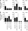
FIG. 1.
FIG. 1.
Effect of PB1-F2 on polymerase activity is virus and cell type specific. Using a luciferase dual-reporter system to examine polymerase activity in vitro, plasmids expressing the PB1 gene segment from PR8, A/Wuhan/359/95 (H3N2), or A/Vietnam/1203/04 (H5N1) were transfected into 293T cells (A) or A549 cells (B), together with PB2, PA, and NP plasmids from PR8 (/PR8) or A/Wuhan/359/95 (/H3N2). Results are the mean activities ± standard deviations (SD) for four to six independent experiments, normalized to transfection efficiency and to the activity of the minigenome containing the WT PB1 within each experiment. An asterisk indicates a significant difference (P < 0.05) compared to all other values within that experiment.
FIG. 2.
FIG. 2.
Full-length PB1-F2 protein colocalizes with PB1. MDCK cell monolayers were infected for 24 h with viruses expressing the WT PR8 PB1 or a PR8 PB1 modified so that PB1-F2 is not expressed (PR8 ΔPB1-F2) or the PB1-F2 from A/Brevig Mission/1/18 (1918 PB1-F2/PR8) or A/Beijing/11/56 (Beij PB1-F2/PR8) is expressed instead. (A) Using confocal microscopy and antibodies specific for PB1 and PB1-F2, the intersection of PB1 and PB1-F2 events was calculated to determine the frequency of colocalization, expressed as the mean % ± SD. (B) Colocalization can be pictured visually in representative fields with the PR8 and 1918 PB1-F2 proteins, but not the Beij PB1-F2 protein. (C) Distributions of PB1-F2 proteins from WT PR8 and 1918 PB1-F2/PR8 viruses are predominantly cytoplasmic with mitochondrial localization, while the truncated Beij PB1-F2, which lacks the C-terminal mitochondrial targeting sequence, is found diffusely throughout the cell. An asterisk indicates a significant difference (P < 0.05) by ANOVA compared to the no-virus control, the PR8 ΔPB1-F2 virus, and the Beij PB1-F2/PR8 virus.
FIG. 3.
FIG. 3.
Expression and distribution of PB1 change over time. MDCK cell monolayers were infected for 2 to 24 h with viruses expressing the WT PR8 PB1 or a PR8 PB1 modified so that PB1-F2 is not expressed (PR8 ΔPB1-F2) or the PB1-F2 from A/Brevig Mission/1/18 (1918 PB1-F2/PR8) or A/Beijing/11/56 (Beij PB1-F2/PR8) is expressed instead. Using confocal microscopy and an antibody specific for PB1, the sum intensities normalized to the 2-h intensity for WT PR8 throughout the cells (A), in the nucleus (B), and in the cytoplasm (C) were determined at 2, 4, 6, 8, and 24 h postinfection. Five fields containing ∼200 cells each were examined at each time point. Due to the large number of measurements, the SD is so small as to not be visible on the graph at this scale. (D) Representative fields for each virus and time point are shown.
FIG. 4.
FIG. 4.
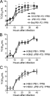
Minimal changes in replication are mediated by PB1-F2 expression. MDCK cell monolayers were infected at an MOI of 0.001 for 48 h with 7:1 recombinant viruses in a seven-gene PR8 background expressing the WT PR8 PB1 (A), PB1 from A/Wuhan/359/95 (H3N2) (B), PB1 from A/Vietnam/1203/04 (H5N1) (C), or PB1 gene segments modified so that PB1-F2 is not expressed (PR8 ΔPB1-F2, H3N2 ΔPB1-F2/PR8, and H5N1 ΔPB1-F2/PR8) or the PB1-F2 from A/Brevig Mission/1/18 (1918 PB1-F2/PR8) or A/Beijing/11/56 (Beij PB1-F2/PR8) is expressed instead. Mean viral titers ± SD at 4, 8, 12, 16, 20, 24, 28, 36, and 48 h were determined in MDCK cells and expressed as log10 TCID50/ml supernatant. An asterisk indicates a significant difference (P < 0.05) by ANOVA for PR8 at that time point compared to the ΔPB1-F2/PR8 and Beij PB1-F2/PR8 viruses.
FIG. 5.
FIG. 5.
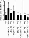
Plaque sizes differ with different PB1-F2 proteins. MDCK cell monolayers overlaid with soft agar were infected for 72 h with 7:1 recombinant viruses in a seven-gene PR8 background expressing the WT PR8 PB1, PB1 from A/Wuhan/359/95 (H3N2), PB1 from A/Vietnam/1203/04 (H5N1), or PB1 gene segments modified so that PB1-F2 is not expressed (PR8 ΔPB1-F2, H3N2 ΔPB1-F2/PR8, and H5N1 ΔPB1-F2/PR8) or the PB1-F2 from A/Brevig Mission/1/18 (1918 PB1-F2/PR8) or A/Beijing/11/56 (Beij PB1-F2/PR8) is expressed instead. The mean plaque area ± SD was calculated from the observed diameters of at least 50 plaques per virus. An asterisk indicates a significant difference (P < 0.05) by ANOVA compared to the other viruses in that grouping.
FIG. 6.
FIG. 6.
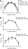
PB1-F2 expression does not affect viral lung loads in mice. Mice were infected with 7:1 recombinant viruses in a seven-gene PR8 background expressing the WT PR8 PB1 (A), PB1 from A/Wuhan/359/95 (H3N2) (B), PB1 from A/Vietnam/1203/04 (H5N1) (C), or PB1 gene segments modified so that PB1-F2 is not expressed (PR8 ΔPB1-F2, H3N2 ΔPB1-F2/PR8, and H5N1 ΔPB1-F2/PR8) or the PB1-F2 from A/Brevig Mission/1/18 (1918 PB1-F2/PR8) or A/Beijing/11/56 (Beij PB1-F2/PR8) is expressed instead. On days 1, 3, 5, 7, and 9 postinfection, lungs were removed from four mice per group, and the mean viral lung load ± SD was calculated by determination of the log10 TCID50 on MDCK cells.

References

    1. Chanturiya, A. N., G. Basanez, U. Schubert, P. Henklein, J. W. Yewdell, and J. Zimmerberg. 2004. PB1-F2, an influenza A virus-encoded proapoptotic mitochondrial protein, creates variably sized pores in planar lipid membranes. J. Virol. 78:6304-6312. - PMC - PubMed
    1. Chen, W., P. A. Calvo, D. Malide, J. Gibbs, U. Schubert, I. Bacik, S. Basta, R. O'Neill, J. Schickli, P. Palese, P. Henklein, J. R. Bennink, and J. W. Yewdell. 2001. A novel influenza A virus mitochondrial protein that induces cell death. Nat. Med. 7:1306-1312. - PubMed
    1. Conenello, G. M., D. Zamarin, L. A. Perrone, T. Tumpey, and P. Palese. 2007. A single mutation in the PB1-F2 of H5N1 (HK/97) and 1918 influenza A viruses contributes to increased virulence. PLoS Pathog. 3:1414-1421. - PMC - PubMed
    1. Dawood, F. S., S. Jain, L. Finelli, M. W. Shaw, S. Lindstrom, R. J. Garten, L. V. Gubareva, X. Xu, C. B. Bridges, and T. M. Uyeki. 2009. Emergence of a novel swine-origin influenza A (H1N1) virus in humans. N. Engl. J. Med. 360:2605-2615. - PubMed
    1. Gibbs, J. S., D. Malide, F. Hornung, J. R. Bennink, and J. W. Yewdell. 2003. The influenza A virus PB1-F2 protein targets the inner mitochondrial membrane via a predicted basic amphipathic helix that disrupts mitochondrial function. J. Virol. 77:7214-7224. - PMC - PubMed

Publication types

Substances

LinkOut - more resources