. 2010 Jan;84(1):503-15.
doi: 10.1128/JVI.01134-09.

Targeting of murine leukemia virus gag to the plasma membrane is mediated by PI(4,5)P2/PS and a polybasic region in the matrix

Affiliations

Targeting of murine leukemia virus gag to the plasma membrane is mediated by PI(4,5)P2/PS and a polybasic region in the matrix

E Hamard-Peron et al. J Virol. 2010 Jan.

Abstract

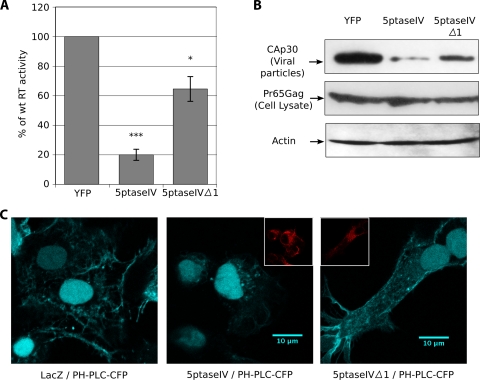
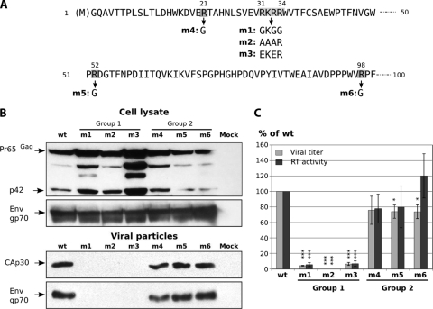
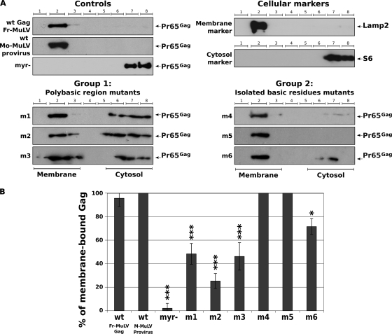
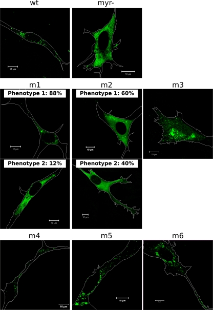
Membrane targeting of the human immunodeficiency virus Gag proteins is dependent on phosphatidylinositol-(4,5)-bisphosphate [PI(4,5)P(2)] located in the plasma membrane. In order to determine if evolutionarily distant retroviral Gag proteins are targeted by a similar mechanism, we generated mutants of the matrix (MA) domain of murine leukemia virus (MuLV) Gag, examined their binding to membrane models in vitro, and analyzed their phenotypes in cell culture. In vitro, we showed that MA bound all the phosphatidylinositol phosphates with significant affinity but displayed a strong specificity for PI(4,5)P(2) only if enhanced by phosphatidylserine. Mutations in the polybasic region in MA dramatically reduced this affinity. In cells, virus production was strongly impaired by PI(4,5)P(2) depletion under conditions of 5ptaseIV overexpression, and mutations in the MA polybasic region altered Gag localization, membrane binding, and virion production. Our results suggest that the N-terminal polybasic cluster of MA is essential for Gag targeting to the plasma membrane. The binding of the MA domain to PI(4,5)P(2) appears to be a conserved feature among retroviruses despite the fact that the MuLV-MA domain is structurally different from that of human immunodeficiency virus types 1 and 2 and lacks a readily identifiable PI(4,5)P(2) binding cleft.


Figures

FIG. 1.
FIG. 1.
Binding of recombinant MoMuLV MA to LUVs measured by a cosedimentation assay. Pellets (P) and supernatants (SN) were migrated on gels and then transferred onto membranes and stained with Coomassie blue for visualization and quantification. (A) Images of the membranes corresponding to the binding of MoMuLV MA to PC/PS (60:40) (top), 95% PC/PI(4,5)P2 (95:5) (medium), and PC/PS/PI(4,5)P2 (75:20:5) (bottom panels). (B) The percentages of MA bound are represented at different acid lipid compositions. The x axis is the accessible (ACC) PI(4,5)P2 concentration. The curves are the least-square fits of equation 1 (see Materials and Methods), which yield the value for the molar partition coefficient, K, from which the affinity constant, Kd (Kd = 1/K), can be deduced. •, MuLV MA nonfitted values (PS). (C) Graph summarizing the Kd values obtained by repeating the same experiments for all the phosphoinositides {data represent means ± standard deviations obtained from at least two independent measurement [four for PI(4,5)P2]}. (D) Graph representing the variation in Kd calculated by comparing the Kd in the presence of PS (LUVs composed of PC/PS/PIP [75:20:5]) and in its absence (LUVs composed of PC/PIP [95:5]). Data were obtained in two independent experiments for PI(4,5)P2 and one experiment for the other phosphoinositides.
FIG. 2.
FIG. 2.
Binding of recombinant MoMuLV MA mutants to LUVs measured by the cosedimentation assay. (A) Mutagenesis of MoMuLV MA basic residues. The first 50 amino acids (total MoMuLV MA consists of 131 residues) are shown. Mutations and clones' names are noted below the sequence. (B) Recombinant wild-type (wt) and mutant MA were subjected to cosedimentation assays as described in the legend of Fig. 1. Images of the membranes correspond to the binding of MoMuLV wild-type, m1, and m3 MA proteins to PC/PS/PI(4,5)P2 (75:20:5) LUVs. Data for the m2 mutant are not shown, as it aggregated. SN, supernatant; P, pellet. (C) The percentage of MA bound is represented. The curve is the least-square fit of equation 1. The results shown here are representative of three independent experiments.
FIG. 3.
FIG. 3.
Particle release upon coexpression with 5PtaseIV. 293T cells were transfected with pY1 (Gag-Pol expression) together with pCS2-Venus (YFP expression [control]), pcDNA 4/T0 5PtaseIV (5PtaseIV expression), or pcDNA 4/T0 5PtaseIV Δ1 (inactive 5PtaseIV expression [control]). Cultures were harvested 24 h after transfection. (A) RT activity of culture supernatants compared to pY1/pCS2-Venus (100%). The statistical significances of differences were calculated by an unpaired t test. ***, P value of <0.0001; *, P value of <0.01. (B) Analysis of viral (MuLV Gag) and cellular (actin) protein contents in cell lysates and viral CAp30 in particles issued from cell culture supernatants by Western blotting, as indicated. (C) 5PtaseIV activity control. 293T cells were transfected with pPH-PLC-CFP (PH domain of phospholipase C fused to CFP) together with pLacZ (LacZ expression [control]), pcDNA 4/T0 5PtaseIV (5PtaseIV expression), or pcDNA 4/T0 5PtaseIV Δ1 (inactive 5PtaseIV expression [control]). Cells were fixed 24 h after transfection, permeabilized, and stained with antibodies against myc and with an anti-mouse antibody coupled to Alexa546. the CFP signal is shown in cyan, while myc staining is represented in red when present (threefold-reduced images).
FIG. 4.
FIG. 4.
Mutations in the matrix domain of Friend MuLV Gag. (A) Mutagenesis of Friend MuLV MA basic residues. The first 100 amino acids (total Friend MuLV MA consists of 131 residues) are shown. Mutations and clones' names are noted below the sequence. (B) Immunoblot analysis for the detection of the MuLV viral proteins Pr65 Gag, CAp30, and amphotropic Env (SUgp70) in cell lysates and in viral particles. (C) NIH 3T3 cells were infected with the wild-type (wt) MuLV vector or with either one of the six MA mutants and tested for GFP expression by FACS analysis 48 h after infection. The graph shows the viral titers and the RT activities of supernatants in comparison with those of wild-type Gag (100%; around 2.2 × 10³ infectious particles per ml for wild-type viral titers). Mock supernatant was taken as the zero value for infectivity and RT activity measurements. Bars show mean values with standard deviations resulting from three independent experiments. Statistical significances of differences between the wild type and mutants were calculated by an unpaired t test. ***, P value of <0.0001; *, P value of <0.01.
FIG. 5.
FIG. 5.
Wild-type (wt) Gag and MA-mutated Gag membrane binding abilities. (A) 293T cells were transfected with either pRR88, pG2Amyr(−), pY1, or MA-mutated pY1 plasmids, as indicated, and 24 h posttransfection, the PNS was submitted to membrane flotation assays (see Materials and Methods). Cellular extracts were loaded at the bottom (fractions 6 to 8) of a discontinuous sucrose gradient. During centrifugation, membranes (and associated materials) float to the interface between 10% and 50% sucrose (fractions 2 and 3). The protein contents of the fractions were revealed by Western blotting, as indicated, for the cellular Lamp2 and S6 and for the viral Gag proteins. (B) The bands in A representative of membrane-bound or cytosolic Gag were scanned by densitometry and quantified by use of MultiGauge software. The percentage of membrane-bound Gag over total Gag (membrane-bound and cytosolic Gag) was calculated as indicated in the graph from data from at least three independent experiments. The significance of the differences observed was assessed by an unpaired t test. ***, P value of <0.0001; *, P value of <0.01. Fr-MuLV, Friend MuLV; M-MuLV, MoMuLV.
FIG. 6.
FIG. 6.
Localization of wild-type and mutated Friend MuLV Gag in murine cells determined by immunoconfocal microscopy. NIH 3T3 cells were transfected with a Gag-Pol expression vector, either the wild type (wt) or harboring G2A myr(−) or MA mutations (m1 to m6, as indicated). Cells were fixed 48 h posttransfection, permeabilized, and stained with antibodies against CAp30 and with an anti-rabbit antibody coupled to Alexa488.
FIG. 7.
FIG. 7.
Electron microscopy of wild-type and FrMuLV MA mutants. 293T cells transfected with Gag-Pol expression vectors were fixed in 4% paraformaldehyde-1% glutaraldehyde, treated, and observed as described in Materials and Methods. Wild-type (wt) MuLV Gag-Pol (A), G2A myr(−) (B), m3 (C), and m6 (D) are shown. Scale bars represent 200 nm.

References

    1. Alfadhli, A., R. L. Barklis, and E. Barklis. 2009. HIV-1 matrix organizes as a hexamer of trimers on membranes containing phosphatidylinositol-(4,5)-bisphosphate. Virology 387:466-472. - PMC - PubMed
    1. Andrawiss, M., Y. Takeuchi, L. Hewlett, and M. Collins. 2003. Murine leukemia virus particle assembly quantitated by fluorescence microscopy: role of Gag-Gag interactions and membrane association. J. Virol. 77:11651-11660. - PMC - PubMed
    1. Basyuk, E., T. Galli, M. Mougel, J. Blanchard, M. Sitbon, and E. Bertrand. 2003. Retroviral genomic RNAs are transported to the plasma membrane by endosomal vesicles. Dev. Cell 5:161-174. - PubMed
    1. Blin, G., E. Margeat, K. Carvalho, C. A. Royer, C. Roy, and C. Picart. 2008. Quantitative analysis of the binding of ezrin to large unilamellar vesicles containing phosphatidylinositol 4,5 bisphosphate. Biophys. J. 94:1021-1033. - PMC - PubMed
    1. Bradford, M. M. 1976. A rapid and sensitive method for the quantitation of microgram quantities of protein utilizing the principle of protein-dye binding. Anal. Biochem. 72:248-254. - PubMed

Publication types

Substances