. 2009 Nov 1;183(9):5738-47.
doi: 10.4049/jimmunol.0901563. Epub 2009 Oct 14.

RANKL is necessary and sufficient to initiate development of antigen-sampling M cells in the intestinal epithelium

Affiliations

RANKL is necessary and sufficient to initiate development of antigen-sampling M cells in the intestinal epithelium

Kathryn A Knoop et al. J Immunol. .

Abstract

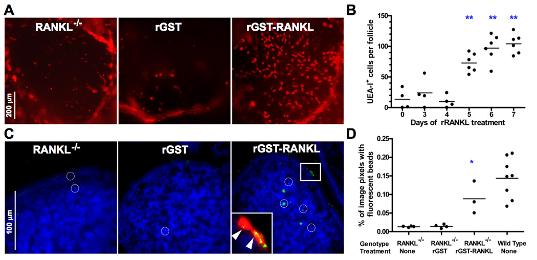
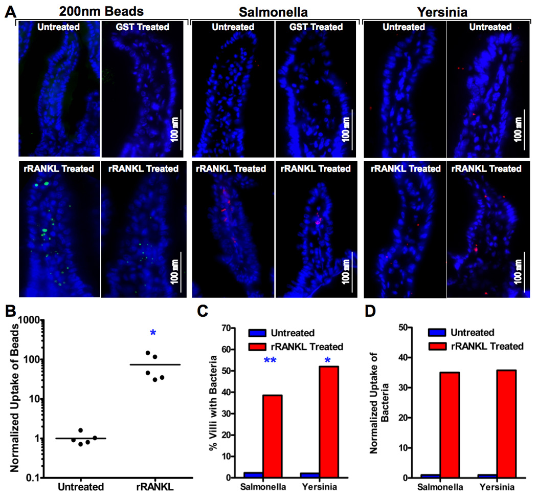
Microfold cells (M cells) are specialized epithelial cells situated over Peyer's patches (PP) and other organized mucosal lymphoid tissues that transport commensal bacteria and other particulate Ags into intraepithelial pockets accessed by APCs. The TNF superfamily member receptor activator of NF-kappaB ligand (RANKL) is selectively expressed by subepithelial stromal cells in PP domes. We found that RANKL null mice have <2% of wild-type levels of PP M cells and markedly diminished uptake of 200 nm diameter fluorescent beads. Ab-mediated neutralization of RANKL in adult wild-type mice also eliminated most PP M cells. The M cell deficit in RANKL null mice was corrected by systemic administration of exogenous RANKL. Treatment with RANKL also induced the differentiation of villous M cells on all small intestinal villi with the capacity for avid uptake of Salmonella and Yersinia organisms and fluorescent beads. The RANK receptor for RANKL is expressed by epithelial cells throughout the small intestine. We conclude that availability of RANKL is the critical factor controlling the differentiation of M cells from RANK-expressing intestinal epithelial precursor cells.


Figures

Figure 1
Figure 1
PP of RANKL null mice contain very few M cells. A, UEA-I staining reveals far fewer M cells in a representative follicle from a PP from a (C57BL/6 X BALB/c)F1 RANKL null compared to a wild type control PP. The number of M cells counted in the follicle is indicated in the lower left hand corner. The follicles shown are from the middle portion of the small intestine. Scale bar, 200 µm. B, FAE from (C57BL/6 X BALB/c)F1 RANKL null mice showed a lack of characteristic M cell features by transmission electron microscopy. The long arrowheads indicate intraepithelial pockets within the M cells of wild type FAE. The short arrowheads point to the shorter microvilli found on the apical surface of M cells. Scale bars, 50 µm. C, Scatter plot summarizing frequency of UEA-I+ M cells in individual PP follicles from mixed background RANKL null and control mice (n=5 mice for both groups). All PP examined were assigned to 1 of 5 groups based on proximal to distal position. **, p ≤ 0.001 compared to control mice by ANOVA.
Figure 2
Figure 2
Administration of rRANKL to RANKL null mice restores PP M cells. A, (C57BL/6 X BALB/c)F1 RANKL null mice were treated i.p. for 7 days with 250 µg/day of GST-RANKL or GST as a control. UEA-I staining of representative follicles from the distal small intestine shows restoration of the normal number and pattern of UEA-I+ M cells by GST-RANKL, but not by GST. Scale bar, 200 µm. B, Reconstitution of UEA-I+ M cells requires 5 days of treatment with GST-RANKL. The results are based on 3 to 6 mixed background mice at each time point and include data from all PP except the most distal PP. ** indicates p ≤ 0.001 compared to untreated mice by ANOVA. C, Uptake of 200 nm diameter fluorescent beads from isolated small intestinal loops into PP of mixed background RANKL null mice 90 minutes after bead injection is restored to near wild type levels by prior treatment with GST-RANKL for 5 days. Merged images of bead fluorescence and DAPI fluorescence from frozen sections of PP are shown with the white circles indicating the location of individual beads or clusters of beads. The inset shows a magnified image of the boxed area additionally merged with the rhodamine-UEA-I signal to show that the clusters of fluorescent beads (indicated by arrowheads) within 2 adjacent UEA-I+ M cells on the surface of the FAE. Scale bar, 100 µm. D, Summary scatter plot showing that GST-RANKL treatment reconstitutes uptake of fluorescent beads as assessed by image analysis of the percentage of pixels containing green fluorescent beads within the area of the PP follicles. *, p < 0.01 compared to untreated RANKL null mice by ANOVA.
Figure 3
Figure 3
Administration of rRANKL induces development of villous M cells on all small intestinal villi. A, Whole mount staining of villous M cells in untreated BALB/c mice and mice treated for 4 days with GST-RANKL or GST (an initial injection of 50 µg i.p. followed by 100 µg s.c. every 24 h) with rhodamine-UEA-I and DAPI. In untreated mice, a few villous M cells in a diffuse pattern are present on occasional villi. GST-RANKL treatment leads to an increased fraction of the villi having M cells and an increase in the number of M cells per villus. The fraction of villi exhibiting both the diffuse and dense patterns of villous M cell distribution increases after GST-RANKL. Scale bars, 200 µm and 500 µm. B, Summary graph showing kinetics of induction of villous M cells in the diffuse and dense patterns of distribution following GST-RANKL administration. C, Scanning electron microscopy reveals the presence of cells with a depressed surface and attenuated and blunted microvilli characteristic of M cells.
Figure 4
Figure 4
RANKL-induced villous M cells are functional for bead and bacteria uptake from the intestinal lumen. A, Wild type (C57BL/6 X BALB/c)F1 mice were treated with 100 µg of GST-RANKL or GST s.c. once a day for 4 consecutive days. On the last two days of injections, the mice and untreated controls also received 1 × 10¹¹ 200 nm fluorescent beads by gavage. One day after the last dose of GST-RANKL or GST, segments of small intestine were harvested and sectioned to check for the presence of green fluorescent beads. Alternatively, isolated small intestinal loops were prepared in anesthetized mice treated for 4 days with RANKL or GST and untreated controls and these loops were injected with paraformaldehyde-fixed Salmonella enterica serovar Typhimurium expressing DsRed-Express or Yersinia enterocolitica labeled with Alexa546. After a 2 h incubation, the tissue was harvested for frozen sections. The merged images show representative villi with DAPI positive nuclei and either green fluorescent beads or red fluorescent bacteria within the villi. B, Uptake of beads was quantitated by image analysis and normalized so that the average uptake in untreated controls was 1.0. *, p < 0.01 by Mann-Whitney test. C, The percentage of villi containing at least one bacterial organism was substantially increased in RANKL-treated mice. **, p ≤ 0.001; *, p < 0.01 (both by Fisher’s exact test). D, Uptake of bacteria was quantitated by counting individual bacteria within villi. The mean number of bacteria found per villus was normalized to a value of 1.0 for the untreated controls.
Figure 5
Figure 5
Treatment of wild type mice with neutralizing anti-RANKL leads to loss of PP M cells. A,B, BALB/c mice were treated i.p. with 250 µg of IKK22-5 mAb or an isotype control rat IgG2a mAb on days 0, 2, 4, and 6. On day 8, isolated bowel loops containing PP were injected with fluorescent beads and the mice euthanized after 90 minutes. Anti-RANKL treatment led to loss of UEA-I+ M cells detected by whole mount staining (A) and a decrease in the uptake of fluorescent beads detected on frozen sections of PP from the bead-injected loops (B). Scale bar, 200 µm in A and 100 µm in B. C, Summary of data from all PP analyzed in A and B for UEA-I+ cells and fluorescent bead uptake. D, Anti-RANKL-induced loss of UEA-I+ M cells detected by whole mount staining begins by 4 days after start of antibody treatment. ** in C and D indicates p < 0.001 compared to untreated mice by t-test (C) or ANOVA (D).
Figure 6
Figure 6
Intestinal epithelial cells express RANK. Frozen sections of a PP from a single wild type BALB/c mouse were stained with rat mAbs to mouse RANK (A, B), mouse RANKL (C), or an isotype control rat IgG2a mAb (D), followed by a biotinylated secondary antibody, streptavidin-peroxidase, and FITC-tyramide plus DAPI as a counterstain. A, RANK expression is localized to epithelial cells in the FAE and on the adjacent villi. Scale bar, 100 µm. B, Higher magnification of boxed area from A showing that RANK is present on both the apical and basolateral surfaces of the FAE. Scale bar, 250 µm. C, Reticular stromal cells concentrated immediately beneath the epithelial layer are the only cells on which RANKL is detected. Scale bar, 200 µm. D, No staining is observed with the rat IgG2a isotype control.
Figure 7
Figure 7
RANKL−/− mice have fewer germinal centers in their PP follicles and lower levels of fecal IgA. A, RANKL−/− PP on a mixed C57BL/6 and BALB/c background have fewer and less developed germinal centers identified by GL7+ cells than control PP. B, Fecal IgA concentrations (mean ± S.D.) measured by ELISA were consistently decreased in RANKL−/− mice compared to littermate controls based on analysis of samples from 5 to 16 mice of each genotype in each age range tested. *, p < 0.01; **, p < 0.001 (compared to control mice by t-test).

References

    1. Fagarasan S, Honjo T. Regulation of IgA synthesis at mucosal surfaces. Curr. Opin. Immunol. 2004;16:277–283. - PubMed
    1. Iweala OI, Nagler CR. Immune privilege in the gut: the establishment and maintenance of non-responsiveness to dietary antigens and commensal flora. Immunol. Rev. 2006;213:82–100. - PubMed
    1. Kraehenbuhl JP, Neutra MR. Epithelial M cells: differentiation and function. Annu. Rev. Cell Dev. Biol. 2000;16:301–332. - PubMed
    1. Pabst O, Bernhardt G, Forster R. The impact of cell-bound antigen transport on mucosal tolerance induction. J. Leukoc. Biol. 2007;82:795–800. - PubMed
    1. Martinoli C, Chiavelli A, Rescigno M. Entry route of Salmonella typhimurium directs the type of induced immune response. Immunity. 2007;27:975–984. - PubMed

Publication types

MeSH terms

LinkOut - more resources