. 2009 Dec;151(4):1773-89.
doi: 10.1104/pp.109.143388. Epub 2009 Oct 14.

The Arabidopsis DCR encoding a soluble BAHD acyltransferase is required for cutin polyester formation and seed hydration properties

Affiliations

The Arabidopsis DCR encoding a soluble BAHD acyltransferase is required for cutin polyester formation and seed hydration properties

David Panikashvili et al. Plant Physiol. 2009 Dec.

Abstract

The cuticle covering every plant aerial organ is largely made of cutin that consists of fatty acids, glycerol, and aromatic monomers. Despite the huge importance of the cuticle to plant development and fitness, our knowledge regarding the assembly of the cutin polymer and its integration in the complete cuticle structure is limited. Cutin composition implies the action of acyltransferase-type enzymes that mediate polymer construction through ester bond formation. Here, we show that a member of the BAHD family of acyltransferases (DEFECTIVE IN CUTICULAR RIDGES [DCR]) is required for incorporation of the most abundant monomer into the polymeric structure of the Arabidopsis (Arabidopsis thaliana) flower cutin. DCR-deficient plants display phenotypes that are typically associated with a defective cuticle, including altered epidermal cell differentiation and postgenital organ fusion. Moreover, levels of the major cutin monomer in flowers, 9(10),16-dihydroxy-hexadecanoic acid, decreased to an almost undetectable amount in the mutants. Interestingly, dcr mutants exhibit changes in the decoration of petal conical cells and mucilage extrusion in the seed coat, both phenotypes formerly not associated with cutin polymer assembly. Excessive root branching displayed by dcr mutants and the DCR expression pattern in roots pointed to the function of DCR belowground, in shaping root architecture by influencing lateral root emergence and growth. In addition, the dcr mutants were more susceptible to salinity, osmotic, and water deprivation stress conditions. Finally, the analysis of DCR protein localization suggested that cutin polymerization, possibly the oligomerization step, is partially carried out in the cytoplasmic space. Therefore, this study extends our knowledge regarding the functionality of the cuticular layer and the formation of its major constituent the polymer cutin.


Figures
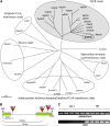
Figure 1.
Figure 1.
Phylogenetic analysis of DCR and isolation of the dcr mutant alleles. A, Unrooted phylogenetic tree of DCR orthologs from various plant species and additional BAHD family acyltransferases. The protein sequences were analyzed using ClustalA 1.81 with a PAM350 matrix. The alignment editing was performed using GeneDoc. Multiple alignment parameters were as follows: gap opening set at 10 (default), gap extension set at 2.0, and the neighbor-joining method was used for calculating the tree. The bootstrapped tree was corrected for multiple substitutions. The scale bar of 0.1 is equal to 10% sequence divergence. The phylogenetic tree was constructed using the TreeView program. Full names and accession numbers of the proteins depicted are described in Supplemental Methods S1. B, Approximate locations of the three different insertions in the DCR locus. Exons are represented by boxes, introns by single lines, and T-DNA insertions by triangles. T-DNA positions in the genomic sequence are indicated as bp downstream of the ATG start codon. C, RT-PCR analysis of the DCR transcript in inflorescence tissues of dcr-1 and wild-type (WT) plants (Nossen). Each lane represents an individual biological replicate. The UBIQUITIN-C gene served as a control for equal cDNA loading. [See online article for color version of this figure.]
Figure 2.
Figure 2.
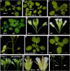
Light microscopy images of different dcr mutant allele phenotypes. A, Phenotypes of 1-month-old dcr-1 versus wild-type (WT; Nossen) plants. B, Phenotypes of 1-month-old dcr-2 and dcr-3 versus wild-type (Col-0) plants. C, Five-week-old dcr-1 plant displaying postgenital fusion between rosette leaves. The site of the fusion is indicated by an arrow. D, TB staining of dcr-1 and wild-type rosettes. E and F, TB staining of dcr-1 and wild-type inflorescences. G, Fusion between flower buds in the dcr-1 inflorescence. H and I, TB staining of a dcr-1 plant complemented with the 35S:DCR construct (H) versus the wild type (I). J, TB staining of dcr-1 versus dcr-1 complemented with the 35S:DCR construct (blue staining in petals is indicated by arrows). K, Flower phenotype of dcr-2 versus the wild type. Folded petals are indicated by arrows. L, Semisterility (indicated by arrows) is obvious in the dcr-1 plant compared with the wild type.
Figure 3.
Figure 3.
Electron microscopy images of dcr mutant epidermal cells. A and B, SEM images of the leaf epidermal cell pattern in dcr-1 (A) and the wild type (WT; Nossen; B). C, A collapsed trichome in dcr-1. The site of collapse is indicated by an arrow. D, A wild-type trichome. E, TEM image of a dcr-1 leaf epidermis. A discontinuous cuticle (cut) is indicated by arrows. F, TEM image of wild-type leaf epidermis. An intact cuticle is indicated by an arrow. G, SEM image of a dcr-1 flower with fused sepals. H, Enlarged image of the sepal fusion area boxed in G. I, dcr-1 sepal adaxial epidermis. Fractures in the epidermis are indicated by arrows. J, Wild-type sepal adaxial epidermis layer. K, dcr-1 sepal abaxial epidermis layer. L, Wild-type sepal abaxial epidermis layer.
Figure 4.
Figure 4.
Electron microscopy images of dcr mutant petals. A, SEM image of dcr-1 petal abaxial epidermis layer. B, SEM image of wild-type (WT; Nossen) petal abaxial epidermis layer. C and D, Enlarged images of abaxial petal epidermal cells of dcr-1 (C) and the wild type (D). Note the cuticular foldings on the surface of wild-type cells (spaghetti-like structures) that are absent in the dcr-1 cells. E, SEM image of dcr-1 petal adaxial epidermis. F, SEM image of wild-type petal adaxial epidermis. G and H, Enlarged images of adaxial petal epidermal cells of dcr-1 (G) and the wild type (H). I and J, TEM images of dcr-1 (I) and wild-type (J) petals. The cuticular foldings in wild-type petals (J) are indicated by black arrows (absent in the mutant cells). K and L, Enlarged images of dcr-1 (K) and wild-type (L) petal abaxial epidermal cells. Note the presence of cuticular foldings in the wild type (black arrows).
Figure 5.
Figure 5.
Seed surface phenotypes in the dcr mutants. A and B, TB staining of dcr-1 (A) and wild-type (WT; Nossen; B) developing seeds. C and D, Shrunken and fused seeds of dcr-1 (C) versus the wild type (D). E and F, SEM pictures of dcr-1 (E) and wild-type (F) mature seeds. G and H, Confocal microscopy pictures of dcr-1 (G) and wild-type (H) mature seeds stained with safranin O (a cell wall-specific dye). H, Visualized columella cells in wild-type seed are marked by small circles. I and J, Water imbibition of dcr-1 (I) and wild-type (J) seeds. Protruded columella cells in wild-type seeds are indicated by arrows. K to M, TEM images of dcr-1 (K and L) and wild-type (M) seed coats. L shows an enlarged image of the boxed region in K; a cuticle-like densely stained structure is indicated by black arrows. The columella cells are marked in K and M by white arrows. N and O, Mucilage release in seeds stained with ruthenium red in dcr-1 (N) and the wild type (O). P and Q, Mucilage release in 0.05 m EDTA-pretreated seeds stained with ruthenium red in dcr-1 (P) and the wild type (Q).
Figure 6.
Figure 6.
Sterility and altered seed characteristics in the dcr mutants. A, Defective male gametophyte in the dcr-1 mutant line. Reciprocal backcrosses show that a normal number of seeds is formed in cases of crossing dcr-1 with wild-type (WT; Nossen) pollen and a significant reduction in seed number per silique when dcr-1 pollen is used for crossing. At least three flowers were used from each genotype for reciprocal crosses. B, Average number of seeds in siliques of dcr-1 and wild-type plants. Seeds from three to five siliques from three to four plants from each genotype were collected for seed counting. C, Weight of 100 seeds of dcr-1 and the wild type. D, Seed width measurement of dcr-1 and the wild type. E, Seed length measurement of dcr-1 and the wild type. Seed pools derived from four to five plants of each genotype were used in C, D, and E. F, Percentage of nongerminating seeds of dcr mutant alleles and the corresponding wild-type ecotypes (Nossen [Nos.] and Col-0) under water-limiting conditions (10% polyethylene glycol 8000). The values indicate means of three biological replicates ± sd. * P < 0.05.
Figure 7.
Figure 7.
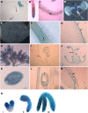
Spatial and temporal expression of a DCR promoter-GUS reporter in Arabidopsis tissues. A, Reporter-GUS expression in a 3-d-old seedling. The expression in a lateral root emergence site and the root cap region is indicated by black arrows. B and C, Close-up views of a root cap region (B) and a lateral root emergence site (C). D, Reporter-GUS expression in a 15-d-old seedling. The expression in the emerging leaves and lateral roots is indicated by arrows. E and F, In the mature leaves (E) and stems (F), GUS expression is clearly observed in trichomes. G, Epidermis expression in the elongating stem. H, GUS expression in inflorescence. I, GUS expression is epidermis specific in anther filaments and sepals (indicated by arrows). J, Expression in anthers and pollen grains (indicated by an arrow). K, Whole mount view of seed-specific GUS expression. L, Torpedo stage seed GUS expression. The outer and inner integumenta layers are indicated by a bracket. M, Bent cotyledon stage seed GUS expression. Expression in the inner integumenta is indicated by an arrow. N, GUS expression in different embryonic developmental stages: i, torpedo; ii, walking stick; iii, bent cotyledon.
Figure 8.
Figure 8.
Subcellular localization of the DCR protein. A to D, Confocal microscopy images through Arabidopsis leaf epidermal cells and protoplasts expressing 35S:DCR-GFP. The images were acquired through a GFP filter (A), chlorophyll filter (B), transmission filter (C), and a merge between GFP, chlorophyll, and transmission filters (D). E and F, A confocal series showing DCR-GFP localization in a protoplast derived from a transgenic plant expressing DCR-GFP. The images with GFP were acquired through a GFP filter (E) and with the plasma membrane marker FM4-64 through a chlorophyll filter (F). G to J, A confocal series showing DCR-GFP localization in Arabidopsis cell culture protoplasts transformed with DCR-GFP (G and I) and with ER (H) and Golgi (J) markers. Golgi stacks are indicated by white arrows.
Figure 9.
Figure 9.
The lipid polyester profiles of dcr-1 and the wild type (WT; Nossen) in flowers and seed coats after BF3 depolymerization. A, The flower cutin polyester profile. Differential monomers are indicated by arrows. B, The seed coat polyester profile. Differential monomers are indicated by arrows. The values indicate means of four biological replicates ± sd. * P < 0.05. For the full names of identified monomers, see Supplemental Table S5. DW, Dry weight.
Figure 10.
Figure 10.
The leaf cutin profile of dcr-1 and the wild type (Nossen) after MeOH/HCl depolymerization. Differential monomers are indicated by arrows. The values indicate means of four biological replicates ± sd. * P < 0.05. For the full names of identified monomers, see Supplemental Table S5. n.d., Not detected. White bars, Ecotype Nossen; black bars, dcr-1.
Figure 11.
Figure 11.
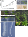
Water loss, root architecture, and tolerance to various stress conditions of the dcr mutants. A, Rate of water loss of dcr-1, dcr-2, and wild-type (WT) plants. Four rosettes per genotype were weighed during the time intervals depicted. The results are derived from three independent experiments and depicted with se of the mean for each time point. The values indicate means of three biological replicates ± se. * P < 0.05. ec. Nos., Ecotype Nossen; ec. Col., ecotype Col-0. B, Salt and mannitol stress experiments. Six-day-old dcr-1 and wild-type (Nossen) seedlings grown on MS agar plates were transferred to MS plates supplemented with 200 mm NaCl or 400 mm mannitol for an additional 2 weeks of growth. The results were documented 6 d after application of the salt stress and 15 d of osmotic stress. C, Water deprivation tolerance experiment. Three-week-old dcr-1, dcr-2, and wild-type plants were exposed to 10 d of water deprivation. Subsequently, seedlings were rewatered once and their appearance was documented after 1 week. D, Increased chlorosis and death in rosette leaves of 8-week-old dcr-1 plants grown in normal laboratory conditions as compared with wild-type plants (damaged leaves marked by arrows). E and F, Increased lateral roots (E) and hair formation (F) in dcr-1 plants compared with the wild type (Nossen).

References

    1. Aharoni A, Dixit S, Jetter R, Thoenes E, van Arkel G, Pereira A (2004) The SHINE clade of AP2 domain transcription factors activates wax biosynthesis, alters cuticle properties, and confers drought tolerance when overexpressed in Arabidopsis. Plant Cell 16 2463–2480 - PMC - PubMed
    1. Baum SF, Rost TL (1996) Root apical organization in Arabidopsis thaliana. 1. Root cap and protoderm. Protoplasma 192 178–188
    1. Beeckman T, De Rycke R, Viane R, Inzé D (2000) Histological study of seed coat development in Arabidopsis thaliana. J Plant Res 113 139–148
    1. Benitez JJ, Heredia-Guerrero JA, Serrano FM, Heredia A (2008) The role of hydroxyl groups in the self-assembly of long chain alkylhydroxyl carboxylic acids on mica. J Phys Chem C 112 16968–16972
    1. Bird D, Beisson F, Brigham A, Shin J, Greer S, Jetter R, Kunst L, Wu X, Yephremov A, Samuels L (2007) Characterization of Arabidopsis ABCG11/WBC11, an ATP binding cassette (ABC) transporter that is required for cuticular lipid secretion. Plant J 52 485–498 - PubMed

Publication types

MeSH terms