. 2009 Dec 10;114(25):5225-35.
doi: 10.1182/blood-2009-05-220400.

Evidence for a novel human-specific xeno-auto-antibody response against vascular endothelium

Affiliations

Evidence for a novel human-specific xeno-auto-antibody response against vascular endothelium

Tho Pham et al. Blood. .

Abstract

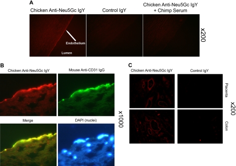
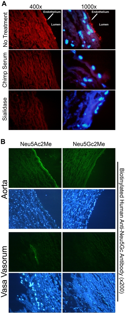
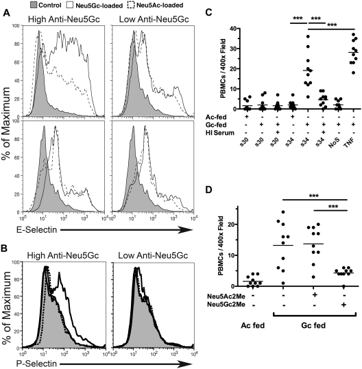
Humans are genetically unable to synthesize the common mammalian sialic acid N-glycolylneuraminic acid (Neu5Gc). However, Neu5Gc can be metabolically incorporated and covalently expressed on cultured human cell surfaces. Meanwhile, humans express varying and sometimes high titers of polyclonal anti-Neu5Gc antibodies. Here, a survey of human tissues by immunohistochemistry with both a monospecific chicken anti-Neu5Gc antibody and with affinity-purified human anti-Neu5Gc antibodies demonstrates endothelial expression of Neu5Gc, likely originating from Neu5Gc-rich foods like red meats. We hypothesized that the combination of Neu5Gc incorporation and anti-Neu5Gc antibodies can induce endothelial activation. Indeed, the incubation of high-titer human sera with Neu5Gc-fed endothelial cells led to Neu5Gc-dependent antibody binding, complement deposition, endothelial activation, selectin expression, increased cytokine secretion, and monocyte binding. The proinflammatory cytokine tumor necrosis factor-alpha also selectively enhanced human anti-Neu5Gc antibody reactivity. Anti-Neu5Gc antibodies affinity-purified from human serum also directed Neu5Gc-dependent complement deposition onto cultured endothelial cells. These data indicate a novel human-specific mechanism in which Neu5Gc-rich foods deliver immunogenic Neu5Gc to the endothelium, giving anti-Neu5Gc antibody- and complement-dependent activation, and potentially contributing to human vascular pathologies. In the case of atherosclerosis, Neu5Gc is present both in endothelium overlying plaques and in subendothelial regions, providing multiple pathways for accelerating inflammation in this disease.


Figures

Figure 1
Figure 1
Detection of Neu5Gc in aortic endothelium of human autopsy samples and microvasculature of colon and placenta. The chicken anti-Neu5Gc antibody (cGcAb) was used to detect the presence of Neu5Gc on the endothelium of autopsy samples of normal-appearing human aorta. Typical representatives of 8 autopsy samples studied are shown. The red Cy3 fluorescence represents labeling of endothelial cells of the aorta. (A) Specificity of the antibody was demonstrated by the lack of signal with the nonimmunized control chicken IgY (middle) and the abrogation of signal by adsorption with Neu5Gc-rich glycoproteins of chimpanzee serum (right). Magnification ×200. (B) Sections were double-stained with anti-CD31 for endothelial cells and counterstained with DAPI to visualize nuclei (magnification ×1000). (C) Sections of placenta (top) and colon (bottom) stain for Neu5Gc along microvasculature endothelial lining with the use of cGcAb. Control IgY (right) demonstrates specificity of signal (magnification ×200).
Figure 2
Figure 2
Affinity-purified human anti-Neu5Gc antibodies bind specifically to Neu5Gc on endothelial cells of aorta sections and vasa vasorum. Affinity-purified human anti-Neu5Gc antibodies were used as a primary reagent on cryosections of autopsy samples of aorta. (A) Sections were incubated with the biotinylated, affinity-purified human anti-Neu5Gc antibodies followed by rabbit anti-biotin and then Cy3-anti-rabbit, with nuclear counterstaining using DAPI (top). Antibody staining was abrogated by adsorption with chimpanzee serum (middle) or by previous sialidase pretreatment (bottom). A representative example of 5 independent experiments is shown. (B) Sections were incubated with biotinylated, affinity-purified human anti-Neu5Gc antibodies followed by mouse anti-biotin and FITC antimouse antibody and nuclear counterstaining with DAPI. Primary antibody staining was abrogated by addition of Neu5Gc2Me but not by Neu5Ac2Me. The staining pattern is appreciated in both aorta and vasa vasorum. A representative example of 3 independent experiments on 8 individuals is shown (magnification ×200).
Figure 3
Figure 3
Human serum antibodies react against Neu5Gc-loaded endothelium. Human sera (S#), diluted 1:5, were assayed for their ability to bind HUVECs in a Neu5Gc-dependent manner. (A) Binding of human serum IgG (top) or IgM (bottom) to loaded HUVECs was assayed by flow cytometry. Neu5Gc-loaded cells (solid line), Neu5Ac-loaded cells (dotted line), and Neu5Gc-loaded cells stained with secondary antibody only (shaded curve). Representative individual human sera are shown. (B) IgG and IgM binding were normalized as a percentage value of the signal obtained with S34. The data are presented as mean ± SEM (n = 3). (C) Binding of anti-Neu5Gc IgG antibodies from S37 to Neu5Gc-loaded HUVEC was inhibited with the alpha-methyl glycoside, Neu5Gc2Me, in a dose-dependent manner and not by Neu5Ac2Me. (D) Classical complement, C3 convertase (C3b), was deposited onto Neu5Gc-loaded HUVEC (solid lines) only when incubated with high-titer anti-Neu5Gc serum (S34, right) and not low-titer anti-Neu5Gc serum (S30, left). Nonspecific complement was deposited onto Neu5Ac-loaded HUVEC (dotted lines) in a variable manner dependent on which sera was used, presumably attributable to unrelated serum factors. As a negative control, heat-inactivated serum was incubated with Neu5Gc-loaded HUVECs as a negative control (shaded curve). (E) Heat-inactivated S34 alone did not deposit complement in Neu5Gc-loaded HUVECs (shaded curve). Supplementation of S30 with heat-inactivated S34 shows Neu5Gc-dependent complement deposition in Neu5Gc-loaded (solid line) and not Neu5Ac-loaded HUVECs (dashed line). This finding indicates that anti-Neu5Gc antibodies are necessary and sufficient to mediate Neu5Gc-dependent complement deposition. All results presented in this figure were performed at least 3 times.
Figure 4
Figure 4
Human serum enhances adhesion molecule expression and PBMC binding on Neu5Gc-loaded endothelium. (A) A representative example of surface E-selectin expression determined on Neu5Ac and Neu5Gc-loaded HUVECs after 5 hours of activation by low-titer serum (S30, right) or high-titer serum (S34) or heat-inactivated forms thereof (bottom). Control peaks (shaded curve) represent E-selectin expression on endothelial cells not activated by serum. (B) As described in panel A, except HUVECs were exposed to serum for 15 minutes only. Heat-inactivated serum controls are included as a negative control (shaded curve). Short serum incubations show a Neu5Gc-dependent expression of P-selectin that requires anti-Neu5Gc antibodies and is sensitive to heat inactivation of serum. This response was most evident in low passage HUVECs (passage # ≤ 3), consistent with the observed (data not shown) and reported passage-dependent loss of P-selectin expression in vitro. (C) HUVECs were cultured on 12-mm glass coverslips (Fisher) in 24-well plates and loaded with either Neu5Ac or Neu5Gc as described previously. Loaded cells were incubated with 50% human sera in EBM-2 for 4 hours at 37°C or with heat-inactivated serum as a control. A negative control was not exposed to human serum (NoS) and a positive control was exposed to TNF-α (10 ng/mL) for stimulation. At 4 hours, 105 PBMCs were added to each well and incubated at 37°C on an orbital shaker at 100 revolutions/minute for 1 hour. Cells were washed thrice in PBS and fixed in PBS containing 3% paraformadehyde. Bound PBMCs were counted in 10 randomly chosen fields at ×400 magnification. Data were averaged and expressed as cells/field. Data are presented as mean with the scatter. This panel is a representative example of 5 similar replicates. ***P < .001. (D) Same as panel C except that the α-methyl-glycosides (1mM) were incubated with the S34 before HUVEC stimulation. All groups were Neu5Gc loaded. We observed inhibition of PBMC binding in S34-stimulated HUVECs with Neu5Gc2Me but not Neu5Ac2Me. ***P < .001. Examples of fluorescent images from the experiment in panel D are shown in supplemental Figure 1. All results presented were performed at least 3 times.
Figure 5
Figure 5
Neu5Gc in the endothelium and subendothelium of atherosclerotic plaques. (A) Expression of Neu5Gc in endothelium overlying human aortic atherosclerotic plaques is shown by double labeling with the cGcAb anti-Neu5Gc antibody (left) the endothelial marker CD31 (center, see merge, right). White arrows indicate subendothelial Neu5Gc within lesions (left). (B) Samples of human aortic atherosclerotic plaques were identified by their characteristic appearance (hematoxylin and eosin, bottom right). Accumulation of macrophages (stained with anti-CD68, top middle) and oxidized-LDL (stained with malondialdehyde, top right) are shown. The cGcAb (top left) was used to detect the presence of Neu5Gc on the endothelium of the plaques. Control IgY and mouse IgG (bottom right and middle) demonstrates specificity of the staining. (C) Human aortic atherosclerotic plaque sections were double-stained with anti-CD68 (middle) for macrophages and cGcAb (left) for Neu5Gc. The merged fluorescent image (right) indicates that macrophages are recruited to the Neu5Gc-lined endothelium. (D) Endothelium was labeled with anti-VWF antibody (center; note that there is also some subendothelial green auto-fluorescence caused by macrophages and/or necrotic foci). In addition to typical endothelial-colocalized Neu5Gc staining, extensive subendothelial labeling with the cGcAb anti-Neu5Gc antibody (red channel, left) is seen in this lesion (see yellow in the merged image, right). All pictures in this figure are representative examples of multiple independent analyses on multiple samples from multiple individuals.
Figure 6
Figure 6
TNF-α augments human antibody reactivity against Neu5Gc-loaded endothelium. Endothelial cells were treated with TNF-α (10 ng/mL) during the last 18 hours of Neu5Gc loading. Antibody binding was assessed for both treated and nontreated cells after incubating with high-titer human serum (S34). Control peaks represent staining with FITC-conjugated secondary antibody alone. The experiment was performed multiple times and representative examples are shown. Several other cytokines studied (IL-1β, IL-4, IL-6, IL-13, and IFNγ) did not show this enhancing effect (not shown).
Figure 7
Figure 7
Affinity-purified human anti-Neu5Gc antibodies bind specifically to Neu5Gc-loaded endothelial membrane glycoproteins and induce complement deposition. Affinity-purified human anti-Neu5Gc antibodies were used as a primary. (A) Biotinylated, affinity-purified human anti-Neu5Gc antibodies were studied by Western blotting against surface membrane glycoproteins of Neu5Ac (Ac)– or Neu5Gc (Gc)–loaded endothelial cells, and the banding pattern was compared with that generated with the cGcAb antibody. HUVECs loaded with either Neu5Ac or Neu5Gc were lifted with 100mM EDTA and sonicated. Membrane fractions were prepared, 3 μg denatured by boiling in SDS separated on 12.5% polyacrylamide mini gels (Bio-Rad), and electrotransferred to nitrocellulose membranes. Membranes were blocked overnight at 4°C with 0.5% Neu5Gc-free cold water fish gelatin (Sigma-Aldrich) in TBST. Primary antibodies were incubated for 5 hours at 4°C with either biotinylated anti-Neu5Gc antibodies purified from individual human sera, diluted 1:100 in TBST, or chicken anti-Neu5Gc antibody (cGcAb), diluted 1:50 000 in TBST. Purification of the cGcAb is described elsewhere. Membranes were washed 4 times for 5 minutes in TBST then incubated with Streptavidin-HRP (Bio-Rad), diluted 1:50 000, or HRP-anti-chicken-IgY (Jackson ImmunoResearch Laboratories), diluted 1:10 000, at room temperature for 45 minutes. Proteins were visualized by chemiluminescence detection (Pierce), followed by exposure to Kodak BioMax XAR film for 5 to 30 seconds. A representative example from 3 independently performed experiments is shown. (B) Anti-Neu5Gc IgG binding (x-axis) and complement deposition (y-axis) were simultaneously assessed on Neu5Ac- and Neu5Gc-loaded endothelium. Affinity-purified anti-Neu5Gc antibodies, low-titer human serum (S30), or both were incubated with Neu5Gc-loaded (top) or Neu5Ac-loaded (bottom) HUVECs. Although the low-titer human serum does not bind HUVECs in a Neu5Gc-dependent fashion (left), the affinity-purified anti-Neu5Gc antibodies are necessary and sufficient to bind Neu5Gc-loaded HUVECs in a Neu5Gc-dependent fashion (middle). Supplementing the low-titer serum with the purified anti-Neu5Gc antibodies allows complement deposition on HUVECs in a Neu5Gc-dependent manner (right). A representative result of 3 independent experiments is shown.

References

    1. Varki A. Glycan-based interactions involving vertebrate sialic-acid-recognizing proteins. Nature. 2007;446(7139):1023–1029. - PubMed
    1. Hayakawa T, Satta Y, Gagneux P, Varki A, Takahata N. Alu-mediated inactivation of the human CMP-N-acetylneuraminic acid hydroxylase gene. Proc Natl Acad Sci U S A. 2001;98(18):11399–11404. - PMC - PubMed
    1. Hedlund M, Tangvoranuntakul P, Takematsu H, et al. N-glycolylneuraminic acid deficiency in mice: implications for human biology and evolution. Mol Cell Biol. 2007;27(12):4340–4346. - PMC - PubMed
    1. Diaz SL, Padler-Karavani V, Ghaderi D, et al. Sensitive and specific detection of the non-human sialic acid N-glycolylneuraminic acid in human tissues and biotherapeutic products. PLoS ONE. 2009;4(1):e4241. - PMC - PubMed
    1. Bardor M, Nguyen DH, Diaz S, Varki A. Mechanism of uptake and incorporation of the non-human sialic acid N-glycolylneuraminic acid into human cells. J Biol Chem. 2005;280(6):4228–4237. - PubMed

Publication types

MeSH terms